




No CrossRef data available.
Article contents
Comparison of four serological tests for the diagnosis of Chagas disease in a Colombian endemic area
Published online by Cambridge University Press: 14 September 2004
Abstract
The performance of 4 serological tests for the diagnosis of Chagas disease was evaluated in Santander, Colombia, a region still presenting active transmission. Serum samples from 638 individuals were submitted to an enzyme immunoassay test (EIA), using total lysate of a local Trypanosoma cruzi strain and 52·5% were positive (335/638). A subset of this group (94 positive individuals and 90 seronegatives) was randomly selected for further serological confirmation. Three additional tests were used – indirect immunofluorescence (IIF) and 2 distinct enzyme-linked immunosorbent assays using total lysate of the Y strain (EIA BM) and a mixture of 2 recombinant antigens (EIA RA). Seventy-nine patients were seropositive in all tests (84·0% – 79/94). The number of positive sera with the IIF, EIA RA and EIA BM was 84/94 (89·4%), 80/94 (85·1%) and 79/94 (84·0%), respectively. In 15 out of the 94 EIA seropositive patients (16·0%), 10 individuals were negative in all 3 tests (10·6% – 10/94). One was negative in the EIA BM and positive in the other two tests (1·1% – 1/94) and 4 patients were positive, solely, in the IIF assay (4·3% – 4/94). Relative to the 90 EIA negative individuals, 89 were confirmed in all other tests (98·9% – 89/90). One individual, although seronegative in the IIF, was positive in both confirmatory EIA tests (1·1% – 1/90). In addition, 120 blood specimens were submitted to PCR amplification. This group consisted of 79 confirmed seropositive cases, 16 individuals with discordant serological results and 25 validated seronegative individuals. The PCR was able to detect the presence of parasite DNA in 67 out of the 79 seropositive patients (84·8%), in 8 individuals with discordant serology (50·0%) and in only one seronegative individual (4·0%). The results pointed to the necessity for performing more than one serological test, preferentially with antigens from autochthonous strains, to achieve a reliable diagnosis of Chagas disease in Colombia.
Information
- Type
- Research Article
- Information
- Copyright
- 2004 Cambridge University Press
INTRODUCTION
The initiative with the most impact on the control of Chagas disease was launched some years ago under the World Health Organization umbrella (WHO, 1996). Interruption of active transmission, promoted both by the elimination of domiciled vectors and control detection in blood-banks, was possible due to the Southern Cone, Andean and Central America control programme supervised by WHO in combination with governmental institutions. The Southern Cone countries were the most successful. Vector and transfusion transmissions of the disease have been controlled in Brazil, Chile and Uruguay (Moncayo, 1997).
Colombia still presents Chagas disease as an endemic entity in 15 out of the 33 Departments. The number of chagasic patients is around 3 million individuals in the country and 20% of the population is at risk of acquiring the infection, if the geographical distribution and anthropophily of the vectors are considered (Guhl, 1999). The vector is Rhodnius prolixus, and the contact with humans is favoured by the low-quality human habitations where the insect colonizes (Angulo, Muñoz & Tarazona, 1993a).
Despite of the fact that Chagas disease is a major public health problem in Colombia, studies in endemic foci are scarce. The knowledge of epidemiological and clinical features may lead to better control activities and consequently, the elimination of active transmission by vectors and blood transfusion, the two most important infection transmission mechanisms (Moncayo, 1997).
The aforementioned was the stimulus to analyse the performance of 4 serological tests using distinct Trypanosoma cruzi antigens, in a population group of Santander, Colombia, a region that has the highest prevalence rates of Chagas disease in the country, with ongoing active transmission (Guhl, 1999). The comparison of 4 methodologies contributes to an understanding of specificities of the serological tests to be used in the Colombian settings in future sero-epidemiological surveys. In addition, polymerase chain reaction (PCR) amplification of a segment of the T. cruzi kinetoplast genome (kDNA) was utilized to detect parasites in the blood of serum donors in the trial.
MATERIALS AND METHODS
Definition of the studied population
The studied area is composed of 3 neighbouring municipalities situated in the Northeast Colombia, in the Department of Santander – Mogotes, SanJoaquin and Gambita (Fig. 1). In these districts, the inhabitants were visited and medical assistance supplied. The criteria used for a patient's inclusion in the study were (i) individuals older than 7 years and (ii) had lived in the area for at least the last 5 years. This investigation was submitted and approved by the ethical committee of the Universidad Industrial de Santander.

Fig. 1. Map of Colombia showing the 3 localities studied in the Department of Santander (1. Mogotes; 2. SanJoaquin; 3. Gambita).
Serology
Enzyme-linked immunosorbent assay CINTROP
Total lysate of the T. cruzi Gonzalo strain, a local stock characterized by enzyme electrophoresis profile as zymodeme 1 in CIDEIM (International Center for Medical Research and Training), Cali, Colombia, was used as antigen for the enzyme immunoassay carried out in Colombia (EIA CINTROP). Microplates, previously sensitized with 30 μg/well of crude antigens at 4 °C overnight, were incubated with 100 μl of diluted sample serum (1/200) in phosphate-buffered saline (PBS) at 37 °C for 2 h. For the removal of unbound antibodies, washes were performed with PBS containing 0·01% of Tween 20 (PBS-T20). The plates were incubated for 1 h at 37 °C with 100 μl of alkaline phosphatase (Sigma®) conjugates. The immune complexes were revealed by the addition of 100 μl of substrate (4-p-nitrophenylphosphate, Sigma®). Optical density (OD) was determined at 405 nm. The cut-off value (CO) was the average OD more than 2 standard deviations, obtained from a serological survey among inhabitants of a non-endemic area for Chagas disease (CO=0·35). Sera with values equal or greater than the established CO were considered reactive.
Immunofluorescence
Indirect immunofluorescence (IIF) assays were performed by conventional techniques (Camargo, 1992) using formaldehyde-fixed epimastigote forms of the T. cruzi Y strain. Serum dilutions of 1/40 or higher, presenting fluorescence signals, were considered reactive.
Enzyme-linked immunosorbent assays: BioManguinhos (EIA BM) and CRA/FRA (EIA RA)
Two commercial EIA kits differing in the antigen used and in the immune complex detection methods were used: EIA Biomanguinhos (EIA BM) using total lysate of the T. cruzi Y strain and EIA-Recombinant Chagas Biomanguinhos (EIA RA) using a mixture of 2 recombinant antigens cloned from a Y strain expression library – cytoplasmic repetitive antigen (CRA) and flagellar repetitive antigen (FRA) (Lafaille et al. 1989; Krieger et al. 1992).
The EIA BM assay was prepared essentially as described by Voller et al. (1975), where sensitized wells were incubated with patient sera at a 1[ratio ]100 dilution. For the EIA RA, the sensitized plates were incubated with 50 μl of undiluted patient sera for 30 min at 37 °C, according to the method of Gomes et al. (2001). The reactions and the CO calculation for each plate, as well as the grey zone, were performed following the manufacturers' instructions. Sera with OD values equal or greater than the CO were considered reactive.
DNA extraction and PCR
Five ml of whole blood were mixed with the same volume of 6 M guanidine hydrochloride in a 200 mM EDTA solution, pH 8·0, immediately after its collection (Avila et al. 1991). After mixing, the tubes were kept at room temperature until used. For decatenation of the mini-circle molecules from the kinetoplast network, the blood lysates were boiled for 15 min (Britto et al. 1993). A sample of 200 μl of the mixture was submitted to phenol[ratio ]chloroform extraction, followed by DNA precipitation with the aid of sodium acetate and ethanol. The DNA pellet was resuspended in 50 μl of TE buffer (10 mM Tris–HCl, pH 8·0; 1 mM EDTA) and stored at −20 °C.
A hot-start PCR to allow amplification of a 330 bp fragment corresponding to the T. cruzi mini-circle variable regions was performed using primers [#121: 5′AAATAATGTACGGG(T/G)GAGATGCATGA3′ and #122: 5′GGTTCGATTGGGGTTGGTGTAATATA3′] (Sturm et al. 1989; Wincker et al. 1994). The amplification reactions were achieved in a volume of 50 μl, consisting of 5 μl of the 10×TaqPlatinum buffer (100 mM Tris–HCl, pH 8·3; 500 mM KCl), 4 μl of a dNTP mixture (200 μM of each), 8 μl of a 25 mM MgCl2 solution, 120 pmol of each primer, 2·5 units of TaqPlatinum (Invitrogen Life Technologies) and 5 μl of the DNA sample. The PCR was carried out using a DNA Thermal Cycler 9600 (Applied Biosystems) using the following conditions: 1 cycle at 94 °C for 1 min, 2 cycles at 98 °C for 1 min and 64 °C for 1 min, 33 cycles at 94 °C for 1 min and 64 °C for 1 min, and a final extension at 72 °C for 10 min.
The same protocol was used for amplification of the human β-globin gene sequences. The primers used were PCO3: 5′ACACAAACTGTGTTCACTAGC3′ and PC04: 5′CAACTTCATCCACGTTTTCACC3′ (Saiki et al. 1985). The amplified products were submitted to a 2% agarose gel electrophoresis and visualized under UV light after ethidium bromide staining.
RESULTS
Population studied and screening serology
In total, 638 individuals were enrolled in the study and submitted to the EIA CINTROP assay, revealing positive results in 52·5% (335/638). A subset of this group (n=184 – 94 seropositive and 90 seronegative) was selected for further serological examination using 3 additional tests: (i) IIF, (ii) EIA BM and (iii) EIA RA.
Confirmatory serology
Seventy-nine patients were seropositive in all 3 serological tests (79/94 – 84%). A strong correlation could be observed among the results of the tests (Kappa index >0·80). The remaining seropositive individuals in the EIA CINTROP (n=15) revealed discordant results in the confirmatory tests: 10 individuals were seronegative in all 3 serological assays (10/94 – 10·6%); 4 patients were positive, solely, in the IIF test (4/94 – 4·3%) and 1 individual was negative in the EIA BM and positive in the other 2 confirmatory tests (1/94 – 1·1%) (Fig. 2). The rates of positive results in the IIF test, EIA RA and EIA BM relative to the 94 EIA CINTROP seropositive patients were, 84/94 (89·4%), 80/94 (85·1%), and 79/94 (84%), respectively (Table 1, Fig. 2).

Fig. 2. Venn-Euler Diagram summarizing the different results obtained with the 4 serological tests performed. The square corresponds to the universe of positive individuals in the ELISA CINTROP (n=94) and the circles, to the confirmatory serological tests. The intersection regions indicate co-positivity. The presented numbers represent positive individuals and the numbers in parentheses correspond to positive PCR results.

Of 90 seronegative individuals in the EIA CINTROP, 89 were also negative in all 3 other tests (89/90 – 98·9%). However, although being negative in the IIF, 1 individual (1/90 – 1·1%) was positive in both confirmatory EIA assays (Table 1).
PCR results
The PCR aimed to detect circulating T. cruzi DNA. Amplified products were detected in 67 out of the 79 seropositive patients (67/79 – 84·8% – Table 2, Fig. 2). The results obtained with the subset of 16 patients that presented discordant results in at least one of the 4 serological tests, revealed positive PCR results in 8/16 (50% – Table 2). In this group, the only individual that was EIA CINTROP and IIF negative and positive in both confirmatory EIA tests, was PCR negative. In addition, 25 seronegative individuals confirmed by the 4 serological assays were also submitted to the molecular test. One of them (1/25 – 4·0%) gave a positive PCR result (Table 2).
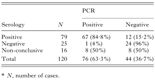
It is important to emphasize that (i) 5 out of 10 individuals that gave positive serological results only in the EIA CINTROP were PCR positive (5/10 – 50%), (ii) 2 out of 4 individuals that were EIA CINTROP and IIF positive and negative in both other EIA tests were PCR positive (2/4 – 50%) and (iii) one patient that was EIA CINTROP, EIA RA and IIF positive and negative in the EIA BM was also PCR positive (Fig. 2).
All samples that were PCR negative for T. cruzi sequences were further tested with primers that amplify a fragment of the human β-globin gene, following the same experimental conditions. The last assay was positive in all cases, demonstrating that the DNA preparation did not present any kind of inhibitor to the reaction.
DISCUSSION
Serological diagnosis of Chagas disease is still a problem. Some of the serological commercial kits for several Latin American countries do not provide reliable results. The commonly used assays (non-recombinant antigens) render false-positives that led to the World Health Organization proposal that at least 2 distinct tests be performed before making a final serodiagnosis. Furthermore, T. cruzi presents 2 major genotypes influencing the antigenic composition of the parasite and consequently in the recognition of epitopes by the patient's antibodies (Andrade et al. 1981; Di Noia et al. 2002; Buscaglia & Di Noia, 2003). The vast majority of commercial serological tests for Chagas disease uses either crude or purified extracts, or even recombinant antigens from T. cruzi II, leading to the possibility of false-negative results in areas where T. cruzi I predominates (Coura et al. 2002).
Colombia faces difficulties in defining the real chagasic patients due to (i) the lack of commercial tests using T. cruzi I antigens, the prevailing parasite genotype, and (ii) the possibility of false-positive results due to cross-reaction with Trypanosoma rangeli that geographically overlaps with T. cruzi (Guhl et al. 1985, 1987; Saldaña & Sousa, 1996; Cuervo et al. 2002).
Using an in-house serological test with T. cruzi I crude extract as antigen (EIA CINTROP), 52·5% positivity was demonstrated in the studied area. This kind of high prevalence has been rarely reported, being restricted to some Bolivian regions (Schmuñis, 1991). This fact, mainly reflects the non-implementation of effective vector control measures in these regions (Guhl, 1999; Moncayo, 1997). Rhodnius prolixus represents the main local vector in rural areas of Colombia (Angulo et al. 1993a,b), infesting 28% of the human dwellings (Angulo et al. 1997).
In order to confirm the EIA CINTROP results, 3 commercial kits (IIF, EIA BM and EIA RA) were used on a subsample composed of 184 individuals (94 patients seropositive to the EIA CINTROP and 90 non-reactive individuals). All these assays have been extensively and successfully tested in several Brazilian settings (Almeida et al. 1990; Krieger et al. 1992; Carvalho et al. 1993; Gomes et al. 2001).
The results showed that the EIA CINTROP detected a higher seropositivity in comparison with the other methodologies. However, the level of concordance between the 4 serological tests was high (Kappa index >0·80). The highest rate of seropositivity by the EIA CINTROP may be a consequence of the use of a T. cruzi I autochthonous strain as antigen, that is compatible with the most common parasite genotype circulating in the region (R.Gutierrez, personal communication), differing from the stocks used as antigen sources by the other 3 tests (Andrade et al. 1981; Moncayo & Luquetti, 1990; Carvalho et al. 1993; Di Noia et al. 2002).
Alternatively, one could hypothesize an over-estimated sensitivity of the EIA CINTROP, resulting in false positives. It is important to emphasize that considering the commercial assays, 10 out of the 94 EIA CINTROP positive individuals would be non-chagasic (negative results in all 3 tests), and 4 would need other confirmatory tests (positive in only one of 3 performed assays).
Moreover, 8 out of 16 cases that revealed a non-conclusive serological diagnosis were confirmed as presenting circulating T. cruzi by the PCR assay, including 5 out of 10 patients that gave positive results only in the EIA CINTROP.
The WHO recommends that serum specimens should be tested by at least 2 conventional assays before being accepted as positive (WHO, 1996). This proposal would not support the detection of 5 EIA CINTROP and PCR positive individuals that presented negative results in the commercial tests, if we have been utilizing only the latter.
A more intriguing situation concerns one negative individual confirmed by all serological tests, who presented a PCR positive result. This amplification reaction was repeated several times, with different DNA preparations, revealing 100% reproduction. Two hypotheses may be raised: (i) a recently asymptomatic acquired infection possible due to active vector transmission in the area (Wincker et al. 1997), or (ii) failure of the patient immune system to mount a detectable immune response against the parasite antigens (Brener & Krettli, 1990).
Regional differences in Chagas disease, due both to the host constitution and the parasite genotype, led to the need of employment of customized tests preferably with the use of antigens from the major genotype circulating in the area. Considering the results reported herein, it is strongly recommended that the performance of more than one serological test, preferentially with antigens from autochthonous strains, is necessary to achieve a reliable serodiagnosis of Chagas disease in Colombia.
This investigation received financial support from the International Agency of Atomic Energy, UNDP/World Bank/WHO Special Program for Research and Training in Tropical Diseases (contract ID. No. 981048), CINTROP-Centro de Investigaciones em Enfermedades Tropicales, Colombia. Constança Britto is fellow of the Conselho Nacional de Desenvolvimento Científico e Tecnológico (CNPq), Brazil.
References
REFERENCES
ALMEIDA, E.,
KRIEGER, M. A.,
CARVALHO, M. R.,
OELEMANN, W. &
GOLDENBERG, S. (1990).
Use of recombinant antigens for the diagnosis of Chagas disease and blood bank screening.
Memórias do Instituto Oswaldo Cruz 85,
513–517.CrossRefGoogle Scholar
ANDRADE, S. G.,
ANDRADE, V.,
ROCHA FILHO, F. D. &
BARRAL NETTO, M. B. (1981).
Antigenic analysis of various strains of Trypanosoma cruzi.
Revista do Instituto de Medicina Tropical de São Paulo 23,
245–250.Google Scholar
ANGULO, V. M.,
MUÑOZ, G. &
TARAZONA, Z. (1993 a).
Factores de riesgo de la vivienda para infestación domiciliaria por triatominos.
Biomedica 12 (Suppl.),
S124–S125.Google Scholar
ANGULO, V. M.,
MUÑOZ, G.,
GÓMEZ, G.,
OCASIONES, R. E.,
GONZÁLES, C. I.,
PABA, J. &
CHÁVEZ, A. M. (1993 b).
Estudio epidemiologico y clinico de um foco de transmision de Trypanosoma cruzi em Santander.
Biomedica 12 (Suppl.),
S132.Google Scholar
ANGULO, V. M.,
ARISMENDI, M. J.,
JOYA, M. I.,
SANDOVAL, C. M. &
GUTIÉRREZ, R. (1997).
Infección natural por tripanosomatidos en triatomíneos (Hemiptera reduviidae) domiciliados en cinco municipios de Santander.
Biomedica 17 (Suppl.),
S161–S162.Google Scholar
AVILA, H. A.,
SIGMAN, D. S.,
COHEN, L. M.,
MILLIKAN, R. C. &
SIMPSON, L. (1991).
Polymerase chain reaction amplification of Trypanosoma cruzi kinetoplast minicircle DNA isolated from whole blood lysates: diagnosis of chronic Chagas' disease.
Molecular and Biochemical Parasitology 48,
211–221.CrossRefGoogle Scholar
BRENER, Z. &
KRETTLI, A. U. (1990).
Immunology of Chagas' disease. In
Modern Parasite Biology: Cellular, Immunologic and Molecular Aspects (ed. Wyler, D. J.), pp.
247–261.
W.H. Freeman and Company,
New York.
BRITTO, C.,
CARDOSO, M. A.,
WINCKER, P. &
MOREL, C. M. (1993).
A simple protocol for the physical cleavage of Trypanosoma cruzi kinetoplast DNA present in blood samples and its use in polymerase chain reaction (PCR)-based diagnosis of chronic Chagas disease.
Memórias do Instituto Oswaldo Cruz 88,
171–172.CrossRefGoogle Scholar
BUSCAGLIA, C. A. &
DI NOIA, J. M. (2003).
Trypanosoma cruzi clonal diversity and the epidemiology of Chagas' disease.
Microbes and Infection 5,
419–427.CrossRefGoogle Scholar
CAMARGO, M. E. (1992).
An appraisal of Chagas' disease serodiagnosis. In
Chagas' disease (American Trypanosomiasis): its Impact on Transfusion and Clinical Medicine (ed. ISBT Brazil 1992), pp.
165–178.
ISBT,
São Paulo.
CARVALHO, M. R.,
KRIEGER, M. A.,
ALMEIDA, E.,
OELEMANN, W.,
SHIKANAI-YASSUDA, M. A.,
FERREIRA, A. W.,
BORGES-PEREIRA, J.,
SÁEZ-ALQUÉZAR, A.,
DORLHIAC-LLACER, P. E.,
CHAMONE, D. F. &
GOLDENBERG, S. (1993).
Chagas' disease diagnosis: evaluation of several tests in blood bank screening.
Transfusion 33,
830–834.CrossRefGoogle Scholar
COURA, J. R.,
JUNQUEIRA, A. C.,
BOIA, M. N.,
FERNANDES, O.,
BONFANTE, C.,
CAMPOS, J. E.,
SANTOS, L. &
DEVERA, R. (2002).
Chagas disease in the Brazilian Amazon: a new cross-sectional study.
Revista do Instituto de Medicina Tropical de São Paulo 44,
159–165.CrossRefGoogle Scholar
CUERVO, P.,
CUPOLILLO, E.,
SEGURA, I.,
SARAVIA, N. &
FERNANDES, O. (2002).
Genetic diversity of colombian sylvatic Trypanosoma cruzi isolates revealed by the ribosomal DNA.
Memórias do Instituto Oswaldo Cruz 97,
877–880.CrossRefGoogle Scholar
DI NOIA, J. M.,
BUSCAGLIA, C. A.,
DE MARCHI, C. R.,
ALMEIDA, I. C. &
FRASCH, A. C. C. (2002).
A Trypanosoma cruzi small surface molecule provides the first immunological evidence that Chagas' disease is due to a single parasite lineage.
Journal of Experimental Medicine 195,
401–413.CrossRefGoogle Scholar
GOMES, Y. M.,
PEREIRA, R. A. V.,
NAKAZAWA, M.,
ROSA, S. D.,
BARROS, D. S. M. N.,
FERREIRA, G. P. A.,
SILVA, D. E.,
OGATTA, Y. F. S.,
KRIEGER, M. A. &
GOLDENBERG, S. (2001).
Serodiagnosis of chronic Chagas infection by using EIE-recombinant-Chagas-Biomanguinhos kit.
Memórias do Instituto Oswaldo Cruz 96,
497–501.CrossRefGoogle Scholar
GUHL, F. (1999).
Estado actual del control de la enfermedad de Chagas en Colombia.
Medicina Buenos Aires 59 (Suppl.),
S103–S116.Google Scholar
GUHL, F.,
HUDSON, L.,
MARINKELLE, C. J.,
MORGAN, S. J. &
JARAMILLO, C. A. (1985).
Antibody response to experimental Trypanosoma rangeli infection and its implication for the immunodiagnosis of South American trypanosomiasis.
Acta Tropica 42,
311–318.Google Scholar
GUHL, F.,
HUDSON, L.,
MARINKELLE, C. J.,
JARAMILLO, C. A. &
BRIDGE, D. (1987).
Clinical Trypanosoma rangeli infection as a complication of Chagas' disease.
Parasitology 94,
475–484.CrossRefGoogle Scholar
KRIEGER, M. A.,
ALMEIDA, E.,
OELEMANN, W.,
LAFAILLE, J. J.,
PEREIRA, J. B.,
CARVALHO, M. R. &
GOLDENBERG, S. (1992).
Use of recombinant antigens for the accurate immunodiagnosis of Chagas' disease.
American Journal of Tropical Medicine and Hygiene 46,
427–434.CrossRefGoogle Scholar
LAFAILLE, J. J.,
LINSS, J.,
KRIEGER, M. A.,
SOUTO-PADRON, T.,
DE SOUZA, W. &
GOLDENBERG, S. (1989).
Structure and expression of two Trypanosoma cruzi genes encoding antigenic proteins bearing repetitive epitopes.
Molecular and Biochemical Parasitology 35,
127–136.CrossRefGoogle Scholar
MONCAYO, A. (1997).
Progress towards the elimination of transmission of Chagas disease in Latin America.
World Health Statistical Quarter 50,
195–198.Google Scholar
MONCAYO, A. &
LUQUETTI, A. O. (1990).
Multicentre double blind study for evaluation of Trypanosoma cruzi defined antigens as diagnostic reagents.
Memórias do Instituto Oswaldo Cruz 85,
489–495.CrossRefGoogle Scholar
SAIKI, R. K.,
SCHARF, S.,
FALOONA, F.,
MULLIS, K. B.,
HORN, G. T.,
ERLICH, H. A. &
ARNHEIM, N. (1985).
Enzymatic amplification of β-globin genomic sequences and restriction site analysis for diagnosis of sickle cell anemia.
Science 230,
1350–1354.CrossRefGoogle Scholar
SALDAÑA, A. &
SOUSA, O. E. (1996).
Trypanosoma rangeli and Trypanosoma cruzi: cross-reaction among their immunogenic components.
Memórias do Instituto Oswaldo Cruz 91,
81–82.CrossRefGoogle Scholar
SCHMUÑIS, G. A. (1991).
Trypanosoma cruzi, the etiologic agent of Chagas' disease: status in the blood supply in endemic and non-endemic countries.
Transfusion 31,
547–557.CrossRefGoogle Scholar
STURM, N. R.,
DEGRAVE, W.,
MOREL, C. M. &
SIMPSON, L. (1989).
Sensitive detection and schizodeme classification of Trypanosoma cruzi cells by amplification of kinetoplast minicircle DNA sequences: use in diagnosis of Chagas' disease.
Molecular and Biochemical Parasitology 33,
205–214.CrossRefGoogle Scholar
VOLLER, A.,
DRAPER, C.,
BIDWELL, D. E. &
BARTLETT, A. (1975).
Microplate enzyme-linked immunosorbent assay for Chagas' disease.
Lancet 1,
426–428.CrossRefGoogle Scholar
WINCKER, P.,
BRITTO, C.,
BORGES-PEREIRA, J.,
CARDOSO, M. A.,
OELEMANN, W. &
MOREL, C. M. (1994).
Use of a simplified polymerase chain reaction procedure to detect Trypanosoma cruzi in blood samples from chronic chagasic patients in a rural endemic area.
American Journal of Tropical Medicine and Hygiene 51,
771–777.CrossRefGoogle Scholar
WINCKER, P.,
TELLERIA, J.,
BOSSENO, M. F.,
CARDOSO, M. A.,
MARQUES, P.,
YAKSIC, N.,
AZNAR, C.,
LIEGEARD, P.,
HONTEBEYRIE, M.,
NOIREAU, F.,
MOREL, C. M. &
BRENIERE, S. F. (1997).
PCR-based diagnosis for Chagas' disease in Bolivian children living in an active transmission area: comparison with conventional serological and parasitological diagnosis.
Parasitology 114,
367–373.CrossRefGoogle Scholar
WORLD HEALTH ORGANIZATION (1996).
Control of Tropical Disease. Chagas' Disease. A Disease Whose Days are Numbered.
World Health Organization,
Geneva.

Fig. 1. Map of Colombia showing the 3 localities studied in the Department of Santander (1. Mogotes; 2. SanJoaquin; 3. Gambita).

Fig. 2. Venn-Euler Diagram summarizing the different results obtained with the 4 serological tests performed. The square corresponds to the universe of positive individuals in the ELISA CINTROP (n=94) and the circles, to the confirmatory serological tests. The intersection regions indicate co-positivity. The presented numbers represent positive individuals and the numbers in parentheses correspond to positive PCR results.

Table 1. Summary of test results obtained with the four different serological assays*
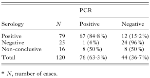
Table 2. Polymerase chain reaction (PCR) results accordingly to the three serological groups*
 You have
Access
- 33
- Cited by