Stress-induced cardiomyopathy was first described in Japan in 1990 by Sato.Reference Sato, Tateishi, Uchida, Kodama, Haze and Hon1 Stress-induced cardiomyopathy, also called apical ballooning syndrome, and, in Japan, “Takotsubo” cardiomyopathy, is an increasingly reported syndrome generally characterised by transient severe systolic dysfunction of the apical and/or mid segments of the left ventricle, which mimics myocardial infarction, but in the absence of coronary artery disease. A recent study showed that electrocardiogram abnormalities at presentation do not correlate with the magnitude of ventricular dysfunction or outcomes and also ST-segment elevation is absent in two-thirds of the patients with this cardiomyopathy.Reference Dib, Asirvatham and Elesber2
The majority of reported cases have been of postmenopausal women.Reference Sato, Tateishi, Uchida, Kodama, Haze and Hon1–Reference Tsuchihashi, Ueshima and Uchida3, Reference Khouri and Imran5 The name of the disorder is taken from the Japanese name for an octopus trap (“takot-subo”), which has a shape that is similar to the apical ballooning configuration of the left ventricle in systole in this disorder. Although the exact aetiology is still unknown, the syndrome appears to be triggered by a significant emotional or physical stress.
Until a recent report,Reference Schoof, Bertram, Hohmann, Jack, Wessel and Mesud Yelbuz6 this phenomenon had not been reported in the young. We wish to report this case in order to highlight its occurrence in a young patient and that a differential diagnosis of Takotsubo cardiomyopathy be considered even in children with apical left ventricular wall motion abnormalities.
Case report
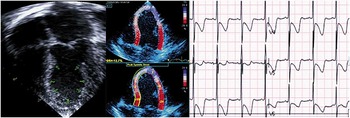
M.D. is a 16-month-old boy who presented to the emergency department with multiple episodes of vomiting. He has a past medical history of cyclic vomiting, episodic systemic hypertension, and failure to thrive. Due to the increasing frequency of his vomiting episodes, the paediatrician admitted the patient for further management. On admission, he had a temperature of 36.3°C, pulse rate of 143, respiratory rate of 32 and blood pressure of 130/83 millimetres of mercury. The next day, he was noted to have a gallop rhythm. His electrocardiogram showed ST-segment depression with T-wave inversion in the inferior and lateral leads. The transthoracic echocardiogram showed diminished left ventricular systolic function with marked dilatation and akinesis of the apical portion of the left ventricle as evidenced by cross-sectional imaging and two-dimensional strain modality (Fig 1). He was transferred to the paediatric intensive care unit for further management. The troponin T was slightly elevated to 0.77 nanogram per millilitre (normal, less than 0.01 nanogram per millilitre). A differential diagnosis of myocardial dysfunction secondary to coronary artery ischaemia or cardiomyopathy/myocarditis was made. He remained haemodynamically stable and was treated with conventional cardiac failure support, milrinone infusion, and diuretics. He underwent cardiac catheterisation and selective coronary angiography that showed normal coronary arteries with no evidence of structural obstruction. The parents refused permission for endomyocardial biopsy.
Echocardiogram at admission shows left ventricular apical akinesis. The two-dimensional strain image confirms the wall motion abnormality. Apical segmental strain of positive seven (normal from minus 14 to minus 20). The electrocardiogram shows ST segment depression and T-wave inversion in precordial and lateral leads.

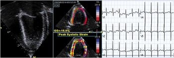
Two weeks later the electrocardiogram and cardiac enzymes were normal and the echocardiogram showed complete recovery of the left ventricle apical akinesis and normalisation of cardiac function (Fig 2).
Echocardiogram performed 2 weeks later shows normalisation of left ventricular motion with normal contractility pattern at the apex by two-dimensional strain. Apical segmental strain of minus 19 (normal from minus 14 to minus 20). The electrocardiogram shows a normal pattern.

Discussion
The classical features of Takotsubo cardiomyopathy from the literature are:
• Age (mean of 58–75 years)
• Female predominance
• Acute onset of symptoms
• Pathology triggered by any form of severe emotional or physical stress
• Myocardial dysfunction affecting the distribution of more than one coronary artery and usually involving the apical and mid segments
• Lack of angiographic evidence of obstructive coronary artery disease or occlusion
• Mild elevation of myocardial damage biomarkers
• S-T segment elevation or T-wave inversion on the electrocardiogram
• Reversal of myocardial dysfunction over several weeks.
The pathogenesis of this disorder is not well understood. Although the clinical presentation simulates that of an acute myocardial infarction,Reference Ako, Takenaka, Uno, Nakamura, Shoji and Iijima7 coronary arteriography consistently shows no critical lesions and only a minority of patients display coronary spasm with acetylcholine provocation.Reference Bybee, Kara and Prasad9 The most commonly discussed possible mechanism for this cardiomyopathy is stress-induced catecholamine release, with paradoxical stunning of the myocardium.
Normal myocardium utilises approximately 90% of its energy from fatty acid metabolism at rest and with aerobic activity. During ischaemia, this pathway is suppressed and, instead, glucose is largely utilised, resulting in impaired function. Patients with Takotsubo cardiomyopathy are found to shift towards the glucose pathway despite relatively normal myocardial perfusion and lack of ischaemia.Reference Tsuchihashi, Ueshima and Uchida3, Reference Abe, Kondo and Matsuoka4 Endomyocardial biopsy of affected patients shows reversible focal myocytolysis, mononuclear infiltrates, and contraction band necrosis.Reference Tsuchihashi, Ueshima and Uchida3, Reference Abe, Kondo and Matsuoka4 The catecholamine theory is gaining momentum as this cardiomyopathy has been induced in rats exposed to physical stress that was prevented when pretreated with α- or β-blockade.Reference Ako, Takenaka, Uno, Nakamura, Shoji and Iijima7 Other supporting evidence for this theory has been shown through myocardial imaging studies using catecholamine analogues that evaluate cardiac sympathetic activity.Reference Lyon, Rees and Prasad8, Reference Bybee, Kara and Prasad9 Ako et al,Reference Ako, Takenaka, Uno, Nakamura, Shoji and Iijima7 by the use of an intra-coronary Doppler wire technique, showed microcirculation impairments in instances of transient left ventricular hypocontraction, suggesting the possibility of myocardial injury from a microvascular spasm.Reference Lyon, Rees and Prasad8 In a mouse model, it has been shown that a high level of epinephrine is negatively inotropic due to a switch from β-2 adrenoreceptor-mediated Gs protein signalling, which is positively inotropic, to Gi protein signalling, which is negatively inotropic. It is speculated that the greater effect at the apical myocardium may be due to a higher density of β-adrenoreceptors at this location.Reference Bybee, Kara and Prasad9 Therefore, the postulated mechanisms include catecholamine excess with paradoxical catecholamine-mediated effects on cardiomyocytes leading to transient left ventricular dysfunction.Reference Lyon, Rees, Prasad, Poole-Wilson and Harding10
Our case describes an example of Takotsubo cardiomyopathy occurring in children. Its diagnosis should be suspected in any patient of any age with acute symptoms of cardiac failure with apical dysfunction and an underlying condition that creates stress and subsequent increased catecholamine load. It is uncommon but may occur in children.
Acknowledgements
We thank Dr Valdes-Cruz for her opinion and critical review of the echocardiogram.