Implications
Despite several advantages, segregated early weaning leads to a post-weaning growth lag. This is due, in part, to the hyper-activation of the immune system. Glucocorticoids and synthetic glucocorticoid receptor agonists (GAs) have anti-inflammatory effects. Thus, we hypothesized that treatments with a GA may moderate the inflammatory response during weaning and consequently improve the growth performance of pigs. Our experiments showed that treatment with a GA improves growth performance by reducing the immunopathology caused by the inflammatory response during weaning. Therefore, GA could potentially reduce the use of other growth-promoting treatments among newly weaned pigs.
Introduction
Segregated early weaning (SEW) is practiced in most commercial swine operations to reduce the vertical transmission of disease and increase sow reproductive efficiency. Despite advantages, early weaning exposes piglets to environmental, immunological and nutritional stresses at an earlier stage in life, leading to an inflammatory response (Campbell et al., Reference Campbell, Crenshaw and Polo2013). Rising evidence indicates that during this response, inflammatory mediators such as pro-inflammatory cytokines, chemokines and anti-viral cytokines produced by the immune system in the periphery cause a central neuroinflammatory response, which affects brain development. The latter is predominantly brought about by chronic activation of brain microglia, which may explain some of the long-lasting clinical effects that subsequently occur. Some of these effects include a reduced ability of the animals to adapt successfully to stressors later in life, such that they experience exaggerated or prolonged physiological and psychological responses to innocuous stressors (Dionissopoulos et al., Reference Dionissopoulos, Dewey, Namkung and De Lange2006; Miguel et al., Reference Miguel, Chen, Van Alstine and Johnson2010; Conrad et al., Reference Conrad, Sutton, Larsen, Van Alstine and Johnson2015).
Although a certain degree of immune response is necessary to prevent disease and maintain homeostasis during the weaning period, the immunopathology caused by inflammation can be detrimental to the animals, as it can exacerbate the compromised digestive physiology and growth performance that is seen post-weaning. The latter can consequently lead to the production of less resilient piglets (Medzhitov et al., Reference Medzhitov, Schneider and Soares2012). The swine industry commonly uses highly palatable and digestible dietary sources and sub-therapeutic antibiotics in weaned pig diets to negate the adverse effects of SEW. The latter has caused concerns over the possible formation of antibiotic resistant bacteria and the former is expensive (Campbell et al., Reference Campbell, Crenshaw and Polo2013).
Glucocorticoids have potent anti-inflammatory effects by inhibiting the signaling of key inflammatory transcriptional regulators, activator protein 1 and nuclear factor kappa-light-chain-enhancer of activated B cells (Coutinho and Chapman, Reference Coutinho and Chapman2011; Handa and Weiser, Reference Handa and Weiser2013). As such, GA such as dexamethasone (DEX) might be expected to partially negate the post-weaning growth lag by moderating the hyper-activated immune system. Dexamethasone is a relatively well-studied synthetic GA typically used to treat inflammation and stress-related conditions in both humans and animals (Wyns et al., Reference Wyns, Meyer, Watteyn, Plessers, De Baere, De Backer and Croubels2013). Two experiments were conducted to evaluate the effects of a GA on overall performance, as well as measures of intestinal integrity and immune function of newly weaned pigs. The first experiment evaluated the effect of three doses of GA on the growth performance and plasma cortisol levels of early-weaned pigs. In the second experiment, we investigated the impact of the GA on measures of immune function and intestinal integrity. We hypothesized that treatment with GA mitigates the negative impact of SEW on the growth performance of newly weaned pigs by partially subsiding the inflammatory response and preventing the immune system from overreacting.
Materials and methods
All experiments were conducted at the Texas Tech University Swine Research Center (New Deal, TX, USA). Dexamethasone (Phoenix Pharm Inc., Catalogue number PHO5731924305) was used as the GA. To avoid the potential effects of circadian rhythms on blood glucocorticoid levels, blood samples were collected between 10:00 am and 11:00 am in both experiments (Ingram and Dauncey, Reference Ingram and Dauncey1985).
Experiment 1
A total of 48 piglets (24 gilts and 24 barrows; BW 4.8 ± 0.7 kg; Sus scrofa domesticus PIC USA Hendersonville, TN, USA) were weaned at 21 ± 1 days of age, and randomly assigned to one of three treatment groups: 0 (control), 0.2 and 0.6 mg GA/kg of BW. Pigs received GA intramuscularly 24 h before weaning. Piglets in the control group (0.0 mg GA/kg of BW) received sterile saline intramuscularly (0.5 ml/pig) to account for any stress caused by injection (Rosen et al., Reference Rosen, Brodin and Eneroth1992). After weaning, pigs were moved into a temperature-controlled nursery and housed in floor-pens (one of each sex per pen; 8 pens per treatment). Pigs were fed conventional corn-soy bean meal (SBM) starter diets for a 28-day period according to a phase feeding program (Table 1; National Research Council (NRC) Reference National Research Council2012). Body weight was measured daily from one day before to 7 days post-weaning, and then weekly until 28 days post-weaning. Feed intake for each pen was measured on a weekly basis and computed as the difference between the weight of the feeder (and feed) at the beginning and the end of each week after accounting for added feed and feed waste. Blood samples were collected from the external jugular vein at 0 and 1 day post-weaning. Blood samples were collected in vacutainers (BD Company, Franklin Lakes, NJ, USA) containing ethylenediaminetetraacetic acid and centrifuged at 1500 × g for 10 min at 4°C. The plasma fraction was then collected, aliquoted, stored at −80°C and used to determine cortisol levels using a commercial Cortisol ELISA kit (Enzo Life Sciences Inc., Farmingdale, NY, USA).
Ingredient composition and nutrient contents of the experimental diets fed to pig

1 Providing the following amounts of vitamins and trace minerals (per kg of diet) for pigs (Sus scrofa domesticus): vitamin A, 10075 IU; vitamin D3, 1100 IU; vitamin E, 83 IU; vitamin K (as menadione), 3.7 mg; d-pantothenic acid, 58.5 mg; riboflavin, 18.3 mg; choline, 2209.4 mg; folic acid, 2.2 mg; niacin, 73.1 mg; thiamin, 7.3 mg; pyridoxine, 7.3 mg; vitamin B12, 0.1 mg; d-biotin, 0.4; Cu, 12.6 mg; Fe, 100 mg; Mn, 66.8 mg; Zn, 138.4 mg; Se, 0.3 mg; I, 1.0 mg; S, 0.8 mg; Mg, 0.0622%; Na, 0.0004%; Cl, 0.0336%; Ca, 0.0634%, P, 0.003%; K, 0.0036%. ME = metabolizable energy. STTD = standardized total track digestibility.
2 Phase 1, 2 and 3 diets were fed 0–7, 7–21 and 21–28 days post-weaning.
3 Amino acids are formulated on a standardized ileal digestible (SID) basis.
Experiment 2
In another experiment 18 gilts (Pig Improvement Company®, USA; BW 5.6 ± 0.85 kg) from six litters were assigned to one of three treatment groups: (i) unweaned (UWS), (ii) weaned treated with GA (WGA; 0.2 mg GA/kg BW) and (iii) weaned plus saline (CON). Siblings from the two litters in the UWS group (n = 3 gilts/litter) were evenly distributed among CON and WGA groups to avoid a possible confounding effect of litter and treatment. Pigs in CON and WGA groups were weaned at 23 ± 2 days of age, while the UWS group remained with sow for the duration of the study. All pigs received either GA or saline one day before and 3 days post-weaning. The CON and UWS pigs received intramuscular injections of sterile saline to account for the stress of injection (Rosen et al., Reference Rosen, Brodin and Eneroth1992). Pigs were euthanized 5 days post-weaning by an intravenous injection of a lethal dose of sodium-pentobarbital (fatal-plus) to minimize the impact of euthanasia on intestinal morphology and mucosal shedding (Gabert et al., Reference Gabert, Jorgensen and Nyachoti2000).
Weaned pigs were moved into a temperature-controlled nursery and housed in floor-pens (three pigs per pen; two pens per treatment). Pigs were fed a conventional corn-SBM-based starter diet for the duration of the study (Table 1, NRC Reference National Research Council2012). Pigs in the UWS group did not receive solid feed, but were nursing. Body weights were measured daily throughout the study. Feed intake for each pen was measured at the end of the 5-day period and computed as the difference between the weight of the feeder (and feed) at the beginning and the end of each week accounting for added feed and feed waste. Blood samples were collected into vacutainer tubes containing sodium heparin via jugular puncture at 0, 1, 4 and 5 days post-weaning. Blood samples were centrifuged at 1500 × g for 15 min at 4°C and the plasma was aliquoted and stored at −80°C until further analysis.
Tissue collection
Immediately after euthanasia, a mid-ventral laparotomy was performed on each gilt to harvest jejunum samples. About 40 cm samples of jejunum were taken at approximately 1 m distal to the gastroduodenal junction and divided into three segments. One segment (~4 cm) was cut longitudinally, stored in a 10% formalin buffer and used for histomorphology analysis. A second segment of the original jejunum sample was washed with phosphate buffered saline and mucosal scrapings were collected (Hu et al., Reference Hu, Xiao, Luan and Song2013). Adherent material was flash-frozen in liquid nitrogen, and stored at −80°C. The remaining portion of jejunum was flash-frozen, stored at −80°C and used for gene expression analysis.
Measures of immune function
The effects of the treatments on immune function were evaluated at 0, 1, 4 and 5 days post-weaning. Plasma haptoglobin levels were measured using a two site-linked immunoassay ELISA kit (GenWay Biotech Inc., San Diego, CA, USA). Plasma IL-1β was measured using a quantikine ELISA kit (R&D Systems Inc., Minneapolis, MN, USA). Plasma total antioxidant capacity was measured by a colorimetric antioxidant assay kit (Cayman Chemical Company, Ann Arbor, MI, USA). Plasma tumor necrosis factor alpha (TNF-α) was measured using an Invitrogen ELISA kit (ThermoFisher Scientific, Waltham, MA, USA).
Morphology of jejunum mucosa
Morphological measures of jejunum mucosal samples were visualized at the Texas Tech University Health Science Center pathology laboratory in Lubbock, TX, USA. Briefly, each jejunum specimen (~2 × 2 cm) was embedded in paraffin wax and cut at 4 µm. Sections were fixed on glass slides and stained with hematoxylin–eosin. For the measurement of villus height and base width, five adjacent villi were randomly selected from each cross-section. The villus height from tip to base, excluding the intestinal crypt, was measured. The width of the villus was measured at the basal part. The crypt depth was defined as the invagination between two villi. A total of 15 villi and crypts from three cross-sections were measured for each pig, and means were calculated.
RNA extraction and gene expression
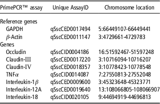
Differential expressions of occludin, claudin-IV, claudin-III, IL-1β, IL-18, IL-12 and TNFα genes were determined in both whole jejunum samples and jejunum mucosal scrapings. Each sample was ground in liquid nitrogen to acquire a small representative sample for RNA extraction. Total RNA was then isolated using an RNeasy mini kit (Qiagen, Hilden, Germany), according to the manufacturer’s instructions. The RNA integrity number and quality were verified using an RNA ScreenTape System (Agilent Technologies, Waldbronn, Germany) and a Nanodrop2000 spectrophotometer (ThermoFisher Scientific, Wilmington, DE, USA), respectively. Samples with an RNA integrity number ⩾7.90 were used for synthesis of complementary DNA (cDNA). Complementary DNA was synthesized using approximately 1000 ng of RNA and an iScript cDNA synthesis kit (Bio-Rad Laboratories, Inc., Hercules, CA, USA). Gene expression quantification was performed by real-time quantitative PCR (CFX Connect, Bio-Rad Laboratories, Inc.) using PrimePCR assays (Table 2) and an ITaq Universal SYBR Green Supermix (Bio-Rad Laboratories Inc.). Samples were cycled using a Bio-Rad CFX manager 3.1 software 96-well SYBR PrimePCR protocol (denaturation for 2 min at 95°C, 39 cycles of 5 s at 95°C followed by 30 s at 60°C, a brief 5 s of 95°C followed by a melt curve from 65°C to 95°C at increments of 5°C for 5 s). Generated quantification cycle (Cq) values were used for the quantification of gene expression. Two housekeeping genes (glyceraldehyde 3-phosphate dehydrogenase, GAPDH and beta actin, β-actin; Table 2) were used as internal references for relative quantification. Treatments did not affect the relative abundance of GAPDH or β-actin genes. Amplification efficiencies of all primers used in the study were approximately equal. The CON group served as the calibrator for comparison to either the GA group or the UWS group, because it represents the common treatment standard for SEW pigs. Whole jejunum samples from the GA or UWS groups were compared only to whole jejunum samples from the CON group. Similarly, mucosal scraping samples from the GA or UWS groups were compared only to mucosal scraping samples from the CON group. The amount of target, normalized to the mean of the reference genes and relative to the CON (calibrator), was defined by the double-delta CT method, as described by Livak and Schmittgen (Reference Livak and Schmittgen2001).
Primer unique assay identification (ID) numbers and chromosome locations1

1 Represents the specific Sus scrofa domesticus PrimePCR™ SYBR® Green assays utilized in the gene expression study, provided in place of a specific gene sequence, by BIO-RAD Laboratories, Inc. GAPDH = glyceraldehyde 3-phosphate dehydrogenase. TNFα = tumor necrosis factor alpha.
Statistical analysis
Statistical analysis of data was carried out using MIXED procedure in SAS (version 9.4, Institute Cary, NC, USA). In Experiment 1, data were analyzed using a completely randomized design with dose of GA, time (day) and interaction (GA dose and time) as fixed effects. Pen was used as the experimental unit, and pig within pen was used as a random effect. Repeated measurements ANOVA was used for the data that was collected over time. In Experiment 2, data were analyzed using a completely randomized design with treatment, time (day) and interaction between day and treatment as fixed effects. Pen was used as the experimental unit, and pig within pen was used as random effect for performance parameters. For time-specific measures of immune function from the blood, individual pigs were used as the experimental unit. In both studies, an appropriate covariance structure was selected for analyses by fitting the model with the structure, which provided the ‘best’ fit, based on Akaike information criterion (AIC) and Schwarz Bayesian criterion (BIC). For parameters measured over time, repeated measurements ANOVA was used. Based on the AIC and BIC statistics and the likelihood ratio test, the autoregressive 1 model provided the best fit for these parameters. For one-time observations, the unstructured model was determined to be the best fit according to the AIC and BIC statistics and the likelihood ratio test. Values were reported as least squares means ± SEM. Means were separated using the Tukey–Kramer test. Treatment effects were considered significant at P ⩽ 0.05. A tendency toward a significant difference between treatment means was also considered at P ⩽ 0.10.
Results
Before and during both the experiments, all pigs were in good health with no clinical signs of illness or injury, and they all readily consumed the experimental diets.
Experiment 1
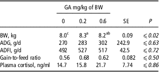
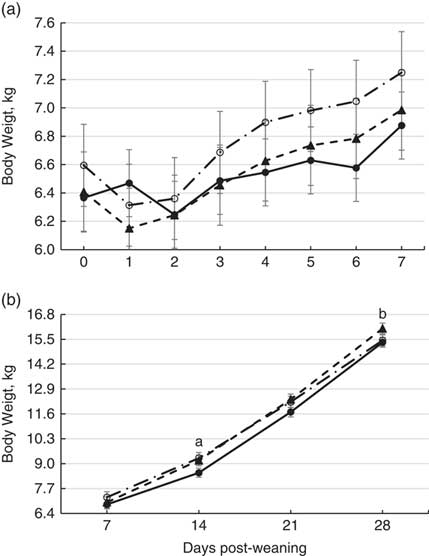
Data are presented in Table 3. Intramuscular injection of 0.2 and 0.6 mg/kg BW GA improved (P <0.02) and tended to improve (P <0.08) the BW of piglets, respectively, during the 28-day course of the study when compared to the control group. No interaction effect between treatment and time (treatment × time) on BW was observed (Figure 1; P = 0.94). Relative to control, both doses of GA numerically increased the overall average daily gain (ADG) and gain-to-feed ratio, but the differences did not reach statistical significance. Weaning increased plasma cortisol levels from 12.1 ± 2.62 to 21.7 ± 2.81 ng/ml (P <0.01). However, the GA had no effect on plasma cortisol levels at 24 h post-weaning (P >0.70). Average daily feed intake (ADFI) was not affected by the treatments.
Effects of a glucocorticoid receptor agonist (GA) on the growth performance of newly weaned pigs (Experiment 1)1

1 Data are the least square mean (± largest SE), and represent the best estimate of mean that were obtained from repeated measurements ANOVA over the 28-day course of the study. A total of 48 piglets (Sus scrofa domesticus) were weaned at 21 ± 1 days and assigned to three treatment groups (two pigs per pen; eight pens per treatment): 0, 0.2 and 0.6 mg/kg dexamethasone. Piglets in each treatment group received GA intramuscularly 1 day before weaning. Piglets in the control group (0.0 mg/kg BW) received 0.5 ml sterile saline. Piglets had free access to water and corn-SBM-based diets in which nutrients met or exceeded the recommendations of NRC Swine (Reference National Research Council2012). For each row, means marked with different superscript letters differ from each other significantly. ADG = average daily gain. ADFI = average daily feed intake.
Effects of three doses of glucocorticoid receptor agonist (GA) treatment on the BW of pigs across 28 days post-weaning (panel A and B; Experiment 1). A total of 48 piglets were weaned at 21 ± 1 days and assigned to three treatment groups (two pigs per pen; eight pens per treatment): 0 (●), 0.2 (○), and 0.6 (▲) mg/kg dexamethasone as GA. Piglets in each treatment group received GA intramuscularly 1 day before weaning. Piglets in the control group (0.0 mg/kg BW) received 0.5 ml sterile saline. Piglets had free access to water and corn-SBM-based diets in which nutrients met or exceeded the recommendations of NRC Swine (Reference National Research Council2012). aGA treatment different from control (P <0.05). bGA (0.6 mg/kg) different from control (P <0.05).

Experiment 2
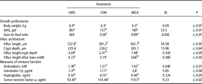
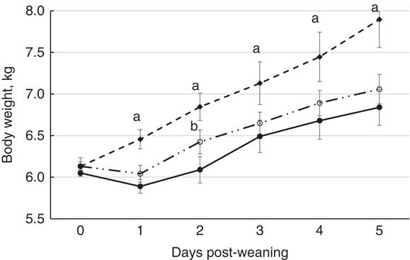
Growth performance and measures of intestinal morphology and immune function data are presented in Table 4. The UWS group that did not experience the stress of weaning had higher BW and ADG than all weaned groups (P <0.01). Treatment with GA improved BW (P <0.04) and the gain-to-feed ratio in weaned pigs (P <0.02) compared with CON. An interaction effect of treatment and time (treatment × time) on BW was observed (Figure 2; P <0.01). Treatment with GA did not improve ADG in weaned piglets at the end of the 5-day study.
Effects of glucocorticoid receptor agonist (GA) treatment on growth performance, jejunum mucosa histomorphology and measures of immune function in pigs (Experiment 2)1

1 Data are the least square mean (± largest SE). Measures of immune function and BW data represent the best estimate of mean that were obtained based on repeated measurements ANOVA. Average daily gain (ADG) and gain-to-feed ratio were determined at the end of the 5-day study. A total of 18 newly weaned piglet were housed in floor pens (three pigs per pen; two pens per treatment) and assigned to three treatment groups: (1) UWS = suckling group treated with 0.5 ml saline, (2) CON = weaned group treated with 0.5 ml saline and (3) WGA = weaned group treated with 0.2 mg dexamethasone/kg of BW. Serial blood samples were taken on days 0, 1, 4 and 5 post-weaning and used to evaluate measures of immune function. For each row, means marked with superscript letters differ from each other significantly.
Effects of glucocorticoid receptor agonist (GA) treatment on the BW of pigs across a 5-day post-weaning period (Experiment 2). Data are the least square mean ± SE. A total of 18 newly weaned piglet were housed in floor pens (three pigs per pen; two pens per treatment) and assigned to three treatment groups: (1) UWS = suckling group treated with 0.5 ml saline (♦), (2) CON = weaned group treated with 0.5 ml saline (●) and (3) WGA = weaned group treated with 0.2 mg dexamethasone/kg of BW as GA (○). aGA and UWS different from CON (P <0.05). bGA different from CON and UWS (P <0.05).

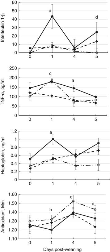
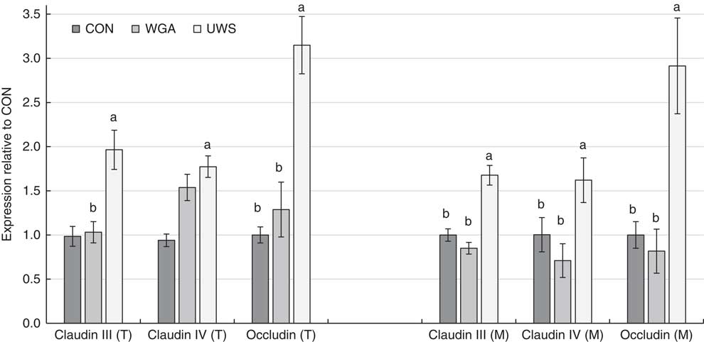
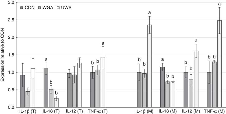
Compared to UWS, both WGA and CON gilts experienced a significant drop in villus height (P <0.01). However, no differences in crypt depth were observed among the treatment groups. Villus height-to-crypt depth ratio was higher in the UWS group relative to the SEW groups (P <0.05). The villus-height-to-villus-base ratio was highest in the UWS group (P <0.02). No differences in villus-height-to-villus-base ratio were observed between CON and WGA groups. No interaction effect between the main effects of treatment and time on plasma IL-1β, TNF-α and antioxidant levels was observed (Figure 3; P >0.14). A tendency for an interaction between treatment and time on plasma levels of haptoglobin was observed (Figure 3; P = 0.09). Plasma levels of haptoglobin and IL-1β in WGA and UWS pigs were lower than those seen in CON pigs (P <0.04). Plasma total antioxidant levels were higher in WGA pigs than in both UWS and CON pigs (P <0.01). Plasma TNF-α levels were higher in CON than UWS pigs (P <0.01) and tended to be higher in the CON group than in the WGA group (P <0.06). Differential gene expressions of selected tight junction proteins in jejunum tissue and in mucosal scrapings at 5 days post-weaning are presented in Figure 4. Claudin-IV was upregulated in jejunum tissue, but not in mucosal scrapings of WGA pigs when compared to CON (P <0.04). Treatment with GA did not affect the expression of claudin-III and occludin in neither the jejunum tissue nor mucosal scrapings of weaned pigs. The expressions of occludin and claudin-III in both jejunum tissue and mucosal scrapings were higher in UWS compared to CON pigs (P <0.02). Relative to CON pigs, expression of claudin-IV was higher (P <0.02) and tended to be higher (P <0.07) in jejunum tissue and mucosal scraping of UWS pigs, respectively. Differential gene expressions of pro-inflammatory cytokines in jejunum tissue and mucosal scrapings at 5 days post-weaning are presented in Figure 5. Expression of IL-18 was downregulated in jejunum tissue and mucosal scrapings of WGA relative to CON (P <0.02). A tendency of decreased expression of IL-1β in jejunum tissue of WGA v. CON (P <0.09) was observed. However, this difference was not observed in mucosal scrapings. Expression of IL-12 and TNF-α was not affected by GA treatment. In general, expression of pro-inflammatory cytokine genes, with the exception of IL-18, was upregulated, some significantly (i.e., TNF-α, IL-1β and IL-12; P <0.05) in jejunum tissue and mucosal scrapings of UWS compared to other treatment groups.
Effects of glucocorticoid receptor agonist (GA) treatment on measures of immune function across a 5-day post-weaning period. Data are the least square mean ± SE. A total of 18 newly weaned piglet were housed in floor pens (three pigs per pen; two pens per treatment) and assigned to three treatment groups: (1) UWS = suckling group treated with 0.5 ml saline (♦), (2) CON = weaned group treated with 0.5 ml saline (●), and (3) WGA = weaned group treated with 0.2 mg dexamethasone/kg of BW as GA (○). aGA and UWS different from CON (P <0.05). bGA different from CON (P <0.05). cGA different from UWS (P <0.05). dGA different from CON and UWS (P <0.05). TNF-α = tumor necrosis factor-alpha.

Effect of glucocorticoid receptor agonist (GA) treatment on the expression of tight junction protein complex genes in jejunum tissue (T) and mucosal scrapings (M). Data are the least square mean ± SE. A total of 18 newly weaned piglet were housed in floor pens (three pigs per pen; two pens per treatment) and assigned to three treatment groups: (1) UWS = suckling group treated with 0.5 ml saline, (2) CON = weaned group treated with 0.5 ml saline, and (3) WGA = weaned group treated with 0.2 mg dexamethasone/kg of BW. Treatments with different superscripts differ significantly from each other (P ⩽ 0.05).

Effect of glucocorticoid receptor agonist (GA) treatment on the expression of interleukin-1β (IL-1β), IL-18, IL-12 and tumor necrosis factor alpha (TNFα) genes in jejunum tissue (T) and mucosal scrapings (M). Data are the least square mean ± SE. A total of 18 newly weaned piglet were housed in floor pens (three pigs per pen; two pens per treatment) and assigned to three treatment groups: (1) UWS = suckling group treated with 0.5 ml saline, (2) CON = weaned group treated with 0.5 ml saline and (3) WGA = weaned group treated with 0.2 mg dexamethasone/kg of BW. Treatments with different superscripts differ significantly from each other (P ⩽ 0.05).

Discussion
Early-weaned pigs experience an inflammatory response that negatively affects their growth, as well as their capability for producing an appropriate immune response following exposure to an antigen later in life (Campbell et al., Reference Campbell, Crenshaw and Polo2013). The anti-inflammatory and immunosuppressive effects of glucocorticoids (i.e., cortisol and its synthetic receptor agonists) have been well documented (Coutinho and Chapman, Reference Coutinho and Chapman2011). High doses of glucocorticoids often have immunosuppressive properties that include reducing or preventing the activity of both the innate and adaptive immune systems, often causing adverse metabolic effects such as increased protein degradation in skeletal muscle (Qin et al., Reference Qin, Dua, Yang, Zhang, Li, Liu, Guan, Hou and An2013). In comparison, lower doses of glucocorticoids often have anti-inflammatory properties that reduce local and systemic inflammation, which reduces immunopathology and has a lesser or no effect on skeletal muscle growth. Therefore, the anti-inflammatory effects of glucocorticoids are often advantageous (Coutinho and Chapman, Reference Coutinho and Chapman2011; Son et al., Reference Son, Lee, Lee, Lee, Jeong, Chung, Park and Kim2015). As such, glucocorticoids might be expected to negate the post-weaning growth lag, since they are part of a negative feedback mechanism that prevents a sustained, high-level immune system response (Coutinho and Chapman, Reference Coutinho and Chapman2011). We hypothesized that if a GA is applied at the right dosage, it can reduce the negative effects of inflammation that usually occur during SEW; hence reducing the post-weaning growth lag and improving the robustness of piglets. The GA utilized in this study, DEX, is a well-studied synthetic glucocorticoid typically used to treat inflammation and stress-related conditions in both humans and animals. Unlike cortisol, which binds to both mineralocorticoid receptors and glucocorticoid receptors, DEX has a high affinity for only glucocorticoid receptors, which are responsible for the anti-inflammatory effects of glucocorticoids. As such, its anti-inflammatory potency is roughly 25 times that of cortisol (Handa and Weiser, Reference Handa and Weiser2013; Vandevyver et al., Reference Vandevyver, Dejager, Tuckermann and Libert2013). In Experiment 2, the treatments were administered at two time points (−1 and 3 days post-weaning) rather than once as in Experiment 1. One can argue that pigs at 3 days post-weaning are still undergoing elevated levels of stress, as shown by the typical duration of the post-weaning growth lag (5 to 10 days). Dexamethasone has a short half-life in pigs (plasma elimination rate of 1.06 h) and has been suggested, in multiple species, to have lasting effects 36 to 72 h post administration (Wyns et al., Reference Wyns, Meyer, Watteyn, Plessers, De Baere, De Backer and Croubels2013). Considering this half-life and the positive results of the first experiment, we chose to administer the GA twice during Experiment 2 to prolong any of its potentially beneficial effects. In addition, in Experiment 2, only gilts were used to avoid any confounding effects on measures of immune function that could potentially be caused by physical castration of male pigs. It has been reported that chronic inflammation is markedly higher in castrated male pigs compared to intact males and gilts (de Kruijf and Welling, Reference de Kruijf and Welling1988). Further studies are needed to evaluate the effects of GA and sex on productivity and measures of immune function in SEW pigs. Repeated injection with GA in Experiment 2 resulted in a higher BW relative to CON pigs at day 5 post-weaning. The interaction between the main effects of treatment and time for BW was significant in Experiment 2 mainly because the BW of UWS pigs was not affected by the process of weaning.
In Experiment 1, the two doses of GA (0.2 and 0.6 mg GA/kg BW) were used to explore whether growth performance response to DEX administration is dose-dependent. We observed an improvement in growth performance of weaned pigs that received a 0.2 mg/kg of BW dose of GA. This improved performance lasted for the 28 days post-weaning (at this point the study ended). The results of this study are not in agreement with the findings of Gómez et al. (Reference Gómez, Angeles and Cuáron1997), who reported no effect of GA on the productivity of SEW pigs (Gómez et al., Reference Gómez, Angeles and Cuáron1997). However, the lack of response to GA treatment in the Gómez study can likely be attributed to the timing of DEX administration relative to the time of weaning. In their study, DEX was administered 3 and 6 days before weaning. Therefore, considering the short half-life of DEX, it may have no longer been present at an effective level when their animals were weaned (Cross et al., Reference Cross, Paul and Goldman2011; Wyns et al., Reference Wyns, Meyer, Watteyn, Plessers, De Baere, De Backer and Croubels2013). The lack of improvement in the growth performance of pigs when they were treated with the 0.6 mg/kg of BW dose of GA suggests that the response to GA was dose-dependent, with higher doses of GA having possibly an immunosuppressive effect. The latter is likely due to the suppressive effects of DEX on leukocyte differentiation and function, as well as its negative effects on muscle tissue accretion. Specifically, immunosuppressive doses of DEX reduce the functional activity of lymphocytes by inducing apoptosis, downregulating adhesion molecules and decreasing proliferative response capabilities (Schwarze and Bartmann, Reference Schwarze and Bartmann1994; Coutinho and Chapman, Reference Coutinho and Chapman2011; Son et al., Reference Son, Lee, Lee, Lee, Jeong, Chung, Park and Kim2015). This idea is consistent with results found by Gatnau et al. (Reference Gatnau, Zimmerman, Nissen, Wannemuehler and Ewan1995), who saw no improvement in growth performance with daily injections of 0.15 mg DEX/kg BW over a period of 4 weeks (Gatnau et al., Reference Gatnau, Zimmerman, Nissen, Wannemuehler and Ewan1995). It is likely, in the aforementioned study, that repeated daily injections resulted in a more immunosuppressive rather than anti-inflammatory effect.
Although a certain degree of immune response is beneficial for animals to ward off dangerous organisms during an infection, an excessive or exaggerated immune response can have a long-term negative effect on the immunocompetency and productivity of pigs (Medzhitov et al., Reference Medzhitov, Schneider and Soares2012). Therefore, in the current study, the improvement in the growth of weaned pigs that received a lower dose of GA may partially be associated with a less severe anti-inflammatory effect of GA (Coutinho and Chapman, Reference Coutinho and Chapman2011). Indeed, in Experiment 2, lower plasma levels of two pro-inflammatory cytokines and the positive acute-phase protein, haptoglobin, were seen in WGA pigs compared to CON pigs. Interleukin-1β and TNF-α are the key pro-inflammatory cytokines that orchestrate an immune response and induce sickness behaviors (i.e., anorexia, lethargy, fever) by modulating the hypothalamic-pituitary-adrenal axis in pigs (Konsman et al., Reference Konsman, Parnet and Dantzer2002). Haptoglobin is a positive acute-phase protein that scavenges hemoglobin and has unique antioxidant properties (Quaye, Reference Quaye2008). Therefore, the reduction of IL-1β, TNF-α and haptoglobin in WGA pigs indicates a lower level of systemic inflammation in these pigs. Furthermore, increases in TNF-α levels in the plasma have been associated with increased intestinal permeability in human and animals (Ulluwishewa et al., Reference Ulluwishewa, Anderson, McNabb, Moughan, Wells and Roy2011). Therefore, in the present study, the observed tendency for lower levels of plasma TNF-α in GA-treated pigs may indicate a lower intestinal permeability in these animals compared to their untreated counterparts. This finding is also in agreement with the 1.54 increase in expression of the claudin-IV gene, an intestinal barrier forming protein, seen in WGA pigs compared to CON pigs. Furthermore, findings of the current study are in line with the findings of Boivin et al. (Reference Boivin, Ye, Kennedy, Al-Sadi, Shepela and Ma2006) who reported an association between elevated levels of plasma TNF-α and decreased transepithelial resistance, a measure of intestinal barrier integrity, in human intestinal epithelium. The observed tendency for an interaction effect between time and treatment on plasma haptoglobin levels can mainly be attributed to the gradual increase in plasma haptoglobin levels in UWS pigs at day 4 and 5 of the study. The increase in total antioxidant levels in WGA pigs compared to CON also provides additional evidence for a lower level of systemic inflammation in these pigs that is most likely due to the decreased production of free radicals, such as reactive oxidative species (Mittal et al., Reference Mittal, Siddiqui, Tran, Reddy and Malik2014). In the current study blood antioxidant capacity was higher among WGA pigs compared to UWS pigs. This difference was likely due to diet and not immune activity, since the starter diet contained higher levels of antioxidants than sow’s milk. Furthermore, in Experiment 2 the expression level of a pro-inflammatory cytokine gene, IL-18, was substantially lower in the jejunum of WGA pigs when compared to CON, suggesting a lower level of intestinal inflammation in GA-treated pigs. Interleukin-18 is a key pro-inflammatory cytokine in the intestine of pigs (Hand, Reference Hand2015). In general, in the current study the expression of pro-inflammatory cytokines, excluding IL-18, were higher in mucosal scrapings of the UWS group compared to other treatment groups. Cytokines IL-1β and TNF-α play important roles in the development of the mucosal layer in neonates (MohanKumar et al., Reference MohanKumar, Namachivayam, Ho, Torres, Ohls and Maheshwari2017). This may explain why, in the current study, the gene expression of IL-1β and TNF-α was higher in the mucosal scrapings of UWS pigs whose continuing intestinal development was not compromised by weaning. Interleukin-18 is not one of the contributing cytokines to mucosal development suggesting that its upregulation in the intestine of weaned pigs can likely be associated with inflammation (MohanKumar et al., Reference MohanKumar, Namachivayam, Ho, Torres, Ohls and Maheshwari2017). This also explains the lower levels of IL-18 expression in both the mucosal scrapings and jejunum tissue of UWS pigs when compared to CON. When considering the higher levels of TNF-α in UWS pigs, it is important to note that TNF-α, like many other cytokines, can have contradicting functions in different tissues and under different physiological circumstances within the animal (Leppkes et al., Reference Leppkes, Roulis, Neurath, Kollias and Becker2014). When elevated in the plasma, TNFα is typically associated with systemic inflammation and has negative effects on intestinal health (Yamamoto et al., Reference Yamamoto, Tanabe, Wakabayashi, Shimazu, Matsumoto and Kitajima2001; Llamas Moya et al., Reference Llamas Moya, Boyle, Lynch and Arkins2008). However, its function within the intestine is more pleiotropic in nature and can sometimes be beneficial to mucosal development and maintenance (Leppkes et al., Reference Leppkes, Roulis, Neurath, Kollias and Becker2014; MohanKumar et al., Reference MohanKumar, Namachivayam, Ho, Torres, Ohls and Maheshwari2017). In Experiment 2, expression of intestinal IL-1β, TNF-α and IL-12 genes was not affected by GA treatment. The latter can partially be attributed to the time of sampling, as it has been shown that expression of these particular cytokines in the mid-small intestine peaks around 1 to 2 days post-weaning, before rapidly returning to pre-weaning levels (Pié et al., Reference Pié, Lallès, Blazy, Laffitte, Sève and Oswald2004). In our study, intestinal samples were collected 5 days post-weaning suggesting that we could have missed the spike in expression of IL-1β, TNF-α and IL-12 when GA treatment would theoretically have the highest impact. Collectively, these results suggest that GA, such as DEX, at lower doses can improve the growth performance of newly weaned pigs by partially suppressing excessive inflammatory responses. In the current study, weaning increased the circulatory cortisol level by 180%, which is consistent with the findings of other workers (Kojima et al., Reference Kojima, Kattesh, Roberts and Sun2008). Previous studies have reported a decrease in the circulatory cortisol levels of pigs, as well as most other mammalian species, when they are treated with a GA due to its negative feedback effect on cortisol (Désautés et al., Reference Désautés, Sarrieau, Caritez and Mormède1999; Cole et al., Reference Cole, Kim, Kalman and Spencer2000). However, in Experiment 1, GA treatment did not have an effect on cortisol levels. It is important to note that in the present study blood samples were collected 48 h post GA treatment. While it has been shown that a GA reduces cortisol production, it is unclear if these effects would still be present 48 h after a single GA treatment at the dosages used in the current study. Most studies reporting the negative feedback effect of a GA on cortisol levels have varied from our study in having a higher GA dose, shorter time of sample collection post GA treatment, or they used repeated treatment administration of GA (Ritchie et al., Reference Ritchie, Belkin, Ranga, Krishnan, Nemeroff and Caroll1990; Cole et al., Reference Cole, Kim, Kalman and Spencer2000). Furthermore, chronic stress can influence the suppressive effect of GA on cortisol, suggesting that the elevated levels of stress in early-weaned pigs may reduce the effectiveness of GA in suppressing cortisol in the present study (Cole et al., Reference Cole, Kim, Kalman and Spencer2000).
The reduction of intestinal villus height and the expression of tight junction protein genes in jejunum tissue and mucosal scrapings in CON pigs compared to UWS is in agreement with other studies showing the negative impact of weaning on intestinal health and integrity (Hu et al., Reference Hu, Xiao, Luan and Song2013). Glucocorticoid receptor agonist treatment had little impact on villus architecture and tight junction protein gene expression compared to the CON pigs. Higher villus length-to-villus base-ratio in GA-treated pigs compared to CON pigs suggests a faster recovery potential from the stresses of weaning in these pigs (Nabuurs et al., Reference Nabuurs, Hoogendoorn, van der Molen and van Osta1993). The latter agrees with the higher growth performance of GA treated pigs at 28 days post-weaning compared to control pigs in Experiment 1. Claudin-IV expression was higher in WGA pigs than CON in whole jejunum tissue, but no differences were observed in the mucosa scraping samples. While higher level of claudin-IV suggested a decreased intestinal permeability, it is also possible that the claudin-IV detected in jejunum tissue is due to the reduction of enteric neurons in the submucosa. Enteric neurons express claudin-IV, but not occludin and claudin-III, in the intestine and that these neurons are negatively impacted as a result of inflammation (Karaki et al., Reference Karaki, Kaji, Otomo, Tazoe and Kuwahara2007; Margolis and Gershon, Reference Margolis and Gershon2016). Taken together these results suggest that the stresses of weaning negatively affect the integrity and barrier functions of the jejunum in pigs. Treatment with GA reduces intestinal inflammation, and consequently improves barrier permeability and possibly accelerates healing. However, we acknowledge that the number of pigs used in Experiment 2 was low and may have precluded discovering differences in villus architecture and the expression of selected genes. Nevertheless, these results indicate that administration of GA can mitigate the impact of early weaning stress on the productivity of nursery pigs.
Collectively, treating SEW pigs with a cortisol analog reduced the post-weaning growth lag by subsiding systemic and intestinal inflammation and likely by improving intestinal barrier function. Indeed, Soler et al. (Reference Soler, Miller, Hummel, Razzazi-Fazeli, Jessen, Escribano and Niewold2016) demonstrated that antibiotics have effects similar to those seen in this study with GA and may also work through anti-inflammatory mechanisms (Soler et al., Reference Soler, Miller, Hummel, Razzazi-Fazeli, Jessen, Escribano and Niewold2016). Therefore, GA may reduce the need for in-feed antibiotics of SEW pigs. The GA that was used in the current study, that is, DEX, has a short half-life of 36 to 72 h and will disappear from the animal’s body well before it reaches slaughter weight. Therefore, unlike in-feed antibiotics, GA presents a lower or no food safety concern (Wyns et al., Reference Wyns, Meyer, Watteyn, Plessers, De Baere, De Backer and Croubels2013). Additionally, GA treatment is likely more cost-effective than in-feed antibiotic treatment, suggesting that cost analysis studies, as well as productivity studies, that directly compare GA to antibiotic treatment during weaning are warranted. However, the underlying mechanisms, such as the expression of tight junction proteins, through which GA improves intestinal integrity and immunocompetence merit further considerations. In addition, the findings of the current study warrant further research to evaluate the longitudinal effects of GAs on the productivity of pigs fed antibiotic-free diets.
Acknowledgements
The current study was presented in part in proceedings of American Society of Animal Science meeting (Midwest section 2015), Des Moines, IA, USA. We thank Whitney McGilvray, Deltora Hewitt, Arlene Garcia, Guilherme Picinin and Glenna Priner for technical assistance. The present study was funded in part by the Animal Biotech and Texas Tech University. This research is a publication from the Department of Animal and Food Sciences, Texas Tech University.
Declaration of interest
Anoosh Rakhshandeh, Hailey Wooten, Mitchell Wachtel, Amanda R. Rakhshandeh and Garrett Thompson have no conflict of interest. John McGlone has an equity interest in Animal Biotech.
Ethics statement
The Texas Tech University Animal Use and Care Committee reviewed and approved all methods and procedures for this experiment (ACUC number 14013).
Software and data repository resources
None of the data were deposited in an official repository.