Background
Anomalous origin of a coronary artery from the contralateral sinus of Valsalva is associated with an increased risk of sudden death and cardiac morbidity.Reference Benson 1 , Reference Davis, Cecchin and Jones 2 Anomalous origin of the right coronary artery from the left sinus of Valsalva or anomalous origin of the left coronary artery from the right sinus of Valsalva are rare congenital anomalies, with a combined incidence ranging from 0.17% in autopsy studies to 1.2% in patients angiographically evaluated.Reference Alexander and Griffith 3 , Reference Engel, Torres and Page 4 However, anomalous origin of the right coronary artery from posterior sinus is extremely rare. To the best of our knowledge, anomalous origin of the right coronary artery from the posterior sinus is extremely rare and has never been reported in a paediatric-age competitive athlete. We report this very rare anomaly in a 14-year-old female competitive athlete diagnosed by cardiac MRI.
Case presentation
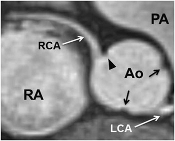
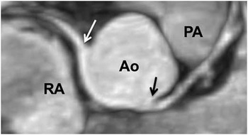
A 14-year-old female patient was referred for evaluation of non-specific chest pain. She is a competitive soccer player and there is no history of presyncope or syncope. An echocardiogram was performed initially for the assessment of chest pain. The origin of the right coronary artery was noted to be further rightward than typical, but it was unclear where the origin was located relative to the right-non commissure. Therefore, the MRI was performed to clarify the origin. Magnetic resonance coronary imaging was performed with respiratory navigation and electrocardiogram gating. Coronary imaging demonstrates that there is no evidence of right or left coronary artery stenosis. The right coronary artery originates from the posterior sinus adjacent to the commissure (Figs 1 and 2). The proximal right coronary artery does not appear narrowed and there is no interarterial course. The left coronary artery originates normally from the left sinus of Valsalva (Fig 1). MRI also confirmed that the aortic valve morphology was normal with three leaflets and that there were no other congenital heart defects. Delayed gadolinium enhancement imaging demonstrates normal myocardial viability without any regions of myocardial fibrosis.
Magnetic resonance coronary angiogram showing anomalous origin of the right coronary artery from the posterior sinus adjacent to the coronary commissure (black arrowhead). The septal and left-non commissures are identified (black arrows). The anomalous RCA ostium is unobstructed and is not interarterial. The LCA originates normally from the left sinus. AO=aorta; LCA=left coronary artery; PA=pulmonary artery; RA=right atrium; RCA=right coronary artery.

Magnetic resonance coronary angiogram showing the origins of the anomalous right coronary artery from the posterior sinus (white arrow) and the normal left coronary artery from the left coronary sinus (black arrow). AO=aorta; PA=pulmonary artery; RA=right atrium.

Discussion
Anomalous origin of a coronary artery from the contralateral coronary sinus of Valsalva is associated with increased risk of sudden death and cardiac morbidity.Reference Benson 1 , Reference Davis, Cecchin and Jones 2 Sudden death, especially in young athletic individuals, is frequently the initial manifestation of these abnormalities.Reference Liberthson 5 , Reference Chaitman, Lespereanc and Salteil 6 Sudden death is thought to be due to a reduction in blood flow in the anomalous vessel, resulting in myocardial ischaemia and/or infarction.Reference Benge, Martins and Funk 7 , Reference Frescura, Basso and Thiene 8 The exact pathophysiology of this decreased blood flow is not fully understood. The mechanism has been described as a tangential acutely angulated origin associated with a slit-like orifice that may be further compressed by aortic dilation during exercise.Reference Frescura, Basso and Thiene 8 We report an extremely rare case of the anomalous right coronary artery arising from the posterior sinus diagnosed by MRI. In our patient, the proximal right coronary artery is widely patent and there is no interarterial course. Although an intramural course cannot be directly assessed or ruled out with cross-sectional non-invasive imaging, in this case the absence of an acutely angulated proximal right coronary artery suggests there is no intramural course. Furthermore, the patient underwent exercise stress testing with nuclear perfusion, which was normal without evidence of ischaemic changes or perfusion defects. Similarly, MRI shows no evidence of myocardial scarring on delayed gadolinium enhancement imaging. In reviewing the literature, there are few reports of this rare anomaly. Vlodaver et alReference Vlodaver, Neufeld and Edwards 9 and Virmani et alReference Virmani, Rogan and Cheitlin 10 report right coronary artery from the posterior sinus associated with significant atherosclerotic disease in two adults aged 72 and 59 years, respectively. Yamanaka and Hobbs’Reference Yamanaka and Hobbs 11 report of identified coronary anomalies in a large catheter angiography series finds four patients with right coronary artery from the posterior sinus, without clinical manifestations or perfusion abnormalities, and considers this anomaly benign until proven otherwise. RobertsReference Roberts 12 reports the one case of right coronary artery he has observed from the posterior sinus in a 19-year-old with a quadricuspid pulmonic valve and cardiomegaly, who suddenly died while playing basketball. Roberts states that, although the cause of sudden death was unclear, it seemed unlikey because of the coronary anomaly, given the significant cardiomegaly. For these reasons, in our patient, the coronary anomaly was considered an incidental finding and the patient was permitted to exercise. Our findings on cardiac MRI support that the anomalous right coronary artery from the posterior sinus should be considered a distinct clinical entity of the anomalous right coronary artery from the left sinus of Valsalva.
Conclusion
Anomalous origin of the right coronary artery from the posterior sinus is extremely rare and has never been reported in a paediatric-age competitive athlete. We report this very rare case of right coronary artery arising from the posterior sinus diagnosed and assessed by cardiovascular MRI.
Acknowledgement
The authors are grateful to the staff of the Cardiovascular Magnetic Resonance Unit at Children’s Hospital of Philadelphia for their support in this work.
Financial Support
This research received no specific grant from any funding agency, commercial, or non-for-profit sectors.
Conflicts of Interest
None.
Ethical Standards
According to Children’s Hospital of Philadelphia Institutional Review Board procedure V, this case report was exempt from IRB approval process.