I. INTRODUCTION
Iron deficiency (ID) affects over 2 billion people worldwideReference Allen, De Benoist, Dary and Hurrell1 and is among 10 leading global risk factors for disease, disability, and death.2 The WHO estimates that 39–52% of women and children in developing countries are anemic,2 with half of the anemia from ID.Reference Zimmermann and Hurrell3 The latter is also common in industrialized countries: in the UK, 21% and 18% of 11–18 years-old and 16–64 years-old females, respectively, are iron deficient,Reference Heath and Fairweather-Tait4 and in the US, iron deficiency anemia (IDA) affects up to 27% of pregnant women.Reference Scholl5 The high prevalence of ID has a substantial negative impact on health and economic prosperity.Reference Zimmermann and Hurrell3 IDA increases risk for maternal death, low birth weight, and infant mortality,Reference Zimmermann and Hurrell3 while in adults, physical work capacity is reduced.Reference Haas and Brownlie6
Similar to ID, zinc deficiency affects approximately 30% of the world population, mainly in Southeast Asia (33% of the population) but also in Western Europe (11%) and US/Canada (10%).Reference Hotz and Brown7 Since zinc is involved in many metabolic pathways, its deficiency results in a wide range of adverse outcomes. During growth, low zinc status can negatively influence growth, sexual development, and skeletal maturation, while the integrity of the immune system may be compromised.Reference Hotz and Brown7
Three main strategies are utilized to reduce nutritional deficiencies: food diversification, food fortification, and supplementation.Reference Allen, De Benoist, Dary and Hurrell1 Supplementation, the provision of additional micronutrients taken in a form other than as foods can be effective, especially if targeted to specific population groups.Reference Wieringa, Berger, Dijkhuizen, Hidayat, Ninh, Utomo, Wasantwisut and Winichagoon8 However, low compliance and in developing countries also ineffective supply are major limiting factors of success.Reference Allen, De Benoist, Dary and Hurrell1, Reference Zimmermann and Hurrell3
In EuropeReference De Jong, Ocke, Branderhorst and Friele9, Reference Marques-Vidal, Pecoud, Hayoz, Paccaud, Mooser, Waeber and Vollenweider10 approximately 15–20% and in the USReference Bailey, Gahche, Lentino, Dwyer, Engel, Thomas, Betz, Sempos and Picciano11 up to 50% of the population consume supplements. Most widely consumed are multivitamin/multimineral preparationsReference De Jong, Ocke, Branderhorst and Friele9, Reference Bailey, Gahche, Lentino, Dwyer, Engel, Thomas, Betz, Sempos and Picciano11 with Ca and Mg either as main componentsReference Webb12 or as single mineral supplements.Reference De Jong, Ocke, Branderhorst and Friele9, Reference Bailey, Gahche, Lentino, Dwyer, Engel, Thomas, Betz, Sempos and Picciano11 Inorganic Ca compounds like CaO, Ca(OH)2, and CaCO3 are approved for supplementation in the USReference Fairweather-Tait and Teucher13 and the European Union.Reference Fairweather-Tait and Teucher13, 14 These compounds are considered to be bioavailable and can contribute to meeting requirements in humans.Reference Flynn, Cashman and Hurrell15
Conventionally prepared multimineral supplements require a homogeneous and stable mixture of different ingredients before pressing tablets. This is often a challenge because size, shape, density, and surface characteristics of each powder influence the mixing process. Especially size differences are a serious cause of segregation in powder mixing.Reference Harnby, Edwards and Nienow16 Mixing nanostructured powders is especially challenging because their strong interparticle forces make it difficult to break up agglomerates.Reference Wei, Dave and Pfeffer17
Nanostructured iron and zinc compounds have promising applications in nutrition for their high bioavailability and low reactivity.Reference Miller18, Reference Zimmermann and Hilty19 Iron from mixed nanostructured Fe/Zn/Mg oxides is as bioavailable in rats as iron from FeSO4, the “gold standard.”Reference Hilty, Arnold, Hilbe, Teleki, Knijnenburg, Ehrensperger, Hurrell, Pratsinis, Langhans and Zimmermann20 The specific surface area (SSA) and thus the particle size is a major determinant of dissolution and bioavailability kinetics.Reference Swain, Newman and Hunt21–Reference Rohner, Ernst, Arnold, Hilbe, Biebinger, Ehrensperger, Pratsinis, Langhans, Hurrell and Zimmermann23 However, even at the nanoscale, chemical composition is still important as shown for calcium-containing iron oxides with low SSA but good dissolution in dilute acid (i.d.a., 0.1 M HCl, pH 1),Reference Hilty, Knijnenburg, Teleki, Krumeich, Hurrell, Pratsinis and Zimmermann24 a good in vitro predictor for in vivo iron bioavailability.Reference Swain, Newman and Hunt21 The addition of Ca or Mg results in solid solutions and mixed oxides with iron oxide (e.g., Ca2Fe2O5 and MgFe2O4), decreasing lattice energy and therewith improving iron dissolution.Reference Hilty, Knijnenburg, Teleki, Krumeich, Hurrell, Pratsinis and Zimmermann24
Here, we use fast-dissolving nano-CaO as a carrier matrix for Fe and/or Zn. These CaO-based nanostructured compounds were prepared by flame spray pyrolysis (FSP).Reference Madler, Kammler, Mueller and Pratsinis25 This simple, scalable one-step process can produce homogeneous powders that are mixed on the nano- or atomic-scale,Reference Strobel and Pratsinis26 avoiding mixing and segregation issues. The relative metal composition in these powders was based on their recommended dietary allowance/adequate intake (RDA/AI) levels for males aged 19–50 years: 1000 mg for Ca, 8 mg for Fe, and 11 mg for Zn.Reference Otten, Hellwig and Mayers27 Because of the higher daily requirement of calcium compared with iron and zinc, dopant levels of the latter are embedded in a calcium-based matrix by forming solid solutions or mixed oxides with CaO. It is shown here that Fe or Zn solubility i.d.a. remains high irrespective of SSA and phase composition. Also elements such as Cu or Mn can be incorporated with potentially high bioavailability. The use of inexpensive nitrate precursors and ethanol-based solvents lowers the manufacturing cost of these nanostructured powders, making them promising for multimineral supplement preparations.
II. MATERIALS AND METHODS
Nanostructured compounds were produced by FSP.Reference Madler, Kammler, Mueller and Pratsinis25 The precursors were mixtures of Ca(II)-nitrate tetrahydrate (puriss. p.a., ≥99%, Fluka, Sigma-Aldrich Chemie GmbH, Buchs, Switzerland), Fe(III)-nitrate nonahydrate (puriss. ≥ 97%, Riedel-de-Haën, Sigma-Aldrich Chemie GmbH, Buchs, Switzerland), Zn(II)-nitrate hexahydrate (purum p.a., ≥99%, Fluka), Mn(II)-nitrate tetrahydrate (purum. ≥ 96.0%, Riedel-de-Haën), and/or Cu(II)-nitrate hemipentahydrate (≥98.0%, Sigma-Aldrich, Buchs, Switzerland) dissolved in a 1:1 mixture by volume of ethanol (abs. denat. 2% 2-butanone, Alcosuisse, Bern, Switzerland) and 2-ethylhexanoic acid (2-EHA, purum >99%, Riedel-de-Haën).Reference Hilty, Arnold, Hilbe, Teleki, Knijnenburg, Ehrensperger, Hurrell, Pratsinis, Langhans and Zimmermann20, Reference Hilty, Knijnenburg, Teleki, Krumeich, Hurrell, Pratsinis and Zimmermann24 The total metal concentration in the precursor was 0.5 M.
The precursor solution was fed into the water-cooled FSP reactor nozzle using a syringe pump (Inotech R232, Inotech Labor AG, Basel, Switzerland) and dispersed by O2 (PanGas, Zurich, Switzerland, purity 99.95%) maintaining a pressure drop of 1.8 bar over the nozzle. The spray was ignited by a premixed methane/oxygen (1/2 L/min) ring-shaped flame.Reference Hilty, Arnold, Hilbe, Teleki, Knijnenburg, Ehrensperger, Hurrell, Pratsinis, Langhans and Zimmermann20 Particles were collected on a glass fiber filter (GF/D Whatman, 257 mm diameter, Whatman Ltd., Maidstone, UK) placed in a water-cooled holder 50–70 cm above the burner. Flame parameters are denoted by x/y, where x is the precursor feed rate (mL/min) and y is the dispersion oxygen feed rate (L/min). All nanostructured powders were produced at x/y = 5/5, unless stated otherwise.
Powder compositions are identified by their Ca:dopant mass ratios. Product particles were characterized by nitrogen adsorption at 77 K in the relative pressure range p/p 0 = 0.05–0.25 (Micromeritics Tristar 3000, Micromeritics Instruments Corp., Norcross, GA) and by x-ray diffraction (XRD, Bruker AXS D8 Advance diffractometer, Bruker Instruments, Billercia, MA).Reference Hilty, Teleki, Krumeich, Buchel, Hurrell, Pratsinis and Zimmermann28 The particle diameter d BET was calculated from the measured SSA by d BET = 6/(ρ·SSA), where ρ is the solid particle density [CaO = 3340 kg/m3, CaCO3 (calcite) = 2710 kg/m3, γ-Fe2O3 = 4880 kg/m3, ZnO = 5600 kg/m3]. The XRD patterns were measured directly after particle production. Crystallite sizes (d XRD) were obtained by Rietveld refinement with TOPAS 4 (Bruker AXS) software using the fundamental parameter approach, calculated from the (100) plane at 2θ = 31.8° and the (002) planeReference Height, Madler, Pratsinis and Krumeich29 at 2θ = 34.4° for ZnO, and at the (200) plane at 2θ = 37.4° for CaO. For calculation of cell constants, peak shifts resulting from sample preparation were eliminatedReference Hilty, Knijnenburg, Teleki, Krumeich, Hurrell, Pratsinis and Zimmermann24 by mixing product powder with metallic Cu (dendritic, 3 μm, 99.7%, Sigma-Aldrich) with known peak positions. TOPAS 4 was used to calculate the unit cell volume of CaO at the (220) plane at 2θ = 53.9°.
In vitro solubility i.d.a. (after 15, 30, and 60 min) and the total metal content of the powders were measured in triplicateReference Swain, Newman and Hunt21, Reference Hilty, Teleki, Krumeich, Buchel, Hurrell, Pratsinis and Zimmermann28 by atomic absorption spectroscopy (SpectrAA-240FS, Agilent Technologies, Santa Clara, CA). The particles were deposited on a carbon foil supported on a copper grid for analysis using (scanning) transmission electron microscopy (S)TEM, performed on an FEI Tecnai F30 (Eindhoven, Netherlands; field emission gun, 300 kV). STEM images were recorded with a high angle annular dark field (HAADF) detector, while the presence of Ca and Fe at selected spots was detected by energy-dispersive x-ray (EDX detector: EDAX) analysis. The total Ca(OH)2 and CaCO3 content of the powders was measured by thermogravimetric analysis (TGA, Netzsch STA 409 coupled with a Netzsch QMS 403C Aëolos mass spectrometer, Netzsch-Gerätebau GmbH, Selb, Germany). Samples were heated to 1100 °C at 20 K/min in an argon stream of 50 mL/min. Hydroxide and carbonate contents were determined from mass losses at 250–500 °C and 500–1000 °C, respectively.
III. RESULTS AND DISCUSSION
A. Iron-containing calcium oxide powders
1. The effect of particle size
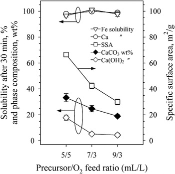
Powders were prepared at the nominal iron content (Ca:Fe = 125) according to the mass ratio of the RDA values of Ca and Fe.Reference Otten, Hellwig and Mayers27 Because of the significantly higher RDA requirement of Ca, Fe is a dopant in the easily soluble CaO matrix. Figure 1 shows the SSA (squares) along with the calcium (circles) and iron solubilities (triangles) of these powders i.d.a. (pH = 1) after 30 min as a function of precursor/O2 FSP-feed ratio. Increasing this ratio results in hotter and longer flamesReference Camenzind, Strobel and Pratsinis30 leading to increased high temperature particle residence times. Furthermore, increasing that ratio increases the particle concentration in the flame leading to increased particle coagulation, sintering, and crystal growth that decrease the SSA from 67 to 30 m2/g (Fig. 1).
SSA (squares), calcium hydroxide (open diamonds), and calcium carbonate contents (filled diamonds) as well as iron (triangles) and calcium (circles) solubilities of flame-made powders at the nominal RDA Ca:Fe mass ratio of 125 for males (19–50 years old) as function of the precursor/O2 feed ratio to the FSP reactor. Increasing that ratio reduces the SSA along with hydroxide and carbonate contents but does not affect the Fe and Ca solubilities i.d.a. in 30 min.

Here, the iron solubility i.d.a. is around 100%, irrespective of SSA, indicating significant in vivo bioavailability. This is in contrast to pure iron oxide (γ-Fe2O3) that exhibits lower solubilityReference Hilty, Knijnenburg, Teleki, Krumeich, Hurrell, Pratsinis and Zimmermann24 i.d.a. (74% after 30 min), despite having significantly higher SSAReference Hilty, Knijnenburg, Teleki, Krumeich, Hurrell, Pratsinis and Zimmermann24 (185 m2/g). Co-oxidation of Fe with Ca during FSP results in mostly CaO/Fe2O3 in solid solution, which improves iron dissolution.Reference Hilty, Knijnenburg, Teleki, Krumeich, Hurrell, Pratsinis and Zimmermann24
Figure 1 shows also that the Ca(OH)2 (open diamonds) and CaCO3 (filled diamonds) contents of these FSP-made powders decrease for increasing precursor/O2 FSP-feed ratio. Calcium hydroxide is readily formed at ambient conditions since CaO has strong moisture affinity.Reference Boynton31 Here, Ca(OH)2 is likely formed on the filter during powder collection. Calcium carbonate may be formed via two routes: in the presence of water (wet carbonation), CaO initially forms Ca(OH)2 and subsequently CaCO3 via dissolution of CO2 in a water layer around Ca(OH)2.Reference Shih, Ho, Song and Lin32 This is a strong function of relative humidityReference Shih, Ho, Song and Lin32, Reference Dheilly, Tudo and Queneudec33 and the dominant mechanism for CaCO3 formation at ambient conditions.Reference Boynton31, Reference Dheilly, Tudo and Queneudec33 In the absence of water, dry carbonation may take placeReference Boynton31 without formation of intermediate Ca(OH)2, dominantly at temperatures above 400 °C. During FSP synthesis of CaO, dry carbonation may take place downstream of the flame where gas temperatures are high, while wet carbonation occurs during sample collection and powder storage at ambient conditions. For both carbonation mechanisms, smaller CaO particles react fast with CO2 and water because of their large exposed surface area. Most calcium carbonate here probably is formed by dry carbonation above the flame because TGA was done directly after synthesis, limiting room temperature carbonation.
As the precursor/O2 FSP-feed ratio increases, the product CaO particles become larger. Higher precursor concentrations lead to larger particlesReference Madler, Kammler, Mueller and Pratsinis25 with less surface available to react with CO2 whose diffusion into CaO is too slow for complete carbonation.Reference Silaban and Harrison34 This is in agreement with Lu et al.Reference Lu, Smirniotis, Ernst and Pratsinis35 who observed the highest CaCO3 content for the smallest precursor/O2 feed ratio for FSP-made Ca-containing powders. They observed <10 wt% Ca(OH)2, suggesting that all CaCO3 was formed during FSP synthesis.Reference Lu, Smirniotis, Ernst and Pratsinis35 In contrast, Huber et al.Reference Huber, Stark, Loher, Maciejewski, Krumeich and Baiker36 observed that the highest carbonate fraction was obtained with the largest precursor/O2 FSP-feed ratio. They had measured, however, the CaCO3 content of their powder hours to days after synthesis. This was verified here by repeating their experimentsReference Huber, Stark, Loher, Maciejewski, Krumeich and Baiker36 and aging such powders for up to 17 days.
The carbonate content here is significantly lower than previous reports.Reference Lu, Smirniotis, Ernst and Pratsinis35, Reference Huber, Stark, Loher, Maciejewski, Krumeich and Baiker36 This can be attributed to the presence of large CaO particles made by droplet-to-particle conversion during FSP of the present ethanol/2-EHA-based precursor solutions (Sec. III. B). Such large particles were absent in powders made by FSP of xylene-basedReference Lu, Smirniotis, Ernst and Pratsinis35, Reference Huber, Stark, Loher, Maciejewski, Krumeich and Baiker36 solvents. In addition, storage of such powdersReference Lu, Smirniotis, Ernst and Pratsinis35, Reference Huber, Stark, Loher, Maciejewski, Krumeich and Baiker36 over time may have increased their carbonate content as well, as will be shown here. It should be noted that the Fe solubility i.d.a. after 30 min was significant despite these changes in phase composition (oxide, hydroxide, or carbonate) and the low SSA of these Ca-based nanopowders.
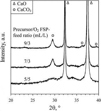
Figure 2 shows the XRD patterns of the FSP-made Fe-doped powders as a function of the precursor/O2 FSP-feed ratio. For short flames (5/5), the Ca(OH)2 and CaCO3 in these powders (Fig. 1) are amorphous. Increasing the FSP-feed ratio increases the crystalline fraction of CaCO3 (calcite) as seen by its peak intensityReference Lu, Smirniotis, Ernst and Pratsinis35 at 29.5°. For the 7/3 and 9/3 flames, the crystalline CaCO3 fraction is 10 wt% for both while the total CaCO3 fraction is 25 wt% and 19 wt%, respectively (Fig. 1), indicating that a large part of CaCO3 is amorphous. In contrast, Lu et al.Reference Lu, Smirniotis, Ernst and Pratsinis35 produced fully crystalline CaCO3 for the same FSP-feed ratios. CrystallizationReference Huber, Stark, Loher, Maciejewski, Krumeich and Baiker36 of CaCO3 takes place around 310 °C. Because of theirReference Lu, Smirniotis, Ernst and Pratsinis35 short (40 cm) burner-to-filter distance (in contrast to 50–70 cm here) and their high enthalpy xylene-based precursor solution, it is likely that during particle synthesis, the temperature at their filter was sufficiently high to fully crystallize the CaCO3. The present d XRD for CaO (CaCO3) made with the 7/3 and 9/3 flames is 68 (11) and 89 (11) nm, respectively. The “goodness-of-fit” figure of merit of the Rietveld refinement was low (<1.3) and was not improved by applying a two-modeReference Rudin, Wegner and Pratsinis37 fit for CaO, indicating that the CaO crystals are unimodal. As these powders fully dissolved i.d.a. within 30 min (Fig. 1), the crystallinity of CaCO3 does not affect Fe dissolution. Both SSA and phase composition of these flame-made Ca/Fe-containing nanopowders do not affect the high solubility of iron i.d.a. pointing out their potential as nutritional supplements.
XRD patterns of Ca-based powders having Ca:Fe = 125 and produced at different precursor/O2 FSP-feed ratios. Increasing that ratio from 5/5 to 9/3 increases the crystalline CaCO3 content of these mostly CaO-containing as-prepared powders. The shortest and coolest flame (5/5), however, has the most CaCO3 and Ca(OH)2 (together 54 wt%, Fig. 1), all amorphous.

2. The effect of Fe-content
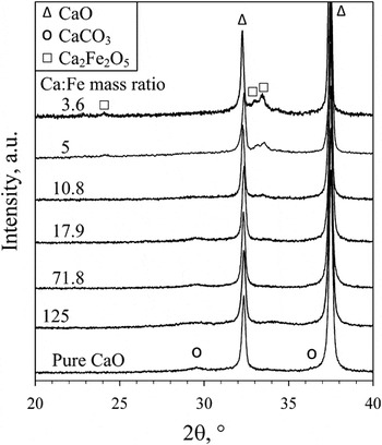
Figure 3 shows the XRD patterns of Ca/Fe-containing powders with varying composition. Pure Ca forms mainly crystalline CaO while the small hump at 2θ = 29.5° indicates amorphous or nanocrystalline CaCO3. Increasing the iron content to Ca:Fe = 17.9, no change is observed in the XRD patterns. For Ca:Fe < 17.9, a mixed iron-calcium-oxide Ca2Fe2O5 is formedReference Hilty, Knijnenburg, Teleki, Krumeich, Hurrell, Pratsinis and Zimmermann24 as indicated by its main peak at 2θ = 33.5°. This is in agreement with the binary phase diagram of CaO–Fe2O3: for Ca:Fe ≥ 0.7 all iron is present as Ca2Fe2O5 in equilibrium with excess CaO.Reference Bergman38 Thus, the high Ca content of the powders prevents the formation of low-solubilityReference Hilty, Knijnenburg, Teleki, Krumeich, Hurrell, Pratsinis and Zimmermann24 crystalline iron oxides.
XRD patterns of as-prepared Ca-based powders (5/5 flame) with varying iron content. For Ca:Fe ≤ 10.8, peaks for crystalline Ca2Fe2O5 become visible while crystalline CaCO3 disappears. Upon aging, however, these powders become predominantly CaCO3 (Fig. 5).

The iron and calcium solubility i.d.a. after 30 min was around 100% (not shown) for Ca:Fe ≥ 10.8. This variation of iron content in the Ca-based powders (Ca:Fe = 10.8–125) does not affect iron dissolution, allowing synthesis of highly soluble compounds tailor-made to specific population groups. For Ca:Fe = 5, however, the iron solubility after 30 min decreased to 91% and to 87% for Ca:Fe = 3.6. Previously, we had shown that iron solubility only decreases for very high iron contents (Ca:Fe < 0.7 or molar Ca:Fe < 1).Reference Hilty, Knijnenburg, Teleki, Krumeich, Hurrell, Pratsinis and Zimmermann24 The low iron oxide content (Ca:Fe = 3.6) of powder made here at x/y = 5/5 has a smaller crystalline iron fraction (33 wt% crystalline Ca2Fe2O5 with d XRD = 24 nm) and a lower SSA of 67 m2/g compared with those made in the pastReference Hilty, Knijnenburg, Teleki, Krumeich, Hurrell, Pratsinis and Zimmermann24 at x/y = 3/7 (Ca:Fe = 0.7 containing 48 wt% crystalline Ca2Fe2O5 with d XRD = 27 nm and an SSA of 98 m2/g). The Ca2Fe2O5 fraction and size may not be responsible for the reduced dissolution at x/y = 5/5, as evidenced by the 100% calcium solubility i.d.a. already after 15 min (not shown). This indicates that in both powders, a fraction of low-solubility and perhaps nanocrystalline iron oxide is present that is not detected by XRD. Increasing the precursor/O2 FSP-feed ratio increases the aerosol concentration and high temperature residence timeReference Strobel and Pratsinis26 that promotes particle/crystal growth.Reference Camenzind, Strobel and Pratsinis30 For such high Fe contents (Ca:Fe ≤ 5), both crystallinity and SSA determine the dissolution rate.
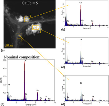
Figure 4(a) shows an HAADF-STEM image of powders containing Ca:Fe = 5 with the corresponding analysis of indicated sample areas 1–3 [Figs. 4(b)–4(d)] by EDX spectroscopy along with the nominal composition [Fig. 4(e)]. Analysis of area 1 [Fig. 4(c)] shows particles that are enriched with Ca: the ratio of Ca:Fe in the EDX signal is stronger than that in the nominal composition [Fig. 4(e)]. For areas 2 and 3, however, the composition [Figs. 4(b) and 4(d)] appears close to the nominal one [Fig. 4(e)]. This difference in EDX between areas 1 and 2/3 indicates that not all iron is homogeneously distributed throughout the powder. This is in agreement with the XRD pattern (Fig. 3) where crystalline Ca2Fe2O5 was observed for that powder.
STEM image (a) of powders containing Ca:Fe = 5 with EDX analysis at indicated positions. Area 1 is enriched with calcium, whereas areas 2 and 3 seem close to the nominal composition of iron and calcium. This demonstrates that iron is rather inhomogeneously distributed inside the CaO matrix, as supported by XRD (Fig. 3) where crystalline Ca2Fe2O5 was found.

3. The effect of storage
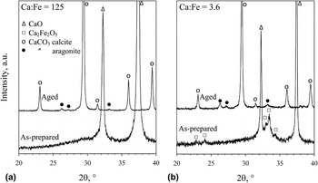
Figure 5 shows the influence of storage time at ambient conditions on the XRD patterns of as-prepared powders with Ca:Fe mass ratio (a) 125 and (b) 3.6 and aged for 225 days. The as-prepared powder with Ca:Fe = 125 [Fig. 5(a)] contains dominantly CaO with a fraction of amorphous Ca(OH)2 and CaCO3 (Fig. 1, 5/5 flame). After 225 days, that powder has changed completely to CaCO3 (circles in Fig. 5) as confirmed by TGA. Then mostly the thermodynamically stable calcite CaCO3 is presentReference Radha, Forbes, Killian, Gilbert and Navrotsky39 with a small fraction (2.8 wt%) of metastable aragonite CaCO3 (filled circles) perhaps due to the presence of Fe. Increasing the iron content to Ca:Fe = 3.6 [Fig. 5(b)] increases the aragonite fraction (12.8 wt%), possibly originating from the initially present Ca2Fe2O5.
XRD patterns of CaO-based powders with Ca:Fe mass ratio (a) 125 and (b) 3.6, as-prepared and aged for 225 days under ambient conditions. The as-prepared materials consist mainly of crystalline CaO with Ca2Fe2O5. Upon storage, both compounds are converted completely to CaCO3 (as confirmed by TGA) by the influence of humidity and CO2 in the atmosphere. Mostly thermodynamically favorable calcite is formed while the presence of iron likely induces formation of metastable aragonite.

The SSA is 67 m2/g for both as-prepared calcium-based powders (Ca:Fe = 125 and 3.6). Upon 225 days storage, their SSA decreased to 19 and 44 m2/g, respectively. The increased iron fraction apparently inhibited SSA reduction by hydration and carbonation. Despite these strong changes in SSA and crystal structure, the iron solubility i.d.a. did not decrease significantlyReference Hilty, Knijnenburg, Teleki, Krumeich, Hurrell, Pratsinis and Zimmermann24: 96% and 84% after 225 days aging of powders containing Ca:Fe = 125 and 3.6, respectively, compared with 97% and 87% for the as-prepared compounds. Quite likely iron is incorporated in the CaCO3 lattice.Reference Fisler, Gale and Cygan40 This favors Fe dissolution because of the lower lattice energy of CaCO3 (2804 kJ/mol)Reference Haynes41 compared with Fe2O3 (14,309 kJ/mol)Reference Haynes41 or Ca2Fe2O5 (7046 kJ/mol).Reference Yoder and Flora42
B. Zinc/calcium oxides
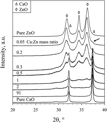
Figure 6 shows the XRD patterns of FSP-made powders with varying Ca:Zn mass ratio. Powders with Ca:Zn = 91 closely correspond to the RDA values (1000 mg and 11 mg for calcium and zinc, respectively).Reference Otten, Hellwig and Mayers27 For low Zn contents (Ca:Zn > 2), the XRD patterns resemble those of crystalline CaO while the small hump at 2θ = 29.5° indicates amorphous CaCO3. Separate ZnO is not detected until Ca:Zn = 1, possibly because ZnO is largely amorphous or nanocrystalline (d XRD ≤ 3.4 nm).Reference Madler, Stark and Pratsinis43 At Ca:Zn = 0.05–1, both CaO and ZnO crystals coexist. The main peak of CaO is visible at 2θ = 37.4° for Ca:Zn = 0.05 (as indicated by an arrow in Fig. 6) while no mixed oxide is formed.
XRD patterns of FSP-made powders (5/5 flame) with varying Ca:Zn mass ratio. For Ca:Zn ≤ 1, the XRD patterns of ZnO and CaO coexist. Crystalline CaO is still detected at Ca:Zn = 0.05 (small peak around 37.4° as indicated by the arrow). No new mixed Ca–Zn oxide appears to be formed.

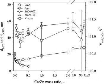
Figure 7 shows the d BET and d XRD of the above powders. Their d BET changes from 15 (pure ZnO) to 28 nm (pure CaO) with a minimum of 12 nm at Ca:Zn = 0.1. The d XRD corresponding to the (002) plane of ZnO decreases from 20 to 5 nm from pure ZnO to Ca:Zn = 0.5, whereas the d XRD of their (100) plane decreases as well from 14 to 5 nm. For Zn-rich materials (Ca:Zn ≤ 0.1), the d BET corresponds well to an average d XRD of the (100) and (002) planes. For Ca:Zn = 0.1–0.5, the d XRD of the two ZnO planes are identical indicative of compact (spherical-like) ZnO crystals, similarReference Height, Madler, Pratsinis and Krumeich29 to Li-doped ZnO. At these compositions, the d BET seems to correspond well to an average of the CaO and ZnO crystal sizes, indicating monocrystalline powders.
CaO unit cell volume (squares), crystal size of ZnO (triangles and inverted triangles) and CaO (diamonds), and BET-equivalent diameter (circles) as function of the Ca:Zn mass ratio. The CaO crystal size is larger than the BET-equivalent diameter quite likely due to the formation of nanosized amorphous Ca(OH)2 and CaCO3 particles that coexist with the larger CaO crystals. For Ca:Zn ≤ 0.1, the two ZnO crystal sizes are in good agreement with the BET-equivalent diameter. The CaO unit cell volume is hardly affected by the Zn addition.

The ZnO crystal size decreases with increasing CaO content similar to SiO2-doping of flame-made ZnO quantum dots,Reference Madler, Stark and Pratsinis43 possibly as a result of slower sintering of mixed oxides compared with pure oxides.Reference Vemury and Pratsinis44 In the case of Si-doping and comparing ionic radiiReference Shannon45 (based on a coordination number of 6), the smaller Si4+ (0.40 Å) compared with Zn2+ (0.74 Å) can enter the ZnO lattice interstitially suppressing ZnO crystal growth.Reference Tani, Madler and Pratsinis46 The Ca2+ ionic radius (1.00 Å) is larger than Zn2+ and is more likely to substitute Zn2+ in the crystal. This may decrease the ZnO crystal size, similar to doping of ZnO with Cd2+ (0.95 Å) by high-pressure solution growthReference Ghosh and Raychaudhuri47 and dopingReference Akhtar, Pratsinis and Mastrangelo48 of TiO2 with Al3+ during its aerosol synthesis in hot wall reactors.
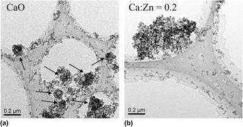
The CaO crystal size is significantly larger than the BET-average diameter for all compositions. To verify whether this is because the CaO crystals are bimodally distributed, the XRD patterns were fitted using the Rietveld refinement with two crystal modes.Reference Rudin, Wegner and Pratsinis37 The goodness-of-fit figure of merit did not decrease significantly compared with a one-mode fit, indicating that both ZnO and CaO form unimodal crystals. A possible explanation for the larger CaO crystal size compared with d BET is the formation of nanosize amorphous CaCO3 as seen by TEM [Fig. 8(a)] and confirmed by TGA. The TEM image of mixed Ca/Zn oxide with Ca:Zn = 0.2 [Fig. 8(b)], however, did not show any large particles even though the d XRD of CaO is larger than the average d BET (Fig. 7): it is quite likely that there are only very few of them. The ZnO does not form a bimodal size distribution since even if large ZnO crystals had been formed, these would decompose rapidly above 1700 °C and nanosized ZnO crystals would form from the gas phase at lower temperatures.Reference Strobel and Pratsinis49
Typical TEM images of (a) pure CaO and (b) Ca/Zn oxide containing Ca:Zn mass ratio of 0.2. Pure CaO forms a bimodal particle size distribution since large particles (indicated by the arrows) coexist with nanosized ones. For Ca:Zn = 0.2, a more homogeneous distribution is found.

The unit cell volume of CaO was calculated (Fig. 7, squares) to explore the formation of CaO–ZnO solid solutions. This volume varies around 111 Å3 (corresponding to a unit cell length of 0.48 nm) so a clear trend is not visible. Incorporation of Zn2+ into the CaO lattice is expected to decrease the CaO unit cell volume because of the smaller radius of Zn2+ compared with Ca2+, as shown for Mg2+ doping.Reference Doman, Barr, McNally and Alper50 The absence of such a trend does not necessarily mean that Zn2+ is not dissolved in CaO. Substitution of Zn2+ for Ca2+ in CaCO3 (calcite) is energetically favorableReference Fisler, Gale and Cygan40 and was found experimentallyReference Reeder, Lamble and Northrup51 to occur at low (<1000 ppm Zn) concentrations. Incorporation of Zn2+ in CaO or CaCO3 reduces the ZnO lattice energyReference Yoder and Flora42 and thus the average Zn–O bond strength, as the lattice energyReference Haynes41 of CaO (3′414 kJ/mol) and CaCO3 (2′804 kJ/mol) is lower than that of ZnO (4′142 kJ/mol). The solubility i.d.a. of pure ZnO (15–25 m2/g), however, is already very highReference Hilty, Teleki, Krumeich, Buchel, Hurrell, Pratsinis and Zimmermann28 (>95%) thus a solid solution cannot further improve Zn dissolution at this pH. Here also the Zn and Ca solubilities i.d.a. after 30 min were already 100%, irrespective of composition (not shown). This shows that the composition of Ca/Zn-containing powders can be tuned freely without sacrificing product performance.
C. Multicomponent doping of calcium oxide
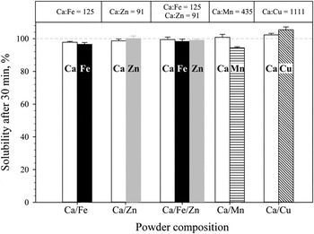
Along with Fe and Zn, Cu and Mn are also essential trace minerals for humans. These elements are present in most multimineral supplement tablets.Reference Webb12 In rodent studies, Mn deficiency was associated with skeletal abnormalities as well as distortion in carbohydrate and lipid metabolism.Reference Shils, Olson, Shike and Ross52 Copper deficiency is associated with anemia, neutropenia, and osteoporosis.Reference Shils, Olson, Shike and Ross52 Because of their importance in human nutrition, we also produced CaO with stoichiometric amounts of copper and manganese at standard FSP conditions; their solubility i.d.a. after 30 min is shown in Fig. 9. The dopant amounts are given above the figure and are based on the respective RDA/AI values of the individual metals for males 19–50 years [Ca (1000 mg), Fe (8 mg), Zn (11 mg), Mn (2.3 mg), and Cu (0.9 mg)].Reference Otten, Hellwig and Mayers27 Here, the solubility of all elements is again high (above 94%) demonstrating that different trace elements and their mixtures at RDA-relevant compositions incorporated in CaO matrices can be produced in one step by scalable flame spray pyrolysis. Even a mixture of Fe and Zn may be incorporated in the CaO with high solubility i.d.a. without affecting each other’s dissolution (Fig. 9). In single meal studies, it has been shown that high concentrations of calcium can negatively influence iron absorption. However, in long-term studies, this effect is often less pronounced or absent (for a review see LynchReference Lynch53). Mineral interactions can be observed in conventional supplementsReference Wieringa, Berger, Dijkhuizen, Hidayat, Ninh, Utomo, Wasantwisut and Winichagoon8, Reference Fairweather-Tait and Teucher13 and are thus not specific for nanostructured compounds.
Solubility i.d.a. after 30 min for the various dopants in the CaO matrix. The Ca:dopant mass ratios (above the figure) are based on the RDA values of each mineral. Apart from iron, also zinc, copper and manganese, or even combinations of elements may be incorporated in the one-step FSP process. For all elements, almost complete dissolution is obtained after 30 min, suggesting these compounds should have high in vivo bioavailability.

IV. CONCLUSIONS
Nanostructured Ca-based supplements with nutritionally relevant amounts of iron and/or zinc were prepared by scalable, one-step flame aerosol technology. Their complete dissolution i.d.a. after 30 min suggests that these nanomaterials should have high in vivo bioavailability. The particle size and composition (oxide/carbonate, mineral composition) do not affect solubility i.d.a. for Ca/Fe and Ca/Zn oxides. Matrix encapsulation of Fe by CaO results in a mixed oxide Ca2Fe2O5 coexisting with mainly crystalline CaO and amorphous calcium hydroxide and carbonate. For Ca:Fe ≥ 10.8, the solubility i.d.a. after 30 min does not depend on crystallinity and SSA. Storage for 225 days under ambient conditions converted the powders to crystalline CaCO3 that hardly affected the Fe solubility. The large amounts of calcium prevent formation of separate, low-solubility iron oxide.
No mixed Ca–Zn oxide was observed and no change in the CaO unit cell volume was measured by Zn doping. Flexibility of the process is demonstrated by variation of powder composition. Also other trace elements such as Mn and Cu may be incorporated, with high solubility i.d.a. and therefore potentially high bioavailability. Thus flame-made nanostructured compounds may offer a promising approach to manufacture tailor-made compounds for supplementation. Since the powders are homogeneous and do not segregate even when multiple phases are present, they may also facilitate the production of mineral supplement tablets and capsules.
ACKNOWLEDGMENTS
This work was supported by ETH Research Grant ETH-06 10-1. (S)TEM investigations were carried out at the Electron Microscopy ETH Zurich.