Congenital tricuspid regurgitation without downward displacement of the leaflet is an extremely rare condition.Reference Aaron, Mills and Lower1, Reference Becker, Becker and Edwards2 Pathological features of congenital tricuspid regurgitation are variable and its surgical management challenging.
Recently, more cases of congenital tricuspid regurgitation have come to light due to the increased use and efficacy of transthoracic echocardiography. There is a renewed interest in this rare clinical entity; however, congenital tricuspid regurgitation has not been discussed in detail, even in extensive studies on aetiology of isolated tricuspid regurgitation.Reference Waller, Bloch, Backer, Roe, Brown and Mahomed3, Reference Hauck, Freeman, Ackermann, Danielson and Edwards4
Only a small number of cases of patients in each individual series have been published so far. The morphologic heterogeneity makes it difficult to derive any conclusions regarding the best strategy for management of this condition. With the above scenario in mind, we analysed the published literature on congenital tricuspid regurgitation using MEDLINE. In addition, the reference sections of all relevant articles were searched to identify additional cases. Hence, the aim of this study was to review all published literature on congenital tricuspid regurgitation, allowing conclusions to be drawn regarding the morphological and clinical spectrum of this rare clinical entity.
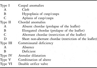
Moreover, a satisfactory and updated classification in view of the anatomical heterogeneities in congenital tricuspid regurgitation is missing. After analysing the published literature, we also propose a new classification for congenital tricuspid regurgitation, which includes the morphological and anatomical variations that can help the surgeon in deciding on the strategy for management.
Patients and methods
Definition of congenital tricuspid regurgitation
There is no definition as such for congenital tricuspid regurgitation, although it is definitely a separate entity characterised by “tricuspid valvular dysplasia”Reference Becker, Becker and Edwards2 and lack of apical displacement of the septal leaflet.
Criterion for selection of patients
We considered congenital tricuspid regurgitation when there was isolated, non-inflammatory, non-traumatic, non-infective tricuspid regurgitation.
Congenital tricuspid regurgitation occurring as a mixed lesion with tricuspid stenosis and double orifice tricuspid valve occurring as a part of Ebstein's anomaly and atrioventicular septal defects were excluded from the study. Tricuspid regurgitation secondary to right ventricular failure was excluded.
Studies published till June, 2009 are included. As far as we could establish, there were 123 cases of Congenital Tricuspid Regurgitation described in 48 publications.
Incidence
The true incidence of congenital tricuspid regurgitation is unknown. It is an extremely rare clinical entity.Reference Aaron, Mills and Lower1–Reference Hauck, Freeman, Ackermann, Danielson and Edwards4 Of the 123 cases of congenital tricuspid regurgitation, 63 were documented during surgery,Reference Aaron, Mills and Lower1, Reference Hauck, Freeman, Ackermann, Danielson and Edwards4, Reference Barr, Celermajer and Bowdler17, Reference Tutarel and Westhoff-Bleck20, Reference Boon, Hazekamp and Hoohenkerk21–Reference Honjo, Ishino and Yoshizumi24, Reference Lynch and Doty56 38 during autopsy,Reference Becker, Becker and Edwards2, Reference Urban5–Reference Tutarel and Westhoff-Bleck20, Reference Klein52, Reference Cascos, Rabago and Sokolowski54, Reference Pignoni, Carraro and Nicolosi55 and 22 were diagnosed by echocardiography or cardiac catheterisation.Reference Green, Ziegler and Mieher19, Reference Tutarel and Westhoff-Bleck20, Reference Boucek, Graham, Morgan, Atwood and Boerth42–Reference Ozkutlu, Gunal, Caglar, Alehan and Gungor44, Reference Goldberg, Schifter, Aron, Shapiro, Krasnow and Stein57
Demography
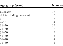
Age was reported in 71 patients, while eight were described as neonates. The mean age was 22.57 years, with a standard deviation of 23.42 years, and the age ranged from 1 day to 80 years, with a median of 16 years The age distribution is shown in Table 1.
Age distribution of the patients having congenital tricuspid regurgitation.

Gender was referred to in 79 cases; these included 47 male patients (59.5%) and 32 female patients (40.5%), such that there was a male predominance, with a male-to-female ratio of approximately 3:2.
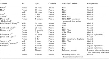
Initial series on congenital tricuspid regurgitation (1904–1972)
These studies (Fig 2) had few features in common:
• Most patients were neonates; adult patients had also presented or were diagnosed in early childhood.
• Clinical presentation was with cyanosis and/or congestive cardiac failure.
• Management was medical therapy or surgical exploration only.
• Course was universally fatal.
• Morphology of lesion was delineated at autopsy or at cardiac catheterisation and angiocardiography.
• Morphology was similar, with short and thickened chordae attached to rudimentary papillary muscles or directly to right ventricular musculature, and fusion of two leaflets leading to a bicuspid valve or thickened and nodular leaflets; however, there was no hypoplasia of leaflets.
• The right ventricle and right atrium were markedly dilated.
Unlike previous studies, two studies by Green et alReference Green, Ziegler and Mieher19 and Kanjuh et alReference Kanjuh, Stevenson, Amplatz and Edwards18 reported hypoplasia of tricuspid leaflets. They even reported a total absence of tricuspid valve leaflets, the so-called “Unguarded” tricuspid valve.
Later studies on congenital tricuspid regurgitation (1975 onwards)
Aaron et alReference Aaron, Mills and Lower1 reported the first successful surgical treatment of congenital tricuspid regurgitation by tricuspid valve replacement with a prosthetic valve. As others also reported success in surgical management of congenital tricuspid regurgitation, surgical treatment became firmly established. Over time, tricuspid valve repair became the preferred treatment, with tricuspid valve replacement being reserved for failed repair (Table 2).
Showing age, sex, absence, or presence of cyanosis and management in Initial publications on congenital tricuspid regurgitation (1904–1972).

ASD = atrial septal defect; PDA = patent ductus arteriosus; PFO = patent foramen ovale
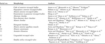
These studies also brought to light various morphologies causing congenital tricuspid regurgitation. These are summarised in Table 3.
Description of various morphological abnormalities in the published literature.

Anomalies associated with congenital tricuspid regurgitation
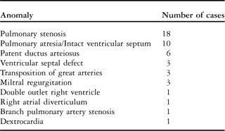
Patent foramen ovale and atrial septal defect are regularly associated with this clinical entity. Additional congenital cardiac malformations are reported in 47 cases (38.2%). More detailed information is provided in Table 4.
Associated congenital malformation associated with congenital tricuspid regurgitation.

Proposed classification
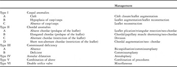
In the absence of an updated classification of congenital tricuspid regurgitation, which takes into account its various morphological variations, we analysed the published literature to propose a new classification for congenital tricuspid regurgitation (Table 5). This not only includes the anatomical variations, but can also help the surgeon in deciding on the best strategy for management. The defect may be one of annular dilatation, defective commissure, defective cusps, abnormal/absent chordae, or a combination of these. The cusp may be anatomically deficient, such as with a cleft or hypoplasia/aplasia. The cusp may be functionally deficient, either prolapsing due to absent/elongated chordae or having restricted movement due to aberrant/short chordae.
Proposed new classification for congenital tricuspid regurgitation.

Discussion
Incidence and clinical presentation
The true incidence of congenital tricuspid regurgitation is unknown. The presentation in neonates is with cyanosis and congestive cardiac failure, in childhood with congestive cardiac failure, while adults present usually with congestive cardiac failure and arrhythmias.
In their review of 32 cases, Lagarde et alReference Lagarde, Garabedian, Coignard, Duret, Piwnica and Droniou46 separated congenital tricuspid regurgitation into two groups according to their clinical contexts and outcome. One group comprised of newborns and infants, the outcome for whom was fatal, because of right ventricular failure, and autopsy confirmed the diagnosis. The other group comprised of patients who decompensated during adult life with right ventricular failure, with a prolonged natural course.
The neonates are of normal birthweight and follow either of two courses. Some are asymptomatic and present later in life, either in childhood or as adults. Some are born with a dilated heart and present as sick neonates with cyanosis and congestive cardiac failure. They may deteriorate if not optimally managed or may even spontaneously improve with time to present later in life.
Investigations
The chest radiograph shows right atrial and right ventricular enlargement and normal pulmonary vascularity.Reference Aaron, Mills and Lower1, Reference Barritt and Urich11, Reference Barr, Celermajer and Bowdler17, Reference Eichhorn, Ritter, Suetsch, von Segesser, Turina and Jenni29–Reference Girard, Nishimura, Warnes, Dearani and Puga40
An electrocardiograph usually shows right axis deviation, right atrial enlargement, right bundle branch block, and first degree atrioventricular block,Reference Aaron, Mills and Lower1, Reference Abrams, Kilner and Till27, Reference Eichhorn, Ritter, Suetsch, von Segesser, Turina and Jenni29 and sometimes prolonged corrected QT interval.Reference Eichhorn, Ritter, Suetsch, von Segesser, Turina and Jenni29 There may be sinus tachycardia, supraventricular extrasystoles, and atrial fibrillation.Reference Barritt and Urich11, Reference Barr, Celermajer and Bowdler17, Reference Eichhorn, Ritter, Suetsch, von Segesser, Turina and Jenni29–Reference Girard, Nishimura, Warnes, Dearani and Puga40
Cardiac catheterisation reveals elevated right atrial pressure, a pronounced “V” wave of tricuspid regurgitation, normal right ventricular pressure, and sometimes a right-to-left shunt at the atrial level.Reference Aaron, Mills and Lower1, Reference Palladino and Kinney10–Reference Sanyal, Bhargava and Saxena15, Reference Montero, Tellez and Simarro33–Reference Yang, Wu, Xu, Shen, Gao and Liu38 However, cardiac catheterisation is not required to make a diagnosis in the present day.
Transthoracic echocardiography can diagnose the severity of tricuspid regurgitation and exclude the presence of increased pulmonary artery pressure and other anomalies, estimate the function of the right ventricle, and also delineate the exact morphological abnormality in the valve. Real-time three-dimensional echocardiography, unlike two-dimensional echocardiography, is able to simultaneously visualise all the three leaflets during the cardiac cycle and their attachment in the tricuspid annulus.Reference Badano, Agricola, Perez de Isla, Gianfagna and Zamorano58 Anwar et alReference Anwar, Geleijnse and Soliman59 showed that a detailed anatomical structure of the tricuspid valve can be delineated by real-time three-dimensional echocardiography, including the annulus size and shape, leaflet shape and mobility, and tricuspid commissural width.Reference Anwar, Geleijnse and Soliman59 It also provides an en-face view of the tricuspid valve from the right atrium – “surgical view” – which can be very helpful to the surgeon in planning the surgery.Reference Badano, Agricola, Perez de Isla, Gianfagna and Zamorano58
Natural history
Severe disease in utero
Congenital tricuspid regurgitation has a high in utero mortality, which is clearly shown by foetal echocardiography.Reference Lang, Oberhoffer and Cook45 According to Lang et al,Reference Lang, Oberhoffer and Cook45 the cause of this is a combination of tricuspid regurgitation with pulmonary hypoplasia.
Severe disease ex utero
Substantial mortality in the neonatal period is also reported in patients with severe tricuspid regurgitation.Reference Aaron, Mills and Lower1, Reference Barr, Celermajer and Bowdler17, Reference Lang, Oberhoffer and Cook45 The pathophysiological basis for this is the presence of poor antegrade pulmonary blood flow due to severe tricuspid regurgitation and high pulmonary vascular resistance. The only source of pulmonary blood in such a situation is a patent ductus arteriosus. This poor pulmonary blood flow as well as right-to-left shunting at the atrial level gives rise to cyanosis. Severe tricuspid regurgitation manifests as congestive cardiac failure. As the ductus arteriosus closes, it would lead to increasing cyanosis and hypoxia and the universally fatal outcome.
The presence of pulmonary stenosis and pulmonary atresia, with intact ventricular septum, adversely impacts outcome by further decreasing pulmonary blood flow and increasing tricuspid regurgitation. Thus, only a few such patients survive beyond the neonatal period.
Moderate disease ex utero
However, in patients with a moderate form of disease, there will be enough antegrade flow tide over the initial crisis. As the pulmonary vascular resistance falls with establishment of the lungs, the pulmonary blood flow increases and tricuspid regurgitation decreases, leading to an improvement in the symptoms.Reference McElhinney, Silverman, Brook, Hanley and Stanger31 Boucek et alReference Boucek, Graham, Morgan, Atwood and Boerth42 also reported such spontaneous resolution, but they failed to attribute it to the natural history of this disease. Over time, the volume-overloaded right ventricle dilates, as does the tricuspid annulus, increasing the degree of regurgitation and presentation during childhood.
Mild disease ex utero
Patients with mild disease are asymptomatic in childhood. The severity of the disease increases over a period of time and they present in adulthood with arrhythmias and right ventricular dysfunction, the risk increasing with age. Death due to fatal arrhythmia is reported.Reference Antia and Osunkoya16
Morphology and management
A combination of functional and anatomical lesions and young age of the patient represents a challenging surgical problem. Initially, most lesions were dealt with by valve replacement. Long-term studies have clearly shown replacement to be a sub-optimal solutionReference Pasque, Williams, Coles, Trusler and Freedom49, Reference Fundaro, Di Mattia, Salati and Santoli50 that is particularly undesirable in children.Reference Castedo, Cañas, Cabo, Burgos and Ugarte51 This is because of the complications and inconvenience of long-term anticoagulation as well as the poor long-term function of prosthetic valves in the tricuspid location. Therefore, tricuspid repair is the ideal surgical treatment for congenital tricuspid regurgitation where feasible. We will now describe the individual defects and the surgical approach to these defects in detail.
Cleft of leaflet
Cleft is most commonly described in the anterior tricuspid leaflet. The incidence is around 0.018% among patients undergoing Doppler echocardiography.Reference Eichhorn, Ritter, Suetsch, von Segesser, Turina and Jenni29 However, two-dimensional echocardiography is the best method to diagnose cleft. The cleft lies close to the area of fusion of the lateral endocardial cushion and right dorsal conus swelling, and thus may be a result of defective fusion.Reference Eichhorn, Ritter, Suetsch, von Segesser, Turina and Jenni29 When it is quite deep, it gives the tricuspid valve a quadracuspid configuration.Reference Motoyoshi, Tofukuji, Sakurai, Ohmi and Koichi23 Some authors have described the cleft of anterior leaflet as the sole cause of congenital tricuspid regurgitation.Reference Motoyoshi, Tofukuji, Sakurai, Ohmi and Koichi23, Reference Okutan, Yavuz, Bigin, Duver and Kutsal26
If the cleft is small and the anterior tricuspid leaflet is well supported by chordae, it is amenable to closure either by interrupted or continuous suture technique with or without annuloplasty. When the cleft is large and the central chordae are absent, leading to prolapse, reconstruction of the leaflet as well as the chordae is required. The repair can be supported with an annuloplasty.
Prolapse of leaflet
Prolapse can also be caused by elongated chordae or absent chordae with or without the absence of the papillary muscle. Elongated chordae can be managed by papillary muscle shortening,Reference Ootaki, Yamaguchi, Yoshimura, Oka, Yoshida and Hasegawa22 neochordae formation,Reference Boon, Hazekamp and Hoohenkerk21, Reference Reddy, McElhinney, Brook, Silverman, Stanger and Hanley39 or by cuspal level chordal shortening.Reference Mantovani, Grossi, Ferrarese and Sala30, Reference Fundaro, Di Mattia, Salati and Santoli50 Absent chordae can be dealt with by neochordae formation using a pericardium or expanded polytetrafluoroethylene.Reference Boon, Hazekamp and Hoohenkerk21, Reference Reddy, McElhinney, Brook, Silverman, Stanger and Hanley39
Restriction of leaflet: aberrant chordae and short non-aberrant chordae
Aberrant chordae are those that are inserted at the clear zone of the tricuspid leaflet, which is usually free of chordal insertion, instead of the usual site of insertion at the free edge or rough zone. They connect to the endocardium instead of the papillary muscle.Reference Kobza, Kurz and Oechslin32 This leads to tethering of the leaflet both anatomically and functionally, resulting in impaired mobility, incomplete coaptation, and apical displacement of the tricuspid leaflet tips during systole. Thus, the origin of the regurgitant jet is displaced towards the apex of the ventricle as opposed to other mechanisms of tricuspid regurgitation. Aberrant chordae must be distinguished from short non-aberrant tendinous chordae as a cause of tricuspid regurgitation as they have implications for surgical repair.Reference McElhinney, Silverman, Brook, Hanley and Stanger31
Short non-aberrant chordae are anatomically in the correct location connecting the papillary muscle to the leaflet-free edge, but are too short to result in complete closure of the tricuspid leaflets. This leads to apical displacement of the leaflet tips from the annulus plane during systole, a defect functionally similar to tethering.
Aberrant tendinous chordae with tethering may occur alone or in combination with short tendinous chordae. These two malformations were found to be associated in 70% of the cases reported by Kobza et al.Reference Kobza, Kurz and Oechslin32
Both entities may affect any of the three tricuspid leaflets. However, distinguishing short chordae as a primary congenital defect from chordae that have become too short in the course of right ventricular or tricuspid annulus dilatation from other causes may be difficult. Interestingly, Kobza et alReference Kobza, Kurz and Oechslin32 did not find short non-aberrant chordae in the patient with the largest right ventricle and tricuspid annulus. Reddy et alReference Reddy, McElhinney, Brook, Silverman, Stanger and Hanley39 have reported short non-aberrant chordae attaching only to the anterior tricuspid leaflet leading to restriction and causing congenital tricuspid regurgitation.
Tricuspid annular dilatation and right ventricular dilatation would cause almost all chordae to appear short and would not cause short chordae to only one of the three leaflets (i.e. anterior tricuspid leaflet).
In theory, the management of aberrant chordae is simple division, but this management has not been reported so far. Short non-aberrant chordae can be managed by chordal augmentationReference Reddy, McElhinney, Brook, Silverman, Stanger and Hanley39 or by neochordae formation and by dividing the short chordae.
Commissural deficiency
There can be two approaches to commissural deficiency. One approach is to recreate the commissure by commissuroplasty. The other is to eliminate the commissure and convert the tricuspid valve into a bicuspid structure. This involves joining two leaflets either with a Teflon or pericardial pleget and supporting them by neochordae formation, or by the other leaflet in the form of a Alfieri stitch.Reference Katogi, Aeba and Ito28, Reference Mantovani, Grossi, Ferrarese and Sala30, Reference Castedo, Cañas, Cabo, Burgos and Ugarte51
Annular dilatation
Annuloplasty should be performed in these patients, which may be limited to annuloplasty at the commissure, DeVegas annuloplasty, or annuloplasty using rings.Reference Katogi, Aeba and Ito28
Unguarded tricuspid valve
Defined by the absence of tricuspid valvular tissue with an existing orifice, it is a very rare malformation often coexisting with pulmonary atresia and intact ventricular septum. The first such case was reported by KleinReference Klein52 and the term “unguarded tricuspid orifice” was coined by Kanjuh et al.Reference Kanjuh, Stevenson, Amplatz and Edwards18
Anderson et alReference Anderson, Silverman and Zuberbuhler53 reviewed 46 cases of pulmonary atresia with intact septum; 17 had associated Ebstein's malformation, while three cases had a congenitally unguarded tricuspid orifice, two with complete absence of valvular tissue. Despite Anderson et alReference Anderson, Silverman and Zuberbuhler53pointing out that the right ventricle was dilated rather than hypoplastic in these cases of unguarded tricuspid orifice, KleinReference Klein52 and Kanjuh et alReference Kanjuh, Stevenson, Amplatz and Edwards18 found a hypoplastic right ventricle in their studies.
Studies by Ozkultu et alReference Ozkutlu, Gunal, Caglar, Alehan and Gungor44 and Kilic et alReference Kilic, Ozkutlu and Gunal43 indicate that echocardiography is sufficient for the diagnosis of unguarded tricuspid orifice. These cases can be managed by tricuspid valve replacement.
Double orifice tricuspid valve
Double orifice tricuspid valve is a rare congenital anomaly.Reference Tutarel and Westhoff-Bleck20 This entity is under-diagnosed because the two orifices are often not seen in standard echocardiographic views and there is a lack of awareness about the condition. There are three types of lesions which constitute a double orifice tricuspid valve:
• Commissural type: the accessory orifice is at the end of a valve commissure and its sub-valvular apparatus – chord and papillary muscle – is the normal one for that commissure, although sometimes accessory papillary muscles may be present.
• Central type: a fibrous band divides the tricuspid orifice into either equal or unequal parts.
• Hole type: the accessory orifice is a hole in the cusp. This form of double valve orifice is to be distinguished from a simple fenestration or cleft that has no sub-valvular apparatus.
It is essential for the identification of duplication of a valve that both orifices should be provided with a sub-valvular apparatus, although this may be rudimentary for one of them.
Successful tricuspid double orifice valve repair of the central type is reported by Lynch et al.Reference Lynch and Doty56 They divided the bridge between the anterior and septal leaflets and closed the regurgitating orifice by approximating the leaflet tissue and by performing a suture annuloplasty. In theory, once the bridge is divided, the tricuspid valve can be repaired as a single orifice valve. However, this repair might leave the valve mildly stenotic.
Valve repair for the commissural type and hole type has not been reported so far in the literature. Theoretically, the hole in the leaflet can be closed with a pericardial patch and the underlying chordae can be severed. Any resulting prolapse can be dealt with as described above. The commissural type can be handled by commissuroplasty, but the practical feasibility of these procedures is merely speculative at present.
Becker's classification and drawbacks
The report of Becker et alReference Becker, Becker and Edwards2 succinctly established the distinction between Ebstein's anomaly and congenital tricuspid regurgitation. In congenital tricuspid regurgitation specifically, there is a lack of apical displacement of the septal leaflet, lack of atrialisation of the right ventricle, and presence of outflow obstruction due to a large anterior leaflet. Becker et alReference Becker, Becker and Edwards2 coined the term “tricuspid valvular dysplasia”, which is characterised by the focal or diffuse thickness of leaflets (Grade I), deficient development of chordae and papillary muscles, improper separation of valve component from the ventricular wall (Grade II), and focal agenesis of valvular tissue (Grade III).
However, this classification does not consider other forms of anomalies like isolated dilated annulus, deficient commissure, and double orifice tricuspid valve as a part of congenital tricuspid regurgitation.
Unlike the description of Becker et al,Reference Becker, Becker and Edwards2 valve thickness is normal in a majority of the cases of congenital tricuspid regurgitation. Similarly, pulmonary stenosis is not a common accompaniment with congenital tricuspid regurgitation as reported by them in 80% of their cases. Our review revealed that out of 123 cases, pulmonary stenosis occurred in 18 patients (14%) and pulmonary atresia with intact interventricular septum in 10 patients (8.1%).
Proposed classification
The classification we propose not only includes the various forms of congenital tricuspid regurgitation but also guides its surgical management (Table 6). For example, cuspal anomalies like cleft can be closed with interrupted fine polypropylene sutures; a hypoplastic leaflet can be augmented with an autologous pericardium or plicated to leave a bicuspid valve. Prolapse of the cusp can be corrected by chordal shortening, at the cusp level or papillary level, or neochordae formation. Restriction can be managed by chordal augmentation or neochordae formation.
The new classification guides surgical management.

Conclusion
Congenital tricuspid regurgitation is an uncommon clinical entity with wide anatomical variations. It is characterised by high in utero and neonatal mortality. The severity of disease dictates the presentation in infancy, childhood, or adulthood. Tricuspid valve repair is the ideal treatment whenever feasible, especially in children.
We propose a new classification for congenital tricuspid regurgitation, which not only includes the anatomical variations, but can also help the surgeon in deciding on the best strategy for management.
Acknowledgement
The authors thank Dr Shiv Kumar Chaudhary, Additional Professor, Department of Cardiothoracic and Vascular Surgery, All India Institute of Medical Sciences, New Delhi, for his valuable suggestions during the compilation of this manuscript.