Implications
Tail biting in growing pigs is a major problem in intensive pig production systems. It has considerable impact on animal welfare as well as on farm economy. This paper is a part of a larger experiment comparing individual characteristics in relation to tail-biting activity and frequency of being bitten. The results show that respiratory disease is associated with victimization, but the mechanism needs to be clarified in further research. Effective measures of prevention of tail biting would have a profound effect on pig welfare.
Introduction
Tail biting in pigs is a behavioural abnormality with considerable impact on animal welfare as well as on farm economy. Despite a fairly good understanding of the predisposing factors at the herd level and widespread use of routine tail docking, the problem is far from solved. Scientists have emphasized a need to identify individual characteristics in both biters and victims in order to understand the mechanism behind tail biting, and to eventually find effective measures of prevention (Edwards, Reference Edwards2006).
Previous research on the basis of epidemiological data has identified correlations between disease and tail-biting prevalence at the farm level, suggesting that unhealthy animals might not resist being bitten, or that discomfort from being sick may cause tail biting (Moinard et al., Reference Moinard, Mendl, Nicol and Green2003). These studies have, however, only noted clinical disease and visible tail damage in living animals. Few attempts have been made to test the latter hypothesis by identifying tail biters (TB).
Tail wounds from biting appear to cause infections elsewhere in the body. Associations between tail damage and other pathological lesions have been reported repeatedly in slaughtered pigs (Elbers et al., Reference Elbers, Tielen, Snijders, Cromwijk and Hunneman1992; Huey, Reference Huey1996). This kind of material, however, only includes acute tail damage in market-sized pigs. Most healed cases as well as severe ones leading to death or culling are not included.
This study aimed to identify health differences in pigs classified according to tail-biting activity. Behavioural observations and sampling were both completed within a few days in order to collect all information during the actual tail-biting outbreak.
Material and methods
Animals and husbandry
The study was conducted on an 800-pig fattening farm in western Finland with a history of substantial tail-biting problems. The farm raised mixed-breed pigs, originating from several different farms, from about 25 to 30 kg to slaughter according to an all-in all-out scheme. Pigs were not tail-docked, as the procedure is prohibited in Finland. The study animals were collected from two consecutive batches between May and October 2009 (Table 1). They were chosen from five rooms with different group size (6 to 20 individuals), pen equipment and feeding method. The animals were fed a standard dry commercial feed for growing/finishing pigs either manually twice a day (two rooms) or ad lib from a feeder (three rooms). All pens were part-slatted with more than half of the floor solid. The dimension of the slats fulfilled Finnish legislation with a maximum of 18 mm spacing between slats of at least 80 mm. No bedding was provided, but a small amount of newspapers or peat was given on the floor once a day, providing enrichment for a short period of time. Details on animals and husbandry are given in Brunberg et al. (Reference Brunberg, Wallenbeck and Keeling2011).
Characteristics of matched quartets of behaviourally categorized tail biters, victims and controls from biting and non-biting pens
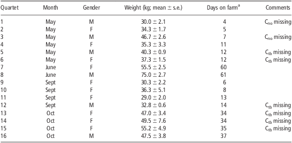
M = castrate; F = female; Cno = control pig in an adjacent pen without tail biting; Ctb = control pig in the biting pen.
aPigs arrived at ∼10 to 12 weeks of age.
Behavioural observations and selection of case–control quartets
The study was designed as a case–control with quartets (n = 16) of animals forming the experimental unit (Table 1). The uniting factors within the quartets were gender, approximate age (the animals arrived on the farm as a batch of equally sized individuals) and room. Six of the quartets consisted of barrows and the other 10 of gilts. The total number of animals was 56, including 16 TB, 16 victims (V) and 10 controls in the same pen referred to as the ‘biting pen’ (Ctb), as well as 14 controls in a pen from the same room but without tail-biting activity (Cno). Six Ctb and two Cno were missing owing to the problems related to identifying suitable non-bitten animals.
The animals were chosen using direct behavioural observations according to a three-stage process, outlined in more detail in Brunberg et al. (Reference Brunberg, Wallenbeck and Keeling2011). The process aimed to identify a sufficient number of quartets to be euthanized immediately upon identification, that is, during the actual tail-biting outbreak. Initially, the whole farm was scanned in order to identify pens with signs of ongoing tail biting and appropriate controls. At this stage, any signs of an outbreak were noted, including restlessness and tails tucked under; however, the most useful information proved to be visible tail lesions and the caretakers’ comments. Second, pen-level observations of behaviour were carried out for 2 × 30 min (in some cases 3 to 4 × 30 min, if needed for clarification of the tail-biting status) during 1 or 2 days. The observations were carried out by one person standing in front of the pen taking notes on paper using all occurrence sampling, according to an ethogram including tail-in-mouth behaviour (taking the tail in the mouth without chewing or biting) and tail biting. The latter was further classified according to the receiver's behavioural reaction as mild (no reaction), moderate (e.g. avoiding, low grunting) or severe (e.g. screaming, running away; for further details see Brunberg et al., Reference Brunberg, Wallenbeck and Keeling2011). The third stage included observations on an individual level in the most promising pens identified in the previous stages, 8 × 15 min per individual during 2 or 3 days. Both the performer and the receiver individuals were noted in order to identify frequent TB and receivers of bites (V), as well as appropriate controls (Ctb and Cno) to form the matched quartets. The observations in stages two and three were conducted by two to four persons per day, and distributed as evenly as possible between 0600 and 2000 h for each subject.
Blood sampling and euthanasia
The animals were euthanized 2 to 12 h after completion of the behavioural observations, one to two complete quartets (three to eight pigs) each morning during 13 days. The pigs were sedated in the home pen with intramuscular injections of midazolam (0.5 mg/kg, Midazolam Hameln R®, Hameln Pharmaceuticals GmbH, Hameln, Germany), butorphanol (0.2 mg/kg, Butordol® 10 mg/ml, Intervet International B.V., Boxmeer, the Netherlands) and ketamine (10 mg/kg, Ketalar®, Pfizer Inc., New York, NY, USA or Ketaminol® vet 50 mg/ml, Intervet International B.V., Boxmeer, the Netherlands). Following loss of voluntary muscular control, 5 to 31 min (median 9 min) later, three 10 ml serum tubes and one EDTA-tube of blood were drawn by jugular venipuncture using 10-ml polypropylene vacuum tubes with 18 G needles. When the signs of deep sedation were fulfilled (loss of palpebral reflex, muscle and jaw tone), an intracardial injection of pentobarbital (20 mg/kg, Mebunat® vet 60 mg/ml, Orion Corporation, Espoo, Finland) was administered. Time of death was 9 to 49 min after sedative injection (median 15 min).
Haematological examinations
All analyses were performed at the Central Laboratory of the Department of Equine and Small Animal Medicine in Helsinki, Finland. The EDTA-tubes were stored in 4°C until the following parameters were determined 12 to 16 h after sampling: platelet, white and red blood cell count (PLT, WBC, RBC), haemoglobin concentration (HGB), haematocrit (HCT) and the erythrocyte indices mean corpuscular volume, mean corpuscular haemoglobin and mean corpuscular haemoglobin concentration (MCV, MCH, MCHC). An automated multiparameter analyzer with software for animal samples (CELL-DYN 3700 System, Abbott Diagnostics Division, Abbott Park, IL, USA) was used.
Serum for biochemical analyses was extracted after centrifugation (1300 × g for 10 min) of serum tubes kept at room temperature for a maximum of 3 h. The sera were stored in −80°C until analysis with a clinical chemistry analyzer (Konelab 30i, ThermoFisher Scientific, Vantaa, Finland). Recommended methods of the International Federation of Clinical Chemistry (IFCC) were used for the determination of alkaline phosphatase (ALP, IFCC, 1983/4), γ-glutamyltransferase (GGT, IFCC, 2002/7), alanine aminotransferase (ALAT, IFCC, 2002/5), aspartate aminotransferase (ASAT, IFCC, 2002/6) and creatine kinase (CK, IFCC, 2002/3) activity in serum. Spectrophotometric methods were used for the determination of serum urea (Gutmann and Bergmeyer, Reference Gutmann and Bergmeyer1974), creatinine (Fabiny and Ertigshausen, Reference Fabiny and Ertigshausen1971), total protein (Weichselbaum, Reference Weichselbaum1946) and albumin (Doumas et al., Reference Doumas, Watson and Biggs1971) concentrations.
Pathological examinations
The head of the pig was detached immediately after death for removal of the brain. The carcass was transported to the University of Helsinki and placed in a cool room 4 to 8 h after death. An autopsy was performed the next day according to standard procedures, including examination of the spinal column. The following tissues were sampled for histological examinations: liver, spleen, kidney, lung, heart, pancreas, both adrenals, cutaneous and glandular part of stomach, jejunum, ileum, colon, joint capsules from the knee and elbow, tail, hypophysis and sacral spinal cord. Histological samples were processed routinely for histopathology, stained with haematoxylin–eosin, and observed using standard techniques.
Tail lesions were classified both macroscopically, by inspection of the tail, and histologically, by cutting the tail end sagittally, as 0 = no, 1 = healed, 2 = mild, 3 = moderate and 4 = severe lesion. The macroscopic classification was defined as 0 = no visible lesion, 1 = scar tissue at the tail tip, 2 = wounds not deeper than subcutis, 3 = wounds deeper than subcutis or moderate infection, 4 = part of the tail missing, severe infection or abscess. Histological findings were classified as 0 = no lesions other than hyperceratosis; 1 = evidence of healed lesion including a rounded and fibrotic tip of the tail with missing dermal hair follicles and possibly abnormal shape of the last observed tail vertebra (e.g. lack of the distal epiphysis); 2 = epidermal lesions, erosion, crusts or pustules and perivascular dermal inflammation; 3 = lesions extending into the deep dermis or the subcutis; and 4 = lesions extending to the muscle layer and/or bone.
Statistical analyses
PASW 18 software (SPSS Inc., Chicago, IL, USA) was used for all statistical analyses. The distribution of continuous and categorical variables was investigated using Kolmogorov–Smirnov and Shapiro–Wilk tests, as well as Q-Q plots. One individual was excluded from the clinicochemical analyses because of haemolysis (category Ctb, Kramer and Hoffmann, Reference Kramer and Hoffmann1997), and one outlier from CK data (category Cno, CK = 9367).
For normally distributed variables, a repeated-measures GLM with category as a within-subjects factor was fitted. If the within-subjects test chosen according to sphericity (Huynh and Mandeville, Reference Huynh and Mandeville1979) was significant (P < 0.05), pair-wise Student's t-tests were conducted. For nonparametric variables, Related-samples Friedman's Two-Way ANOVA by Ranks were applied to test the null hypothesis that the observations in the four categories came from the same distribution. A P-value of < 0.05 was selected to reject the null hypothesis and allow for pair-wise testing of differences between the groups using the Wilcoxon Matched-Pairs Signed-Rank Test. Category effects on dichotomous variable were tested using Cochran's Q-test, and if significant (P < 0.05), McNemar's test was used for pair-wise comparisons.
Results
Categorization and general characteristics of the animals
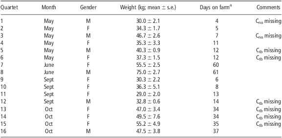
The animal's BW ranged from 23.0 to 82.5 kg at autopsy. Characteristics of the quartets are given in Table 1 and of the categories of animals in Table 2. BW did not differ between the categories (Table 2.)
BW and tail-biting activity in pigs categorized by tail-biting behaviour

1During 8 × 15 min of individual observation; given as median (min–max).
a,b,cMeans with different superscripts differ significantly (P < 0.05) in pair-wise comparisons.
Evaluation of tail lesions
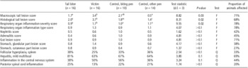
The distribution of histologically classified tail lesions is given in Table 3 and category averages for histological and macroscopic scoring in Table 4. Histological and macroscopic tail damage scores were highly correlated (r = 0.90, P < 0.001, n = 47). In the five cases where there were histological signs of healed tail trauma, this was also visible macroscopically. In the two pigs with macroscopic evidence of healed trauma, additional deep acute lesions were identified only upon histological examination.
Distribution of histological tail lesions in pigs categorized by tail-biting activity

aNumber of pigs (percentage within the category).
Prevalences of and category effects on macroscopic tail lesions, and histological findings in pigs categorized by tail-biting activity
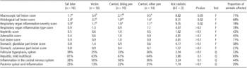
Test: F = Friedman; Q = Cochran's Q.
Tail lesion scores: 0 = no lesion, 1 = healed trauma, 2 = mild, 3 = moderate, 4 = severe lesion.
Respiratory organ inflammation severity scores: 0 = no lesion, 1 = mild, 2 = moderate, 3 = severe.
Respiratory inflammation type, nephritis and adrenalitis scores: 0 = no lesion, 1 = acute, 2 = chronic.
Gut: 0 = no lesion, 1 = reactive lymph tissue, 2 = enteritis.
Stomach, glandular part: 0 = no lesion, 1 = hyperceratosis, 2 = acute gastritis, 3 = chronic gastritis.
Stomach, cutaneous part: 0 = no lesion, 1 = hyperaemia, 2 = hyperceratosis, 3 = acute gastritis, 4 = chronic gastritis.
Note: Values are given as category means or percentage of affected animals.
a,b,cMeans with different superscript letters differ significantly (P < 0.05) in pair-wise comparisons.
The number of observed bites on the tail correlated better with histologically than with macroscopically evaluated tail damage (r = 0.62, P < 0.001, n = 51 v. r = 0.50, P < 0.001, n = 48, respectively). Looking at the behavioural reaction by the bitten pig, the histological score correlated best to the number of received bites, causing a mild reaction (RTB1 (received tail bites); r = 0.61, P < 0.001), followed by severe reaction (RTB2; r = 0.46, P = 0.001) and no reaction (RTB0; r = 0.39, P < 0.01). The macroscopic tail damage score was uncorrelated to RTB0, but moderately correlated to RTB1 and RTB2 (r = 0.48, P = 0.001 for both).
Haematology
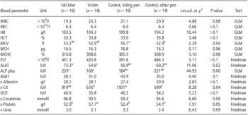
Results of the statistical analyses on blood parameters and clinical chemistry are given in Table 5. MCV, ALAT, ALP, CK and serum protein concentration differed significantly between categories (P < 0.05).
Category effects on blood parameters and clinical chemistry in pigs categorized by tail-biting activity

r.m.s.d. = root mean squared deviation for the within-subjects effect in the GLM; χ 2 = chi-squared statistic for Friedman's test with d.f. = 3; WBC = white blood cell count; l = litre; GLM = repeated measures general linear model with category as within-subjects factor; RBC = red blood cell count; HB = haemoglobin concentration; g = gram; HCT = haematocrit; MCV = mean corpuscular volume; f = femto; MCH = mean corpuscular haemoglobin; p = pico; MCHC = MCH concentration; PLT = platelet count; ALAT = alanine aminotransferase; IU = international units; ALP = alkaline phosphatase; ASAT = aspartate aminotransferase; s = serum; CK = creatine kinase; GGT = γ-glutamyltransferase; m = milli.
Note: Values are given as category means.
a,b,cMeans with different superscript letters differ significantly (P < 0.05) in pair-wise comparisons.
A HGB below 100 was chosen as a limit for anaemia (Friendship et al., Reference Friendship, Lumsden, McMillan and Wilson1984). Accordingly, of the animals, three TB (21% of animals in the category), five V (33%), five Ctb (63%) and four Cno (29%) were anaemic. No category effect was present (P > 0.1, Cochran's Q-Test). In order to characterize the types of anaemia, individual haematology values were compared with reference limits for feeder pigs given by Friendship et al. (Reference Friendship, Lumsden, McMillan and Wilson1984). Significant microcytosis or macrocytosis was not identified, but all animals had low MCHC (<320 g/l).
Pathology
Only one animal had no findings in the pathological examinations. Most individuals had inflammatory lesions in multiple organs suggestive of generalized activation of the immune system, possibly because of systemic spread of an infection. Most findings were histologically classified as mild. Abscesses, previously repeatedly associated with tail lesions (Elbers et al., Reference Elbers, Tielen, Snijders, Cromwijk and Hunneman1992; Huey, Reference Huey1996), were rare and found in the lung (one V and two TB), the diverticle of the urinary bladder (one TB) and in the tail (one V).
Pathological lesions are summarized per category in Table 4. Lesion prevalences or severity scores differed significantly between categories only in tail lesions and respiratory infection severity. The latter was more severe in the victims than in all other categories.
Discussion
This study aimed to identify health differences in pigs classified according to tail-biting activity. Disease and tail lesion prevalences have previously been found to correlate at the farm level (Moinard et al., Reference Moinard, Mendl, Nicol and Green2003), indicating that sickness-induced discomfort may eventually lead to tail-biting behaviour (Fritchen and Hogg, Reference Fritchen and Hogg1983). This did not seem to be the case on the present farm, as TB did not suffer from more or different pathological conditions than control pigs. Nevertheless, disease cannot be ruled out as a cause for tail biting on this farm. Pathological lesions were also very common in biters. Individual factors may interact with sickness to induce tail-biting behaviour. Moreover, it is uncertain if the chosen biters were the actual initiators of the behaviour. Tail biting is known to spread visually (Blackshaw, Reference Blackshaw1981), and characteristics of followers may differ from those of the instigators.
A high infection pressure seemed to be present on the farm, as suggested by both pathological findings and a high prevalence of anaemia characterized by low MCHC, which is an indication of chronic inflammation (Friendship and Henry, Reference Friendship and Henry1992). Although this may have masked some differences associated with tail-biting activity, several measures indicated that the health status of the victims was worse as compared with all the other categories of pigs. Tail and respiratory organ lesions were more severe, serum protein concentration higher and WBC counts numerically higher, indicating that V were more severely challenged by (bacterial) infections (Odink et al., Reference Odink, Smeets, Visser, Sandman and Snijders1990). As freedom from disease is assigned high priority when assessing animal welfare (e.g. Duncan and Fraser, Reference Duncan and Fraser1997), these findings emphasize the detrimental effects of being tail bitten.
Pathological examinations in the present animals indicated that respiratory organ disease was, at least in many cases, secondary to infected tail lesions. The most common route of bacterial spread was venous (Sihvo et al., Reference Sihvo, Simola, Munsterhjelm and Syrjä2011). Previously, macroscopic lung infections have been reported to associate with tail lesions in pig carcasses examined at the slaughterhouse (Elbers et al., Reference Elbers, Tielen, Snijders, Cromwijk and Hunneman1992; Huey, Reference Huey1996).
Respiratory organ disease may also be a predisposing factor for being bitten, as suggested by Moinard et al. (Reference Moinard, Mendl, Nicol and Green2003). The mechanism may be a reluctance or inability of ill pigs to avoid tail biting. Chronic immune activation – obviously frequent in this study – will elicit a non-specific ‘sickness response’ behaviourally manifested by, for example, fatigue, increased pain sensitivity, depressed activity and anorexia (Hart, Reference Hart1988).
Serum activity of ALAT and ALP were higher in TB than V and Ctb, and also higher in Cno than V. Levels of ALP were within the reference range given by Friendship et al. (Reference Friendship, Lumsden, McMillan and Wilson1984), whereas ALAT activity was considerably higher. The significance of the high activity is uncertain. Comparison with reference values is complicated by a number of factors, including that the values may be out of date and therefore no longer applicable for modern breeds (Lumsden, Reference Lumsden1998). In the present animals, sedation (Evans, Reference Evans1994) and freezing of the serum (Kramer and Hoffmann, Reference Kramer and Hoffmann1997) are further possible sources of systematic errors.
The reason for the observed differences between the categories of pigs in ALAT and ALP is also not clear. Both enzymes are liver-specific, although ALAT activity in pigs is low (Boyd, Reference Boyd1983), and the bone is the primary source of ALP in growing animals (Kramer and Hoffmann, Reference Kramer and Hoffmann1997). There might be a weak correlation with hepatitis prevalence, as category averages of enzyme activities seem to follow a similar pattern. Alternatively, pathological processes may play a role as reported by Odink et al. (Reference Odink, Smeets, Visser, Sandman and Snijders1990), who found decreasing ALAT and ALP activity (among others) with increasing severity of inflammatory lesions in slaughter pigs.
The link between being a victim and decreasing ALP activity may also be decreased feed intake (Baetz and Mengeling, Reference Baetz and Mengeling1971). Inappetence is associated with being tail bitten (Niemi et al., Reference Niemi, Sinisalo, Valros and Heinonen2011) and a part of the ‘sickness response’ (Hart, Reference Hart1988). In the present animals, BW did not differ between categories. Nevertheless, growth may have been temporarily decreased in V because of respiratory disease or tail biting, as reported by Wallgren and Lindahl (Reference Wallgren and Lindahl1996) and Niemi et al. (Reference Niemi, Sinisalo, Valros and Heinonen2011).
A few differences between Ctb and Cno indicate that environmental effects on health existed in tail-biting pens. CK activity was higher in Ctb than Cno and V. Category averages for ASAT seemed to follow a similar pattern to CK. A combination of elevated CK and ASAT, previously reported in pigs by Friendship et al. (Reference Friendship, Lumsden, McMillan and Wilson1984), suggests that exercise and/or tissue damage was the cause (van der Meulen et al., Reference van der Meulen, Kuipers and Drukker1991), and that the damage was not acute (Tennant, Reference Tennant1997). The pathological examinations gave no obvious reason for the findings. Nevertheless, restlessness is associated with tail biting and may explain the findings.
Erythrocyte volume (MCV) was larger in Ctb than Cno, but still within reference values by Friendship et al. (Reference Friendship, Lumsden, McMillan and Wilson1984). Inflammatory processes may decrease MCV (Odink et al., Reference Odink, Smeets, Visser, Sandman and Snijders1990), whereas increasing environmental disease pressure or erythrocyte regeneration in anaemia may increase it (Friendship and Henry, Reference Friendship and Henry1992). Pathological findings did not explain the difference in MCV between the two control categories.
The pathological investigations indicated that the appearance of (bitten) tails might not be the most accurate measure of tail-biting activity or tail lesions. We noted, in accordance with Simonsen et al. (Reference Simonsen, Klinken and Bindseil1991), that severe inflammatory changes may exist under healthy-looking skin.
In conclusion, no associations between health status and tail-biting activity could be established on this farm with widespread tail biting and apparently high infection pressure. Being a victim of tail biting was associated with severe inflammatory lesions in the respiratory organs. Deep infections may exist under healthy skin in bitten tails.
Acknowledgements
This research was funded by The Nordic Joint Committee for Agricultural Research. The authors wish to thank the farm, the non-author researchers and Susanna Takaluoma for help and hard work during the experiment, and Satu Sankari for carrying out the blood work. Above all, we would like to acknowledge Anne Larsen for very professional technical assistance during the long hours of data collection.