Introduction
Many insects are infected with maternally inherited intracellular bacteria. These are divided into the primary symbionts present in all individuals and the secondary symbionts (SS) facultatively associated with their insect hosts (Chiel et al., Reference Chiel, Gottlieb, Zchori-Fein, Mozes-Daube, Katzir, Inbar and Ghanim2007). Many of the different SS organisms have been shown to influence the biology, ecology and evolution of their hosts. One of the most widely studied SS is Wolbachia, which is known to occur in many arthropod species. This endosymbiont can alter the reproduction of its hosts principally through parthenogenesis induction and cytoplasmic incompatibility (Werren, Reference Werren1997). In addition, Wolbachia can have both positive and negative influences on host fitness. Some Wolbachia improve the reproduction, hence fitness, of their hosts (e.g. Hariri et al., Reference Hariri, Werren and Wilkinson1998; Vavre et al., Reference Vavre, Girin and Boulétreau1999), while others reduce it by reducing fecundity (e.g. Min & Benzer, Reference Min and Benzer1997; Wenseleers et al., Reference Wenseleers, Sundström and Billen2002), life span (McMeniman et al., Reference McMeniman, Lane, Cass, Fong, Sidhu, Wang and O'Neill2009), ability to disperse (Silva et al., Reference Silva, van Meer, Roskam, Hoogenboom, Gort and Stouthamer2000), adult survival and locomotor performance (Fleury et al., Reference Fleury, Vavre, Ris, Fouillet and Boulétreau2000). Wolbachia has also been implicated in affecting competitive ability and host evolution (Heddi et al., Reference Heddi, Grenier, Khatchadourian, Charles and Nardon1999; Zchori-Fein & Brown, Reference Zchori-Fein and Brown2002) and is associated with host invasiveness (Shoemaker et al., Reference Shoemaker, Ross, Keller, Vargo and Werren2000; Tsutsui et al., Reference Tsutsui, Kauppinen, Oyafuso and Grosberg2003; Chu et al., Reference Chu, Cong, Zhang, Xu, Wu and Zhu2005). Several other SS organisms have also been shown to play an important role in host fitness (Koga et al., Reference Koga, Tsuchida and Fukatsu2003; Zabalou et al., Reference Zabalou, Riegler, Theodorakopoulou, Stauffer, Savakis and Bourtzis2004), host nutrition, adaptation to plant hosts (e.g. Douglas, Reference Douglas1998; Leonardo & Muiru, Reference Leonardo and Muiru2003; Tsuchida et al., Reference Tsuchida, Koga and Fukatsu2004), resistance to parasitoids (Oliver et al., Reference Oliver, Moran and Hunter2005) and to heat stress (Montllor et al., Reference Montllor, Maxmen and Purcell2002).
The sweet potato whitefly, Bemisia tabaci (Gennadius) (Hemiptera: Aleyrodidae), is an important agriculture pest worldwide (Oliveira et al., Reference Oliveira, Henneberry and Anderson2001). It is considered to be a species complex composed of at least 24 recognizable ‘species’ based on mitochondrial cytochrome oxidase I (mtCOI) maker, which (where data exist) have been shown to be either reproductively isolated or produce hybrid progeny that are considerably less fit than their parents (Dinsdale et al., Reference Dinsdale, Cook, Riginos, Buckley and De Barro2010; Xu et al., Reference Xu, De Barro and Liu2010). Among them, two B. tabaci species, referred as Mediterranean (commonly known as biotype Q) and Middle East Asia Minor 1 (biotype B) (Dinsdale et al., Reference Dinsdale, Cook, Riginos, Buckley and De Barro2010), have become globally invasive.
Over the past decade or so, six SS, viz. Wolbachia, Arsenophonus, Rickettsia, Hamiltonella, Fritschea and Cardinium, have been identified from various members of the B. tabaci species complex (e.g. Zchori-Fein & Brown, Reference Zchori-Fein and Brown2002; Nirgianaki et al., Reference Nirgianaki, Banks, Rrohlich, Veneti, Braig, Miller, Bedford, Markham, Savakis and Bourtzis2003; Thao et al., Reference Thao, Baumann, Hess, Falk, Ng, Gullan and Baumann2003; Weeks et al., Reference Weeks, Velten and Stouthamer2003; Chu et al. Reference Chu, Cong, Zhang, Xu, Wu and Zhu2005; Chiel et al., Reference Chiel, Gottlieb, Zchori-Fein, Mozes-Daube, Katzir, Inbar and Ghanim2007). Among these SS, the distribution of Wolbachia and its role in the B. tabaci has been the most studied. The term supergroup of Wolbachia has been employed to designate the closely related groups based on gene sequences (Lo et al., Reference Lo, Casiraghi, Salati, Bazzocchi and Bandi2002). Previous studies have shown that B. tabaci were infected by Wolbachia belonging to supergroup B, but that infections were not found in all members of the species complex (Zchori-Fein & Brown, Reference Zchori-Fein and Brown2002; Nirgianaki et al., Reference Nirgianaki, Banks, Rrohlich, Veneti, Braig, Miller, Bedford, Markham, Savakis and Bourtzis2003; Chu et al., Reference Chu, Cong, Zhang, Xu, Wu and Zhu2005; Ruan & Liu, Reference Ruan and Liu2005). In the case of biotype B, several studies (Zchori-Fein & Brown, Reference Zchori-Fein and Brown2002; Chu et al., Reference Chu, Cong, Zhang, Xu, Wu and Zhu2005; Ruan & Liu, Reference Ruan and Liu2005) failed to detect the presence of Wolbachia using both the wsp or 16s rDNA genes, whereas another molecular study (Nirgianaki et al., Reference Nirgianaki, Banks, Rrohlich, Veneti, Braig, Miller, Bedford, Markham, Savakis and Bourtzis2003) detected infections in one out of 12 collections using the wsp gene. Similarly for biotype Q, no infections were found using the wsp gene in four collections from China, Israel and Spain (Chu et al., Reference Chu, Cong, Zhang, Xu, Wu and Zhu2005), whereas a more recent study using the 16 s rDNA gene revealed five out of ten populations in Israel to be infected (Chiel et al., Reference Chiel, Gottlieb, Zchori-Fein, Mozes-Daube, Katzir, Inbar and Ghanim2007). In contrast, collections belonging to species other than those containing biotypes B and Q appear more likely to be infected with Wolbachia (Li et al., Reference Li, Lin and Guo2007).
The removal of some SS including Wolbachia and/or reduction of the primary endosymbiont using antibiotics may produce both favourable and unfavourable effects on the fitness of B. tabaci (Ruan et al., Reference Ruan, Xu and Liu2006). In individuals belonging to the species associated with Asia that were infected with Wolbachia and Arsenophonus, treatment of adults with the antibiotics tetracycline or ampicillin trihydrate lead to an increase in the rate of development of their offspring, but did not significantly affect their survival, whereas treatments with another antibiotic, rifampicin, significantly retarded development and reduced progeny survival. The lack of detection of Wolbachia in some species belonging to the B. tabaci species complex suggests that it may not play a role; however, advances in DNA technology have demonstrated considerable increases in the level of sensitivity, suggesting that previous negative findings may have been false (Li et al., Reference Li, Lin and Guo2007; Qu et al., Reference Qu, Cong, Chu and Dong2009).
Recent studies have shown that SS may play a major role in the biology of B. tabaci. A recent study of the distribution of SS in Israeli populations of B. tabaci biotypes B and Q found both Hamiltonella and Rickettsia in biotype B and Arsenophonus, Wolbachia and Rickettsia in biotype Q (Chiel et al., Reference Chiel, Gottlieb, Zchori-Fein, Mozes-Daube, Katzir, Inbar and Ghanim2007). Fritschea and Cardinium were not found in either biotype (Chiel et al., Reference Chiel, Gottlieb, Zchori-Fein, Mozes-Daube, Katzir, Inbar and Ghanim2007). Quantitative real-time PCR and FISH analyses showed that proliferation of Rickettsia is strongly induced when the B. tabaci was parasized by the parasitic wasp Eretmocerus mundus (Mercet) (Hymenoptera: Aphelinidae), which suggests that it may be involved in the insect's resistance to various environmental stresses (Mahadav et al., Reference Mahadav, Gerling, Gottlieb, Czosnek and Ganim2008). For example, Rickettsia in biotype B increased its susceptibility to several insecticidal compounds (Kontsedalov et al., Reference Kontsedalov, Zchori-Fein, Chiel, Gottlieb, Inbar and Ghanim2008), while double infections Rickettsia-Arsenophonus or Wolbachia-Arsenophonus had a significant influence on the survival of biotype Q exposed to the insecticides thiamethoxam, imidacloprid, pyriproxyfen and spiromesifen (Ghanim & Kontsedalov, Reference Ghanim and Kontsedalov2009).
China has several indigenous members of B. tabaci complex whitefly (Qiu et al., Reference Qiu, Coats, Ren, Idris, Xu and Brown2007), but in recent years both biotypes B and Q have successfully invaded and spread across large parts of the country (Luo et al., Reference Luo, Yao, Wang, Yan, Hu and Zhang2002; Chu et al., Reference Chu, Zhang, Brown, Cong, Xu, Wu and Zhu2006). It appears that while biotype B invaded China in the mid-1990s (Luo et al., Reference Luo, Yao, Wang, Yan, Hu and Zhang2002), biotype Q was a more recent arrival being first detected in Kunming, Yunnan Province, in 2003 and subsequently in the Beijing, Henan and Shandong Provinces (Chu et al., Reference Chu, Zhang, Brown, Cong, Xu, Wu and Zhu2006, Reference Chu, Jiang, Liu, Jiang, Tao, Fan, Zhou and Bi2007). Biotype B showed higher fecundity (Muniz & Nombela, Reference Muniz and Nombela2001; Nombela et al., Reference Nombela, Beitia and Muniz2001) and competitive ability than biotype Q under laboratory conditions (Pascual & Callejas, Reference Pascual and Callejas2004), while biotype Q was found highly resistant to many insecticides, such as the neonicotinoid group of pesticides (Nauen et al., Reference Nauen, Stumpf and Elbert2002; Rauch & Nauen, Reference Rauch and Nauen2003; Horowitz et al., Reference Horowitz, Kontsedalov, Khasdan and Ishaaya2005). Recent studies have suggested that biotype Q has begun to displace biotype B (Chu et al., Reference Chu, Jiang, Liu, Jiang, Tao, Fan, Zhou and Bi2007, Reference Chu, Zhang and Wan2010). A possible contributor to this successful displacement could be the presence of SS; and, thus, the determination of distribution of SS within biotypes B and Q should provide useful baseline information with which to examine the causes of this displacement.
The infection status of B. tabaci in China, with respect to SS, is poorly known, with studies focusing only on Wolbachia (Chu et al., Reference Chu, Cong, Zhang, Xu, Wu and Zhu2005; Ruan & Lui, Reference Ruan and Liu2005; Lin & Li, Reference Lin and Li2008). It would be useful to know the extent of infections and whether infections may have been derived locally or were present in the invaders before they established in China (Chu et al., Reference Chu, Jiang, Liu, Jiang, Tao, Fan, Zhou and Bi2007, Reference Chu, Zhang and Wan2010). The aim of the present study is to clarify the infection status of Wolbachia, Arsenophonus, Rickettsia, Hamiltonella, Fritschea and Cardinium in biotypes B and Q in China over the period 2005–2009. We then focus more closely on those SS that have been associated with insecticide resistance, Wolbachia, Rickettsia and Arsenophonus (Kontsedalov et al., Reference Kontsedalov, Zchori-Fein, Chiel, Gottlieb, Inbar and Ghanim2008; Ghanim & Kontsedalov, Reference Ghanim and Kontsedalov2009) and compare their infection levels in biotype Q from China and its home range (Mediterranean countries).
Materials and methods
Whitefly samples and determination of B. tabaci biotypes
A total of 93 Chinese collections of B. tabaci on different hosts were made across seven sites (Liaocheng, Dezhou, Jinan, Zizo, Shouguang, Zaozhuang and Linyi) in Shandong Province of China between 2005 and 2009. At each location in each year, the collections were made from a range of host plants. For host plants, a collection involved sampling whitefly from every second available plant until at least 100 adults had been collected. A further 14 collections were provided by researchers from different Mediterranean countries (Bosnia-Herzegovina (one collection), Greece (two collections), Croatia (three collections), Cyprus (one collection), Egypt (one collection), Italy (one collection), Morocco (two collections), Spain (two collections)) and USA (one collection). The adults were collected live and placed into tubes containing 95% ethanol and then stored at –20°C. Individual adults were subjected to lysis and DNA extraction following the procedure of Frohlich et al. (Reference Frohlich, Torres-Jerez, Bedford, Markham and Brown1999). The identity of B. tabaci from China determined, based on the CAPS (cleavage amplified polymorphic sequence) of mtCOI (mitochondrial cytochrome oxidase I) with the restriction endonucleases VspI and StuI. The mtCOI fragment (∼620 bp) amplified by a set of PCR primers C1-J-2195 (5′-TTGATTTTTTGGTCATCCAGAAGT-3′) and R-BQ-2819 (5′-CTGAATATCGRCGAGGCATTCC-3′) was first cleaved by VspI; uncut products were then cleaved by StuI. In the regions of China, all of the mtCOI cut by VspI have previously been shown to belong to biotype Q, whereas those cut by StuI were biotype B (Chu et al., Reference Chu, Zhang and Wan2010). The identity of B. tabaci from Mediterranean countries was determined through sequencing of the COI (Chu et al., Reference Chu, Zhang, Brown, Cong, Xu, Wu and Zhu2006). Identity of the sequences was determined by comparing these unknown sequences with the 24 recently published consensus sequences (Dinsdale et al., Reference Dinsdale, Cook, Riginos, Buckley and De Barro2010).
Determination of secondary symbionts
Wolbachia 16 s rDNA was amplified using the primers 16s315f (5′-GCATGAGTGAAGAAGGCC-3′) and 16s628r (5′-AGATAGACGCCT TCGCCA-3′) in Li et al. (Reference Li, Lin and Guo2007). Five μl of PCR product was electrophoresed on 2% agarose gels. To confirm the presence of Wolbachia, the 314 bp product was digested with NheI following the methodology of Li et al. (Reference Li, Lin and Guo2007). Only Wolbachia 16 s rDNA product is cut by this enzyme and produces two bands (118, 196 bp). Digestion with VspI was done using 5 μl of PCR product in 10 μl reaction according to the manufacturer's instructions (Qu et al., Reference Qu, Cong, Chu and Dong2009). Sequencing of this fragment (Qu et al., Reference Qu, Cong, Chu and Dong2009) has confirmed it as being Wolbachia 16 s rDNA. Digestion with VspI followed product separation using agarose gels revealed three banding patterns: (i) one band (314 bp) is expected for Wolbachia belonging to supergroup A; (ii) three bands (164, 94, 56 bp) for supergroup B Wolbachia; and (iii) four bands (314, 164, 94, 56 bp) created by double infection. The presence of Hamiltonella, Rickettsia, Arsenophonus, Fritschea and Cardinium was determined using primers from Chiel et al. (Reference Chiel, Gottlieb, Zchori-Fein, Mozes-Daube, Katzir, Inbar and Ghanim2007). At least ten adults were taken from each collection, DNA extracted (Frohlich et al., Reference Frohlich, Torres-Jerez, Bedford, Markham and Brown1999) and the product then amplified using the methodology described in Chiel et al. (Reference Chiel, Gottlieb, Zchori-Fein, Mozes-Daube, Katzir, Inbar and Ghanim2007). The amplified products were then sequenced and their identity confirmed by comparing the sequences against those in GenBank using a standard BLAST comparison.
Statistical analysis
To determine the difference in the frequency of each SS in biotypes B and Q, all data for biotype B from China were pooled, respectively, and then independent sample t-tested to test the difference with those from the collections made from the Mediterranean region. The percentage of SS in each collection was arcsine transformed prior to analysis. We repeated the analysis for the comparison of the percentage infection of each SS species in biotype Q from China and those from the Mediterranean region.
The difference in the frequency of infection for each SS in biotype B across different host plants was analysed using one-way analysis of variance (ANOVA). Percentage data were arcsine transformed, host plants was regarded as factor and the percentage of each SS in each collection was the dependant variable. Biotype Q was analysed in the same way.
Results
The presence of different SS in biotypes B and Q from China and their changes in percentage infection during 2005–2009
In total, 373 individuals belonging to biotype B and 1830 to biotype Q were used to screen for the presence of SS. Biotype B was found to be the most common and widely distributed of the two biotypes found in China from 2005 to 2006 and biotype Q from 2007 to 2009. All SS species except Fritschea were detected in both biotypes B and Q. For biotype B, the percentage infection of Hamiltonella was highest (92.0%) followed by Rickettsia (70.2%), with Arsenophonus the lowest (1.1%). For biotype Q, the percentage infection of Hamiltonella was again highest (73.3%), with Arsenophonus again the lowest (0.7%). The percentage infections by Wolbachia, Rickettsia and Hamiltonella in biotype B were each significantly higher than those in biotype Q (P<0.05). However, the percentage infection with Cardinium in biotype B was significantly lower than that in biotype Q (P<0.05) (table 1).
Percentage infections for the different SS in Bemisia tabaci biotypes B and Q.

N a number of individuals tested from biotypes Q and B populations.
b based on the method in Li et al. (Reference Li, Lin and Guo2007).
W, Wolbachia; R, Rickettsia; A, Arsenophonus; H, Hamiltonella; C, Cardinium; –, undetected.
Among the five SS, Wolbachia in the infected individuals of both biotypes B and Q belonged to supergroup A. Of the collections examined, 33.3% of collections containing biotype B and 12.5% of those with biotype Q were infected with Wolbachia. In terms of individual frequencies, 12.6% of biotype B and 2.1% of biotype Q individuals were found to be infected (table 1).
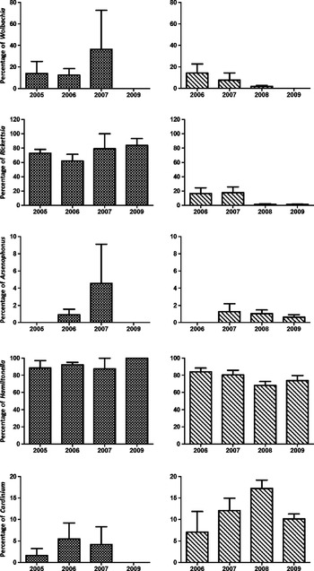
The percentage infections in biotypes B and Q varied over the period 2005–2009 (fig. 1). The percentage of biotype B infected with Wolbachia ranged from 5.9% to 36.4% during 2005–2008 and declined to zero in 2009. The percentage of biotype Q infected with Wolbachia ranged from 1.0% to 8.4% during 2005–2008 and also declined to zero in 2009. The percentage of biotype B infected with Rickettsia ranged from 66.3% to 83.3% during 2005–2009, whereas for biotype Q it ranged from 14.3% to 16.2% in 2006 and 2007 before declining to almost zero in 2008 and 2009. Arsenophonus was nearly always <5.0% in both biotypes B and Q. Hamiltonella was the most frequently encountered infection maintaining levels above 68.3% in both biotypes B and Q over the period. Cardinium maintained a low presence of <6.5% in biotype B, while in biotype Q ranged between 7.6% and 17.3% (fig. 1).
The distribution of the different symbionts within B. tabaci biotypes B and Q from China during 2005 to 2009 ( , biotype B;
 , biotype Q).

The difference of percentage infection of SS in biotypes B or Q from different locations and host plants in China
The percentage infection with Wolbachia in biotype B from DeZhou (68.2%) was the highest, while in all other locations was <13.1%. The percentage infection of Arsenophonus in biotype B from most locations was higher than 53.3% except at ShouGuang where only 9.4% of individuals were infected. The percentage infection of Rickettsia in biotype B from all locations was lower than 4.6%, while those for Hamiltonella exceeded 68.8%. The percentage infection with Cardinium in biotype B from LiaoCheng (18.8%) was the highest, while at other locations was lower than 4.6%.
The percentage infection with Wolbachia, Arsenophonus, Rickettsia and Cardinium in biotype Q from all locations was <16.9%. The percentage infection with Hamiltonella in biotype Q from all locations exceeded >29.3% and was usually >83.4%. The difference of the percentage infection for each SS in biotypes B and Q did not differ significantly across the different host plant species (P>0.05).
Percentage infections of Wolbachia, Rickettsia and Arsenophonus in biotype Q from Mediterranean regions and China
A total of 177 individuals from Bosnia-Herzegovina, Crete, Croatia, Cyprus, Egypt, Italy, Morocco, Spain and a non-Mediterranean population from the USA were examined for the presence of Wolbachia, Rickettsia and Arsenophonus. Only biotype Q from Cyprus was found to be infected by all three symbionts, viz., Wolbachia (85.0%), Rickettsia (100.0%) and Arsenophonus (30.0%). Wolbachia in the infected individuals from Cyprus belonged to supergroup A. Biotype Q from Italy was infected by Rickettsia (9.1%) and Arsenophonus (9.1%). Rickettsia was also found in biotype Q from Croatia and the USA with percentage infections of 70.0% and 100.0%, respectively. Arsenophonus was also found in biotype Q from Morocco and Spain.
Based on the analysis of mtCOI obtained from biotype Q individuals in each of the collections used in this study (not shown), two distinct subgroups within biotype Q were identified and found to be consistent with previously published research (Chu et al., Reference Chu, Wan, Tao, Liu, Fan and Bi2008). Biotype Q from Bosnia-Herzegovina, Crete, Croatia (Croatia (1) and Croatia (2)), Egypt, Italy, Morocco, Spain and China belonged to one subgroup (I) while those from Cyprus, Croatia (Croatia (3)) and USA belonged to the second subgroup (II). The percentage infections of Wolbachia, Rickettsia and Arsenophonus in biotype Q (subgroup I) from China and from the Mediterranean region was <4.1%. While percentage infections with Wolbachia, Rickettsia and Arsenophonus in biotype Q (subgroup II) from Cyprus, Croatia (Croatia (3)) and USA was 36.2%, 90.4% and 12.8%, respectively. The percentage infection of Rickettsia in biotype Q from Cyprus, Croatia (Croatia (3)) and USA was always >70.0%.
Discussion
The distribution of the SS in the biotypes B and Q
Our study has demonstrated the presence of the endosymbionts Wolbachia, Arsenophonus, Rickettsia, Hamiltonella and Cardinium in both B. tabaci biotypes B and Q from China. This is a considerable departure from previously published data (Chiel et al., Reference Chiel, Gottlieb, Zchori-Fein, Mozes-Daube, Katzir, Inbar and Ghanim2007; Ahmed et al., Reference Ahmed, Shatters, Ren, Jin, Mandour and Qiu2009). In the earlier study, only Rickettsia (64.0%) and Hamiltonella (100.0%) were detected in biotype B from Israel and only Wolbachia (33.0%), Rickettsia (74.0%) and Arsenophonus (87.0%) were detected in biotype Q from Israel (Chiel et al., Reference Chiel, Gottlieb, Zchori-Fein, Mozes-Daube, Katzir, Inbar and Ghanim2007). Based on these findings, the authors speculated on the possible roles of SS. However, our study showed that percentages of infection in biotype B were similarly high for Rickettsia and Hamiltonella, whereas percentages of infection by the remaining four SS were always below 25.7% with Arsenophonus and Cardinium being particularly low.
We also found Wolbachia, Rickettsia and Arsenophonus in biotype Q from China. However, the percentage of individuals in China infected with Wolbachia, Rickettsia and Arsenophonus were all very much lower than the percentages reported for Israel (Chiel et al., Reference Chiel, Gottlieb, Zchori-Fein, Mozes-Daube, Katzir, Inbar and Ghanim2007), and the percentage of Wolbachia was much lower than that for China (42.5%) (Ahmed et al., Reference Ahmed, Shatters, Ren, Jin, Mandour and Qiu2009). Furthermore, in China infections with Hamiltonella and Cardinium were detected, whereas in Israel they were absent. In addition, our study showed that percentage infections with Wolbachia, Rickettsia, Hamiltonella and Cardinium differed significantly for both biotypes B and Q from China, whereas those for Arsenophonus did not. Our findings, thus, present quite a different picture to that provided by previous studies (Chiel et al., Reference Chiel, Gottlieb, Zchori-Fein, Mozes-Daube, Katzir, Inbar and Ghanim2007) and suggests that much more work is needed to analyse the roles played by SS. Why diversity of SS infections in China should differ so markedly with those from Israel is unclear; there may be underlying biological explanations, but, equally, the patterns observed may reflect different levels of sampling effort.
The change in percentage infection of SS in biotypes B and Q from China during 2005–2009 and their influential factors
A key finding from this study is that the relative abundance of several of the SSs remain extremely fluid over time, suggesting that existence of complex ecological/genetic/physiological relationships that have yet to be understood. For both biotypes B and Q, the percentage infection varied across the period 2005–2009, but there was no obvious association with different host plants. This is a departure from earlier work (Chiel et al., Reference Chiel, Gottlieb, Zchori-Fein, Mozes-Daube, Katzir, Inbar and Ghanim2007); however, we are reluctant to draw too many conclusions from this as sampling effort was quite different between the two studies. In our present study, the percentage infections of Hamiltonella and Arsenophonus were consistent in Q over the period 2005–2009. Hamiltonella was always the most commonly detected SS, while infections with Arsenophonus remained at <5% over this period. Interestingly, the other two SSs, Wolbachia and Rickettsia, decreased during 2008–2009, whereas infections with Cardinium decreased in 2009. Wolbachia become undetectable in 2009 and Rickettsia declined to almost zero. The process(es) driving these widespread changes in infection frequencies is/are unknown, but the findings reveal that infection levels show considerable instability over time rather than being fixed. The data also suggest that considerable changes in infection profiles over quite short time periods may be expected. This adds a further complication to comparisons across different studies.
For biotype B, the percentage infections of all SSs varied over the period 2005–2009. Hamiltonella and Rickettsia were the most frequently encountered SS. In contrast, infections with Wolbachia, Arsenophonus and Cardinium were low during 2005–2007 and each was absent in 2009.
In a recent study of insecticide resistance in field populations of biotypes B and Q from Shandong Province, we found that the biotype Q had higher levels of resistance to many insecticides compared to those in biotype B (Chu et al., Reference Chu, Zhang and Wan2010). These observations reflect those of earlier work by Kontsedalov et al. (Reference Kontsedalov, Zchori-Fein, Chiel, Gottlieb, Inbar and Ghanim2008), who showed an association to exist between the presence of Rickettsia with increased susceptibility to acetamiprid, thiamethoxam, spiromesifen and especially pyriproxyfen insecticides. Similarly, another study (Ghanim & Kontsedalov, Reference Ghanim and Kontsedalov2009) showed that the insecticides thiamethoxam, imidacloprid, pyriproxyfen and spiromesifen were all more effective against biotype Q with Rickettsia-Arsenophonus and Wolbachia-Arsenophonus infections than those with only Arsenophonus infections. Our study similarly suggests that infections with Wolbachia and Rickettsia may increase susceptible to insecticides and that the loss of these particular SS infections leads to increased resistance (Ghanim & Kontsedalov, Reference Ghanim and Kontsedalov2009). Hence, our data is consistent with the hypothesis of Ghanim & Kontsedalov (Reference Ghanim and Kontsedalov2009), although more work is required to confirm this apparent relationship.
The potential role of Wolbachia in biotypes B and Q
Wolbachia is known to affect a range of biological processes in arthropods (Ros et al., Reference Ros, Fleming, Feil and Breeuwer2009), but their role in B. tabaci is unknown (Chiel et al., Reference Chiel, Gottlieb, Zchori-Fein, Mozes-Daube, Katzir, Inbar and Ghanim2007). Phylogenetic reconstructions using the ftsZ gene have been used to identify eight supergroups (A to H) within Wolbachia (Lo et al., Reference Lo, Casiraghi, Salati, Bazzocchi and Bandi2002; Ros et al., Reference Ros, Fleming, Feil and Breeuwer2009). Of these, all but C and D have been detected in arthropods. A and B are the most widely distributed across arthropods.
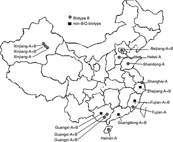
In China, Wolbachia infections in both invader and indigenous populations belonged mostly to supergroup A as a single infection, whereas the indigenous populations tended to carry Wolbachia from supergroup B as a co-infection with no single infections with supergroup B being observed (Li et al., Reference Li, Lin and Guo2007; Guo & Li, Reference Guo and Li2008; Lin & Li, Reference Lin and Li2008) (fig. 2). Their data showed that the Wolbachia infections were closely related to the genetic group structure of B. tabaci, and that infection percentages were much higher than those reported previously (Li et al., Reference Li, Lin and Guo2007; Guo & Li, Reference Guo and Li2008; Lin & Li, Reference Lin and Li2008). Bemisia tabaci populations sensu lato are known to have a complex genetic structure (De Barro et al., Reference De Barro, Driver, Trueman and Curran2000; Boykin et al., Reference Boykin, Shatters, Rosell, McKenzie, Bagnall, De Barro and Frohlich2007), and it has recently been shown that at 3.5% mtCOI divergence there is a distinct break in the frequency distribution (Dinsdale et al., Reference Dinsdale, Cook, Riginos, Buckley and De Barro2010). If the observed genetic variation is due to differentiation among populations of the one species, it would be expected that there would be no distinct gaps and that variation, although clumped, would be relatively continuous. This was not the case. Further, Xu et al. (Reference Xu, De Barro and Liu2010) has shown that, while the data is patchy, sampled populations show marked levels of reproductive isolation between the groups identified at this level of sequence divergence with either no female progeny produced, or progeny with reduced fitness relative to their parents. One possible driver behind this structure is between genetic group cytoplasmic incompatibility caused by Wolbachia.
Wolbachia infection status of B and non-B/Q biotype B. tabaci using PCR-RFLP analysis of 16 s rDNA in China based on references (Li et al., Reference Li, Lin and Guo2007; Guo & Li, Reference Guo and Li2008; Lin & Li, Reference Lin and Li2008). Note: A, B, A+B indicates Wolbachia supergroup A, B, both A and B, respectively.

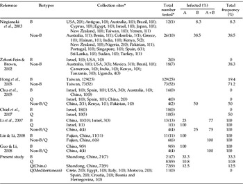
Several early studies (Zchori-Fein & Brown, Reference Zchori-Fein and Brown2002; Nirgianaki et al., Reference Nirgianaki, Banks, Rrohlich, Veneti, Braig, Miller, Bedford, Markham, Savakis and Bourtzis2003; Chu et al., Reference Chu, Cong, Zhang, Xu, Wu and Zhu2005; Hong et al., Reference Hong, Ko and Wang2005) showed that B. tabaci infected with Wolbachia belonged primarily to genetic groups other than those to which biotypes B and Q belonged (tables 2 and 3). The data, however, are quite variable.
Percentage of infection with Wolbachia within collections of Bemisia tabaci biotypes B and Q.

* The number of infected populations is indicated in parentheses.
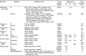
Percentage of infection with Wolbachia within Bemisia tabaci biotypes B and Q individuals.

* The number of infected individuals is indicated in parentheses.
More recent studies (Chiel et al., Reference Chiel, Gottlieb, Zchori-Fein, Mozes-Daube, Katzir, Inbar and Ghanim2007) found no biotype B individuals to be infected but that a third of biotype Q individuals in half the collections were (tables 2 and 3). In a series of recent studies from China (Li et al., Reference Li, Lin and Guo2007; Guo & Li, Reference Guo and Li2008; Lin & Li, Reference Lin and Li2008), all collections of biotype B contained individuals with infection levels ranging from 98.4–99.0% (tables 2 and 3). Our results are in agreement with these but suggest that levels of infection were much higher than previously recorded. It seems, then, that levels of infection are increasing. Even so, Li et al. (Reference Li, Lin and Guo2007) showed, using their revised method for detecting Wolbachia infections, that increased detection sensitivity and, so, the observed increase is more likely to be due to an improvement in methodology. According to Li et al. (Reference Li, Lin and Guo2007), co-infection within biotype B individuals was lower than that for non-B/Q biotype individuals (table 2). Furthermore, Guo & Li (Reference Guo and Li2008) and Lin & Li (Reference Lin and Li2008) observed only single supergroup A infection in B. tabaci biotype B. Our study reveals similar levels of infection in biotypes B and Q. Yet our percentage of infected biotype B individuals, 12.6%, is far less than the 98.4% observed in Guo & Li (Reference Guo and Li2008) and Lin & Li (Reference Lin and Li2008). Undoubtedly, then, there is considerable variability in infection status of B. tabaci across China.
In conclusion, a review of past studies plus the new data generated by this study showed that in China (i) Wolbachia infections in biotype Q are less common than infections in biotypes B and non-B/Q B. tabaci; (ii) Wolbachia infections within biotype Q belong primarily to supergroup A as a single infection; (iii) Wolbachia infections in biotype B are more widespread and are primarily single infections belonging to supergroup A; (iv) to date, there are no records of single infection by supergroup B in either biotype B or Q; and (v) single infection with supergroup B or coinfections with supergroups A and B are mainly confined to the non-B/Q biotype B. tabaci. Phylogenetic analysis shows that biotypes B and Q are closely related (Boykin et al., Reference Boykin, Shatters, Rosell, McKenzie, Bagnall, De Barro and Frohlich2007; Dinsdale et al., Reference Dinsdale, Cook, Riginos, Buckley and De Barro2010) but different species (Dinsdale et al., Reference Dinsdale, Cook, Riginos, Buckley and De Barro2010). Together, biotypes B and Q belong to a cluster of African, Mediterranean, Middle East and Asia Minor species which are quite distinct from those occurring in Asia, Australia and the New World (Dinsdale et al., Reference Dinsdale, Cook, Riginos, Buckley and De Barro2010). While the data is not yet unequivocal, supergroup B infections are far more common in these latter groups, which mean that Wolbachia may well have played a role in the past in the speciation process of the B. tabaci cryptic species complex.
Acknowledgements
We thank Ian Denholm (Rothamsted Experimental Station, UK) for providing whitefly samples from Cyprus for the experiments. This work was funded by the Outstanding Youth Science Foundation of Shandong Province (JQ200811), the National Basic Research and Development Program (2009CB119200), and the National Natural Science Foundation of China (30771410; 31071747).