Case report
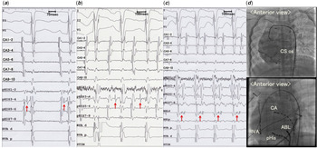
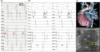
Atrioventricular re-entrant tachycardia involving twin atrioventricular nodes has been described in right atrial isomerism. However, little is known about the targets for catheter ablation when anterograde conduction through the atrioventricular node is absent. A 2-year-old male patient after Glenn operation for atrial situs inversus and atrioventricular discordance {I,D,D}, an endocardial cushion defect, a double outlet right ventricle and pulmonary stenosis with right atrial isomerism was referred for supraventricular tachycardias. Incremental and single-extra-stimulus pacings at the high anterior (a 12 o’clock position viewed in the anterior projection), anterolateral (an approximately 10 o’clock position), anteromedial (an approximately 2 o’clock position) and low posterior (a 6 o’clock position) sites of the common atrium showed a single QRS morphology with a superior axis. In addition, historical surface electrograms and rhythm strips performed on this patient had never demonstrated a 2nd discrete non-pre-excited QRS complex. We detected a His-bundle electrogram only at the posterior side of the common atrioventricular valve during sinus rhythm (Figs 1 a, d and 2c). Elaborate mapping around the anterior atrioventricular ring of the common atrium could not detect an anterior His-bundle electrogram even at the timing of posterior atrioventricular nodal block, suggesting that the anterograde atrioventricular conduction was only through the posterior atrioventricular node. On the other hand, ventricular pacing unmasked two ventriculoatrial pathways with decremental conduction. Both atrioventricular jump and ventriculoatrial jump over the posterior atrioventricular node were observed, though we could not confirm the AH jump because of our failure of His-bundle recording during the atrial pacing. Programmed stimulations induced two types of supraventricular tachycardia. Both tachycardias showed the same narrow QRS complex of a superior axis with the posterior His-bundle electrogram preceding the ventricular potential, indicating anterograde conduction over through the posterior atrioventricular node (Figs 1b and 2 a: tachycardia 1 and 2, respectively). The tachycardia 1 was induced by the atrial extra stimulus pacing following the prolongation of atrioventricular interval. Moreover, the ventricular extra stimulus pacing also induced the tachycardia 1 following the ventriculoatrial jump. Therefore, we diagnosed the tachycardia 1 as slow–slow (or fast–slow) atrioventricular nodal re-entrant tachycardia mediated by the posterior atrioventricular node. We detected the earliest atrial potential with a quasi-slow pathway potential during tachycardia 1 near the coronary sinus ostium, which was remote from the recording site of the posterior His-bundle electrogram (pHIS 1–2 and 3–4 in Fig 1c and d). Radiofrequency ablation using a 4-mm tip catheter (NaviStar, Biosense Webster, Diamond Bar, California, United States of America; 20–25 W aimed at a temperature of 55 °C) for this site terminated the tachycardia 1. After the termination of tachycardia 1, we continued the application by paying attention to the AH interval not to be prolonged during sinus rhythm. The accelerated junctional rhythm did not occur during the application of radiofrequency energy.
( a ) Surface and intracardiac electrocardiograms during sinus rhythm. His-bundle electrogram (a red arrow) was detected by the catheter placed at the posterior atrioventricular node. ( b ) Surface and intracardiac electrocardiograms during tachycardia 1. His electrogram precedes the ventricular potential (red arrow). ( c ) Surface and intracardiac electrocardiograms during tachycardia 1. The earliest electrogram resembling slow pathway potential of ventriculoatrial conduction was detected by the ablation catheter (red arrow). ABL: the ablation catheter placed at the site near the coronary sinus ostium (CS os); Common atrium (CA): the catheter placed at the CA via an inferior vena cava; Posterior His (pHIS): the catheter placed at the posterior of the common atrium for the recording of the posterior His bundle potential; RV apex (RVA): the catheter placed at the right ventricular apex via an aorta. ( d ) (Upper) Fluoroscopic images of the return phase of coronary angiography in the anterior view. (Lower) Fluoroscopic images in the anterior view.

( a ) Surface and intracardiac electrocardiograms during tachycardia 2. His electrogram precedes the ventricular potential (red arrow). ( b ) Surface and intracardiac electrocardiograms during ventricular pacing at the cycle length of 450 ms (left panel) and 380 ms (right panel), respectively. Shortening of the pacing cycle length prolonged the interval between ventricular activation and the sharp potential (red arrow), suggesting the decremental conduction property. ABL: the ablation catheter placed at the anterior of the common atrium via an inferior vena cava; Common atrium (CA): the catheter placed at the common atrium via an inferior vena cava; Common AV valve: the yellow circle indicated the site of the common atrioventricular valve; Posterior His (pHIS): the catheter placed at the posterior of the common atrium for the recording of the posterior His-bundle potential; RV apex (RVA): the catheter placed at the right ventricular apex via an aorta. ( c ) (Upper) The three-dimensional computed tomography shows a whole image of the patient’s heart. (Lower) Fluoroscopic images in the anterior view.

The anterior wall of the common atrium activated earlier during the tachycardia 2 (Fig 2 a and c). All the atrial electrograms were orthodromically entrained by the pacing from the right ventricular apex with slight decremental ventriculoatrial conduction and the difference between the first post-pacing interval and the tachycardia cycle length was 14 ms. Therefore, the possible diagnosis of the tachycardia 2 could be atrioventricular re-entrant tachycardia with twin atrioventricular nodes (posterior/anterior) that had anterograde conduction through the posterior atrioventricular node and retrograde conduction through the anterior atrioventricular node, though atrioventricular re-entrant tachycardia with a decremental accessory pathway was not fully excluded. During the mapping of retrograde ventriculoatrial conduction, we detected a sharp potential resembling the His-bundle electrogram around the presumed anterior atrioventricular nodal area. This sharp potential exhibited decremental property (Fig 2b, red arrow). During ventricular pacing, radiofrequency catheter ablation targeting for the sharp potential eliminated the anterior ventriculoatrial conduction. We confirmed the elimination of both supraventricular tachycardias and disappearance of retrograde (ventriculoatrial) conductions except for the emergence of ventriculoatrial conduction over the posterior atrioventricular node during isoproterenol challenge. The supraventricular tachycardias have not recurred for more than 5 years including the perioperative period of total cavopulmonary connection operation since this catheter ablation.
Discussion
This is the first report demonstrating that a retrograde sharp potential resembling the His-bundle electrogram could be an indicator for the successful elimination of the atrioventricular re-entrant tachycardia with twin atrioventricular nodes (posterior/anterior), especially in a patient with concealed conduction through an anterior atrioventricular node. Usually, the target of ablation for atrioventricular re-entrant tachycardia with twin atrioventricular nodes is an atrial side of the His-bundle electrogram with a non-dominant atrioventricular node. In this case, the target was a possible anterior atrioventricular node with only retrograde conduction. In such a case, we would recommend the mapping for the sharp potential indicating the possible retrograde His-bundle electrogram rather than the earliest atrial electrogram during ventricular pacing or tachycardia.
The present case raises two issues. First, we could not exclude the possibility of a decremental accessory pathway that produced the sharp potential, because we had no evidence of discrete 2nd non-pre-excited QRS complex by any atrial stimulus pacing. We should have tried (a) the atrial pacing at the sharp potential recording site exhaustively to elicit the 2nd discrete narrow QRS complex and (b) the adenosine test during ventricular pacing. Second, even if the sharp potential represented retrograde His-bundle conduction over the anterior atrioventricular node, we could not exclude the existence of Mönckeberg sling. Reference Wenink1–Reference Bessiere, Mongeon, Therrien and Khairy3 The finding that two different His-bundle potentials preceded the ventricular activation during the tachycardia is a unique and definite evidence of the sling connection between the two atrioventricular nodes. Reference Bae, Noh and Choi4 The short post-ventricular pacing interval following the orthodromic entrainment in our case may rule out the involvement of Mönckeberg sling in the re-entrant circuit. However, no paper has been published regarding the post-ventricular pacing interval during the tachycardia involving twin atrioventricular nodes. The post-pacing interval depends on the pacing site, and the pacing applied near the fascicle may result in the short post-pacing interval. In addition, Takeuchi et al reported that the earliest ventricular-to-retrograde His interval was prolonged with entrainment pacing from the ventricle. Reference Takeuchi, Shoda, Takahashi and Nakanishi5 This indicates a decremental property of the re-entrant circuit. Whether or not the retrograde His potential precedes the ventricular activation may depend on the sling conduction and anterograde HV interval during the tachycardia.
Despite the limitations, a sharp potential with the decremental property as shown in this case report could be a useful indicator for the successful elimination of this atypical tachycardia. This is because a retrograde sharp potential can be mapped without inducing the tachycardia, thereby avoiding haemodynamic collapse in patients with complex congenital heart disease.
Acknowledgements
None.
Financial Support
This research received no specific grants from any funding agency or from commercial or not-for-profit sections.
Conflict of Interest
None.
Ethical Standards
The authors assert that all procedures contributing to this study comply with the ethical standards of relevant guidelines on human experimentation and with the Helsinki Declaration of 1975, as revised in 2008. Our Institutional Review Board has confirmed that no ethical approval is required because this is a case report.