INTRODUCTION
During their development, many human and animal helminth parasites have an obligatory pathway through the lungs of their hosts, resulting in a pronounced pulmonary inflammation with eosinophilia. It has been proposed that the lung can act as an important site of retention of larvae (Capron and Capron, 1994; Silveira et al. 2002; Wilkinson et al. 1990).
Trichinella spiralis has a tissue-migratory larval stage, the newborn larva (NBL), which passes through the lung microvascular system on its way to the skeletal muscles (Despommier, 1983; Soulé, 1991). It is known that NBL are destroyed in the presence of cells and specific antibodies (Abs) (Gansmüller et al. 1987; Wang and Bell, 1992; Venturiello et al. 1993). Based on in vivo studies, some investigators suggest that this organ is the principal retention and destruction site of NBL (Bruschi et al. 1992; Gansmüller et al. 1987), and as a consequence only those larvae capable of evading the attack mechanisms of the immune system would accomplish muscle invasion (Wang and Bell, 1986; Wranicz et al. 1999). However, the immunological mechanisms involved at the lung level during a T. spiralis infection have not yet been established.
Due to the invasion of the intestinal epithelium by the muscle larva (ML), the first antigenic stimulus, the intestinal mucosa plays an important role at the onset of the host's immune response against T. spiralis. After reaching its first habitat, ML moult into adult worms which, once gravid, start to shed the NBL which enter the bloodstream, pass through the lungs and arrive to striated muscles. The invasion of the gut epithelium and the parasite expulsion are associated with intestinal pathology, which is a T-cell dependent phenomenon (Manson-Smith et al. 1979; Wakelin, 1997; Garside et al. 2000).
The mucosal immune system is an integrated system where all mucosae-associated lymphoid tissues (MALT) are involved and where the immunization in one site induces protective immunity in another (Lamm and Phillips-Quagliata, 2002; Roux et al. 2003; Holmgren and Czerkinsky, 2005). The MALT represents a compartmentalized immune system functioning independently of the systemic immune system. Taking into account these data and the mentioned response of the intestinal mucosa in trichinellosis, we might speculate that the parasite's antigenic stimulus in the gut-associated lymphoid tissue (GALT) should trigger alterations at the lung level, particularly in the bronchus-associated lymphoid tissue (BALT).
The aim of the present study was to characterize the histological and immunological modifications taking place in the bronchial mucosa and their putative relevance in the protection of the host against T. spiralis. To attain these objectives, studies were carried out in rats at 2 time-points post-infection (p.i.) important in the life-cycle of T. spiralis, i.e. on day 5 p.i., when adult worms start depositing the NBL which begin migrating, and on day 14 p.i. when all the adult worms have been rejected from the gut and the migratory phase has finished (Despommier, 1983; Soulé, 1991).
MATERIALS AND METHODS
Animals and infection
Groups of 4–6 two-month-old female Wistar rats belonging to a closed colony from the breeding unit and kept at the animal facilities of the laboratory were orally infected with 700 μl of a suspension containing 2800 ML/ml per rat employing a gastric canule. The ML were obtained by the digestion method (Larsh and Kent, 1949). Other groups of 4–6 animals remained uninfected as controls. Animals were allowed to have water and food ad libitum. During all the experiments animals were exposed to 12 h light-darkness cycles and room temperature was kept at 21±1 °C.
Tissue sections
Rats were sacrificed on days 5 or 14 p.i., and the lower respiratory tract was removed and subjected to the Sainte-Marie's technique (Sainte-Marie, 1961). Briefly, tissues were fixed in 95% ethanol pre-cooled at 4 °C, dehydrated in pre-cooled absolute ethanol, clarified with pre-cooled xylene and embedded in paraffin at 56 °C. Tissue sections (4–5 μm thick) were cut and placed on glass slides.
Immunohistochemical studies
For immunofluorescence (IF), paraffin from tissue sections was removed by gently shaking the slides in 4 consecutive baths of xylene, followed by 2 washes in pre-cooled absolute ethanol, 2 baths of pre-cooled 95% ethanol and 3 baths of saline solution. Cell labelling was done by incubating tissue sections with the following Abs: for IgE+ cells, a goat anti-rat IgE serum (Bethyl Laboratories Inc., Montgomery, TX, USA) followed by a fluorescein isothiocyanate (FITC)-conjugated anti-goat IgG serum (Sigma, St Louis, MO, USA) were used; for IgA+ cells a FITC-conjugated anti-rat IgA serum was used (Serotec Ltd, Kidlington, Oxford, UK); for CD4, CD8α, CD8β, CD5 cells, the corresponding monoclonal Abs (mAb; Pharmingen, San Diego, CA, USA) followed by a FITC-conjugated F(ab)’2 anti-mouse IgG serum (ICN, Cappel, Aurora, OH, USA) were used. For detection of CD5 cells, the mAb secreted by the clone OX-19 was used. Tissue sections were examined with an Olympus (Japan) epi-illumination microscope.
The number of cells per 15 fields (magnification ×1000) in the lamina propria of the bronchus-associated lymphoid tissue (BALT) was recorded in each section by 2 independent observers.
Histochemical studies
Paraffin from pulmonary tissue sections was removed as above and sections were stained to visualize the pulmonary inflammatory response: H&E was used to study the BALT and the presence of eosinophils, May-Grünwald-Giemsa was used to evaluate the presence of mast cells and PAS-haematoxylin was employed to analyse mucus-secreting goblet cells in the BALT epithelium.
Preparation of lung cell suspensions
To obtain lung cell suspensions from control and infected rats (day 5 p.i.), animals were anaesthetized, bled by heart puncture and sacrificed by cervical dislocation. Tissues were perfused by injecting 30 ml of phosphate-buffered solution pH 7·4 (PBS)-heparin (10 IU/ml, Sigma) into the heart right ventricle until the lungs turned to white colour. Then, lungs were removed and cell suspensions were prepared by cutting the tissues in small pieces that were digested in RPMI medium (Gibco, Grand Island, NY, USA) supplemented with collagenase A (0·5 mg/ml, Roche, Penzberg, Germany), DNAse (0·1 mg/ml, Roche), L-glutamine (0·29 mg/ml, Sigma) and streptomycin-penicillin (100 μg/ml–100 IU/ml, Sigma) for 30 min at 37 °C with ocassional shaking. Tissues were then dissociated using a tissue homogenizer and cell suspensions were passed through a nylon net to remove tissue debris. Remaining erythrocytes were lysed with ammonium chloride buffer. Cell suspensions were suspended and washed twice with PBS plus EDTA 5 mM and 3% fetal bovine serum (FBS, Gibco) and counted in a haemocytometer employing Trypan blue. Cell viability was invariably greater than 95%. Finally, cell suspensions were adequately resuspended in Dulbecco′s modified Eagle's medium (DMEM, Gibco) supplemented with streptomycin-penicillin (100 μg/ml–100 IU/ml, Sigma), L-glutamine (0·29 mg/ml, Sigma) and 5% FBS.
ELISPOT assay
In order to detect the presence of IgE-secreting lung cells, ELISPOT assays were performed as described previously (Czerkinsky et al. 1983) with some modifications. Briefly, flat-bottomed 96-well nitrocellulose plates (Millipore Co, Bedford, MA, USA) were coated overnight at 4 °C with sheep anti-rat IgE specific polyclonal Abs (10 μg/ml; Bethyl). After washing with sterile PBS, plates were blocked with 10% FBS in DMEM for 1 h at 37 °C. After washing, 8×10⁵ lung cells/well from control or infected rats were added in triplicate and incubated for 24 h at 37 °C in a moist atmosphere with 5% CO2. Thereafter, wells were washed with PBS and PBS plus 0·05% Tween 20 (PBST) and incubated overnight at 4 °C with horseradish peroxidase-conjugated anti-rat IgE serum (Bethyl) suitably diluted in PBST supplemented with 5% FBS. After washing with PBST, spots were developed with 3-amino-9-ethylcarbazole-H2O2 (Sigma) incubating at room temperature. The reaction was stopped by the addition of tap water. After drying the plates, spots were enumerated using an Immunospot reader (CTL, Cleveland, USA). Results were expressed as number of IgE-secreting cells per 8×10⁵ lung cells.
Collection of cells and fluid from bronchoalveolar lavage (BAL)
Cells and fluid from BAL were collected as described previously (Egwang et al. 1984) on days 5 and 14 p.i. Briefly, BAL was obtained by tracheal intubation using a Portex tube (Scientific Laboratory Supplies, Newcastle, UK). Lavages were done with 4 ml of saline solution with protease inhibitors (Complete, Boehringer, Mannheim, Germany). The BAL fluid (BALF) was obtained by centrifugation and the supernatant was aliquoted and kept at −20 °C until used. Cell pellets were resuspended and washed twice with Eagle's Minimum Essential medium with Earle's salts (MEM, Gibco). Cells were counted in a haemocytometer employing Trypan blue. Cell viability was invariably greater than 95%. Relative percentages of cell types in cell pellets were determined by preparation of smears and stained with May-Grünwald-Giemsa.
Quantification of total IgE and IgA in BALF
Total IgE levels in BALF samples were determined by capture ELISA. Briefly, flat-bottomed PolySorp polyvinyl microtitre plates (Nunc, Roskilde, Denmark) were coated with an anti-rat IgE serum (Bethyl) at a concentration of 10 μg/ml in carbonate buffer, pH 9·6. After 2 h of incubation at 37 °C and between all subsequent steps, plates were washed 3 times with PBST. Plates were then blocked at 37 °C with a 10% dried non-fat milk solution in PBS (PBSM). After washing, each BALF sample was added undiluted. After incubating for 1 h at 37 °C and overnight at 4 °C, plates were washed and incubated for 1 h at 37 °C with a biotin-conjugated anti-rat IgE Ab (Serotec) suitably diluted in PBST. Plates were washed and a pre-formed macromolecular complex of avidin and biotinylated peroxidase (Vector Laboratories Inc., Burlingame, CA, USA) was added. After incubating, plates were washed and the reaction was developed by addition of tetramethylbencidine-H2O2 (Sigma). The reaction was stopped by addition of 4 N H2SO4. Optical densities (OD) were read on an ELISA reader (Metertech Σ960) at 450 nm. Standard curves of rat IgE (Serotec) were run in each plate in order to determine the IgE concentration in each sample. Results were expressed in ng/ml. Experiments were done in duplicate.
Total IgA levels in BALF samples were determined using a commercial kit (Bethyl) according to the manufacturer's instructions. BALF samples were tested at a dilution of 1/100 in PBST. Standard curves of rat IgA were run in each plate in order to determine the IgA concentration in each sample. Results were expressed in μg/ml. Experiments were done in duplicate.
Collection of NBL
NBL were obtained as previously described (Dennis et al. 1970). Briefly, adult worms were recovered from the intestine of rats orally infected with 7000 ML 6 days before. Worms were cultured at 37 °C in an atmosphere containing 5% CO2 in MEM supplemented with antibiotics (penicillin 50 IU/ml, streptomycin 50 μg/ml, Sigma) plus 5% FBS (Gibco). NBL were collected after 2 h and preserved in liquid nitrogen until used in IF tests or used immediately in the cytotoxicity assays where all larvae were alive and in good condition as judged by their motility.
Detection of specific IgE, IgA and total antibodies (IgGAM) anti-NBL surface in BALF
Anti-NBL Abs were detected using the indirect IF assay on slides containing methanol-fixed NBL. Slides were incubated for 30 min at 37 °C with 15 μl of BALF samples diluted 1/5 for IgE, IgA and IgGAM. Reaction was then continued by the addition of 15 μl of a goat anti-rat IgE or a goat anti-rat IgA serum (Bethyl) suitably diluted in PBST followed by a FITC-conjugated anti-goat IgG serum (Vector) or a FITC-conjugated anti-rat IgGAM serum (Jackson Immunochemicals, West Grove, PA, USA) suitably diluted in Evans blue. All steps were carried out at 37 °C with 3 washes with PBST in between. Slides were then air-dried and mounted using buffered glycerin. BALF samples were considered positive when fluorescence of parasite surface was observed using a fluorescence microscope (Olympus). Positive and negative controls were included in each assay. BALF from rats infected 45–55 days before and BALF from non-infected animals were employed as positive and negative controls respectively. Experiments were done in duplicate.
Cytotoxicity assays
To study the cytotoxic properties of BAL cells, the test was performed in flat-bottomed microwell modules (Nunc). A 30 μl volume of a larval suspension in MEM containing approximately 50 NBL was placed in each well and the exact number of larvae was counted. One hundred μl of BAL cell suspension containing 4×10⁵ cells/ml from infected and control rats and 10 μl of an anti-T. spiralis cytotoxic rat serum (CS) or a normal serum, were added to each well. Reactions were kept for 4 h or 24 h at 37 °C in an atmosphere of 5% CO2 in air.
The CS employed was a serum obtained from a rat orally infected with ML of T. spiralis 45 days before and that was able to mediate the NBL death in antibody-dependent cell cytotoxicity (ADCC) assays.
To study the cytotoxic activity of anti-NBL Abs in BALF, the same test was performed employing BAL cell suspensions of control rats and replacing the CS by BALF of infected (day 14 p.i.) and control rats.
The NBL death was assessed by direct microscopy by 2 independent observers in a blind fashion and the mortality percentage calculated according to the following formula:

where NBLi and NBLf are the numbers of NBL counted at the beginning and at the end of reaction respectively. Two independent experiments were done employing samples in duplicate.
Statistics
Tests employed in the statistical analysis as well as significance levels are indicated in each experiment. In all cases, the results were expressed as mean±S.E.M.
RESULTS
Histochemical studies
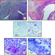
BALT hyperplasia was observed on days 5 and 14 p.i. (Fig.1A). A marked pulmonary inflammatory reaction containing eosinophils and mast cells together with other inflammatory cells scattered over the pulmonary parenchyma was observed on both days studied (Fig. 1C). Goblet cell hyperplasia was observed at the BALT epithelium level on days 5 and 14 p.i. These goblet cells contained a large amount of mucus glycoprotein. A large amount of mucin was also observed in the alveolar spaces (Fig. 1D).

Fig. 1. Histopathology of lungs during the early phase of Trichinella spiralis infection in rats. Sections of lung tissue obtained from infected (5 or 14 days p.i.) and control rats. Panel A (H&E): BALT hyperplasia in infected rats. Panel B (H&E): BALT from control rats. Panel C (May-Grünwald-Giemsa): inflammatory infiltrate, mast cells scattered together with eosinophils in the lung parenchyma from infected rats (ε and μ indicate eosinophils and mast cells respectively). Panel D (Haematoxylin-PAS): goblet cell hyperplasia in the BALT epithelium and mucus secretion in infected rats (γ indicate goblet cells). Panel E (Haematoxylin-PAS): goblet cells (γ) from uninfected rats.
Immunohistochemical evaluation of BALT cell phenotype
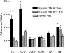
The phenotype of cells present in the BALT lamina propria of T. spiralis infected rats is shown in Fig. 2. Significant differences in the number of cells per 15 fields between infected rats on day 5 p.i. and control animals were observed in the following cell populations and subpopulations: CD4+ cells: 528±65 vs 185±18 (P<0·001); CD5+ cells: 684±77 vs 333±19 (P<0·002) and IgE+ cells: 401±8 vs 259±30 (P<0·002). On day 14 p.i., CD4+ and CD5+ cell numbers returned to baseline values; only IgE+ cell counts remained increased (375±23 vs 259±30, P<0·002). No significant differences were found in the number of IgE+ cells between days 5 and 14 p.i. The number of IgA+, CD8α+ and CD8β+ cells in the BALT lamina propria of infected animals did not differ significantly when compared to controls on both 5 and 14 days p.i.

Fig. 2. Cell phenotype in the lamina propria of the bronchus-associated lymphoid tissue during the early phase of Trichinella spiralis infection in rats. Data are expressed as mean±S.E.M. of 5 animals per group. Significant differences *(P<0·002) between infected and control rats were observed in the following BALT cell populations and subpopulations: CD5+ cells and IgE+ cells on day 5 p.i. and IgE+ cells on day 14 p.i. Significant differences **(P<0·001) between infected rats on day 5 p.i. and control animals were observed in the CD4+ cell population. Student's t-test.
IgE-secreting lung cells
IgE-secreting cells were detected in the lung cell suspensions of infected rats on day 5 p.i. finding 18±2 IgE-secreting cells per 8×10⁵ total cells. No spots were found in wells where lung cells of control animals were added.
Parasite-specific Abs and total IgE and IgA levels in BALF
Specific Abs against the NBL surface were detected in BALF on day 14 p.i. in the IgGAM (60% of animals), IgE isotype (40% of animals) and IgA isotype (40% of animals). Results are shown in Table 1. Significant differences were found only in the levels of total IgE in BALF of infected rats on day 14 p.i. when compared to the control group (18·0±3·5 vs 4·1±0·4 ng/ml, P<0·005). No significant differences were found in total IgA values between groups on both 5 and 14 days p.i. (Table 1).
Table 1. Total and specific immunoglobulins in bronchoalveolar lavage fluid during the early phase of Trichinella spiralis infection in rats (Data are expressed as mean±S.E.M. of results obtained from 2 independent experiments performed with groups of 5 animals each. Values in parentheses indicate the percentage of positive animals. Significant differences *(P<0·005) for total IgE between infected rats on day 14 p.i. and control animals. Student's t-test.)

Biological activity of cells and Abs in BAL
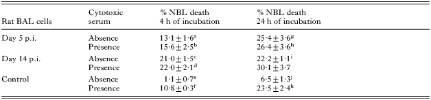
Results of the biological activity of BAL cells are shown in Table 2. BAL cells from normal rats were able to induce NBL death only in the presence of CS achieving 23·5±2·4% of mortality after 24 h of incubation. BAL cells from infected rats were able to induce NBL death on both 5 and 14 days p.i. even in the absence of CS. This phenomenon was observed as early as at 4 h of incubation.
Table 2. Cytotoxic activity of bronchoalveolar lavage cells during the early phase of Trichinella spiralis infection in rats (Data are expressed as mean±S.E.M. of results obtained from 2 independent experiments performed with groups of 4 animals each. Significant differences (P<0·01) between a vs e; c vs e; g vs j; i vs j; d vs f; b vs h; e vs j; f vs k. Significant differences (P<0·05) between a vs c. Significant differences (P<0·001) between a vs g, e vs f, j vs k. Unspecified combinations are not statistically different. In specific death, percentages in the absence of cells (NBL only or NBL plus CS only) were 6·0±5·5%. Three-way ANOVA with repeated measures of 2 factors.)
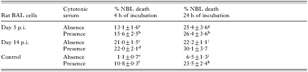
The maximum mortality percentage reached by BAL cells from infected rats obtained on day 5 p.i. was observed at 24 h of incubation in either the presence (26·4±3·6%) or the absence (25·4±3·6%) of CS. On the other hand, when BAL cells from infected rats obtained on day 14 p.i. were used, the maximum percentage mortality of NBL was reached as early as at 4 h of incubation in either the presence (22·0±2·1%) or the absence (21·0±1·5%) of CS. However, when NBL were incubated with BALF from infected rats obtained on day 14 p.i. (which presented Abs against NBL surface) and BAL cells from normal rats, neither cell adherence nor NBL mortality were observed, being the mortality percentage (10·0±1·2%) similar to that observed in control reactions employing BALF from normal rats (8·4±2·0%).
BAL cell populations from infected rats did not differ from controls. Mean percentages found in both groups were: 1·5±0·5% neutrophils, 1·0±0·6% eosinophils, 0% basophils, 3·0±1·0% lymphocytes, 80·5±1·5% monocytes and 14·5±0·5% macrophages.
DISCUSSION
Our results demonstrate that histopathological changes of the Th2 type take place in the lungs of infected rats during the early stages of the infection with T. spiralis. On day 5 p.i., when larvae start migrating from the intestine, on their way to the skeletal muscles, they find and go through a lung which presents a marked eosinophilic inflammatory process with the presence of mast cells, goblet cell hyperplasia with mucus production and BALT hyperplasia with an increase in the number of CD4+, CD5+ and IgE+ cells. These histopathological changes were observed from day 3 p.i. on (data not shown).
Since the mAb employed in our experiments recognizes only CD5 of T-lymphocytes (Vermeer et al. 1994) our observations suggest that the increase of such a cell population is due to an increase in CD4+ T-cells. The presence of IgE on the surface of BALT cells would indicate that these T-cells have a Th2 phenotype. Besides, the IgE+ cells observed in the BALT are B-lymphocytes, for no other IgE-bearing accessory cells (e.g. eoninophils, mast cells) were found in such lymphoid tissue. This idea is supported by the fact that IgE-secreting lung cells were detected, thus demonstrating a local pulmonary synthesis and secretion of IgE. The mentioned histopathological changes were maintained until the end of the larval migratory phase (day 14 p.i.) when specific Abs, as well as an increment in the total IgE levels in BALF, were detected.
On evaluating the biological activity of BAL cells, it was observed that cells from infected animals were able to kill the NBL even in the absence of adherence and specific Abs. This effect was observed shortly after the NBL were placed in contact with the BAL cells. This rapid killing of parasites would indicate that BAL cells are activated for this effector mechanism. The differences obtained in NBL death between BAL cells from control and infected rats could not be accounted for by differences in the effector cell number since BAL cell populations were not significantly different. These results demonstrate the role of the pulmonary response in parasite death, suggesting that the histological changes observed are important for the protection of the host against T. spiralis infection. In vivo studies are currently being undertaken in our laboratory to corroborate this hypothesis.
It is known that the mucosal immune system is an integrated system where immunization in one site induces protective immunity in another (Roux et al. 2003; Holmgren and Czerkinsky, 2005). The communication between the BALT and distal mucosae via intermucosal cell traffic involves predominantly a ‘gut to bronchus’ flow of cells from Peyer's patches (Scicchitano et al. 1984; Bienenstock and Clancy, 2005). Furthermore, it is known that the intraepithelial localization of Trichinella in the small intestine stimulates a strong immune-mediated inflammatory response that leads to the expulsion of the AW out of the intestine (Manson-Smith et al. 1979; Wakelin, 1997; Garside et al. 2000). In agreement with the above-mentioned statements, our results suggest that the host's defence mechanisms against T. spiralis at the pulmonary level would develop as a response to a dual antigenic signalling. The first signal would be originated in the gut due to the penetration of ML in the intestinal epithelium, initiating the immune response against T. spiralis and causing the alterations observed in the lung. The second signal, generated in situ, would correspond to the continuous antigenic stimulation by the NBL during their passage through the lung and would be responsible for the increase or persistence of the characteristic parameters found in helminth infections such as IgE+ cells, eosinophils, mast cells and the presence of IgE in fluids.
Furthermore, some authors have demonstrated that the entrapment of parasites or the presence of Sephadex particles in the lung vasculature constitute a sufficient stimulus to trigger the recruitment of eosinophils due to the release of chemotactic factors (Laycock et al. 1986; Culley et al. 2002). As the size of the NBL is similar to that of the pulmonary capillary vessels it would not be surprising that this entrapment phenomenon is also taking place in our model.
The lack of in vitro biological activity of the BALF in parasite death is probably due to an insufficient level or a lack of effector Abs to mediate ADCC, a well-known attack mechanism against the NBL (Kazura, 1981; Wang and Bell, 1992; Venturiello et al. 1993).
The presence of IgA and IgE anti-NBL in infected animals demonstrates the simultaneous secretion of both isotypes in the bronchial mucosa during the infection with T. spiralis, which has been previously demonstrated in the intestinal mucosa (Negrão et al. 1999; Nuñez et al. 2003).
In summary, our results demonstrate that in rat trichinellosis there is an early marked inflammatory reaction in the lung which is involved in the effective defence mechanism against the migratory stage of the parasite. This inflammatory reaction would provide this organ the necessary effector cell populations to destroy the parasite. This study reinforces the hypothesis of other authors who have postulated the lung as an organ of parasite entrapment and destruction (Wang and Bell, 1986; Bruschi et al. 1992). This concept would imply that only those larvae capable of escaping the attack mechanism (Jungery et al. 1983; Gansmüller et al. 1987) would be able to travel through the lung and accomplish invasion of striated muscles in a primary infection.
Finally, our work shows that the early pulmonary response observed should also be considered in further investigations of the host's defence against the NBL of T. spiralis.
The present work was supported by grants B073 from the University of Buenos Aires and PIP No 02858 from the National Research Council of Argentina (CONICET).