I. INTRODUCTION
Nanomaterials have been the subject of enormous interest nowadays. In the past, many people focused on how to produce less-agglomerated and low-dimensional nanoparticles. Gradually, people have turned their attention to different shapes of nanomaterials, such as nanorods, nanocube, nanodisks, nanowires, and nanoplates, etc., because their properties could be tuned with the shapes. Reference Xiong and Xia1–Reference Lee, Choi, Jang, Kim, Ju, Min and Ahn3 Among the nanomaterials, silver nanowires (Ag NWs) are attractive due to their high electrical and thermal conductivity, and have many applications. They can be utilized as highly transparent and flexible electrodes in polymer light-emitting diodes or polymer solar cells, and highly stretchable electrodes in muscle-like transducers. Reference Hu, Kim, Lee, Peumans and Cui4–Reference Lee, Lee, Lee, Yeo, Hong, Nam, Lee, Lee and Ko7 They also can be used in catalysis, chemical nano-sensors, surface-enhanced Raman scattering, infrared signature reduction, and plasmonic circuitry. Reference Alia, Duong, Liu, Jensen and Yan8–Reference Singh, Raghuwanshi and Kumar12
The methods of producing Ag NWs can be roughly divided into two types, template-used, and template-free. The templates are cylindrical membrane pores or standing cylindrical rods within which particles are confined, such as porous anodic aluminum oxide, Reference Yang, Sui, Gong and Qu13 carbon nanotubes, Reference Yu, Peng, Zhai, Li and Wei14 and sonoself-reduction templates. Reference Malandrino, Finocchiaro and Fragala15 In the template-free method, capping agents are required to attach to the preferring face of silver to make the anisotropic growth of silver. Xia and coworkers have demonstrated a polyol process for the synthesis of Ag NWs with poly(vinyl pyrrolidone) (PVP) as a capping agent and PtCl2 as an additive. Reference Sun, Yin, Mayers, Herricks and Xia16 It was discovered that Cl− ions are very helpful in the synthesis of Ag NWs, because they catch Ag+ to form silver chloride. Reference Tsuji, Matsumoto, Jiang, Matsuo, Tang and Karnarudin17 The silver chloride is a hard to dissolve salt, so it can control the concentration of Ag+ in the solution. Other anions which also form hard to dissolve silver salts are also investigated, such as S−. Reference Chen, Zhu, Zhu, Qiao and Chen18 For the study of capping agents, Becker used aniline, pyridine, or 3-aminopyridine as surfactants to grow Ag NWs. Reference Becker, Soderlind, Liedberg and Kall19 Yang used citrate as the reduction agent and capping agent to synthesize Ag NWs successfully. Reference Yang, Qian, Chen and Anker20 Recently, Lee's group produced very long Ag NWs in which the longest length is about 200 μm in the presence of Cl−. Reference Lee, Lee, Lee, Lee, Lee and Ko21
Xia and coworkers also synthesized Ag NWs by the polyol process but without Cl− upon LaMer's model. Reference Wiley, Sun, Mayers and Xia22 Chou developed the process window for the scale up in the polyol process without Cl−. Reference Lu, Chou and Nogami23 It is feasible to synthesize Ag NWs without any hardly soluble salts. In our study, we tried to synthesize high aspect ratio Ag NWs in a one-pot method via syringes without the present of any hardly soluble salts.
There are two points that are clear about the synthesis of Ag NWs. First, the nuclei of Ag NWs are decahedrons, and one decahedron comprises 5 tetrahedrons. Reference Chen, Zhang, Chen and Sun24 Second, PVP chains attach to the (100) face of nuclei, which makes Ag+ unable to be reduced and to be accumulated on the (100) face. This forces Ag+ turning to the (111) face only on both ends, and results in anisotropic growth along 〈110〉. Reference Sun, Yin, Mayers, Herricks and Xia16 However, from the dynamics perspective, it is difficult to accumulate Ag+ only on both ends, because the increase in aspect ratio of Ag NWs decreases the ratio of the surface of (111) to the surface of (100), which lowers the probability of Ag+ colliding to both ends. Several questions arise in the growth of Ag NWs, such as the synthesis of very long wires, width consistency in a wire, and deciding factors of the wire widths. In our research, we accidentally discovered some interesting phenomena during the growth of Ag NWs. Therefore, we have developed a model to explain the questions mentioned above in the research.
II. EXPERIMENTAL PROCEDURE
A. Materials
Silver nitrate (AgNO3, 98%) was purchased from Showa Chemicals (Saitama, Japan). PVP (MW = 58,000) was obtained from ICN Biomedical, Inc (Costa Mesa, California). Ethylene glycol (EG, 95%) was obtained from Sigma–Aldrich Corporation (St. Louis, Missouri).
B. Synthesis of Ag NWs
In a representative synthesis, 20 mL of EG was added in a 250 mL four-necked flask, and heated to 160 °C. Then, 50 mL of 0.05 M AgNO3 in EG and 50 mL of 0.075 M PVP in EG were syringing (in a rate of 0.5 mL/min) into the preheated EG separately at the same time. After syringing, the solution was kept at 160 °C for half an hour, and cooled to room temperature naturally. The grey-colored suspension was centrifuged at 6000 rpm. The precipitate was washed with deionized water and acetone twice to remove EG and PVP for further characterization. We have also tried to use different syringe rates of 1 and 1.5 mL/min in the experiment.
To further study the concentration effect, we have used higher concentrations of AgNO3 (0.25 and 1 M) and PVP (0.375 and 1.5 M) in the experiment. The syringe rate was fixed at 1 mL/min.
C. Characterization
The UV–visible (UV–VIS) spectra of Ag NWs suspended in water were recorded by Hitachi U-2001 UV–VIS spectrophotometer (Hitachi, Tokyo, Japan). High resolution field emission scanning electron microscope (JEOL JSM-6700; JEOL Ltd., Peabody, Massachusetts) and transmission electron microscope (TEM; JEOL-2100F, JEOL Ltd.) were performed for the morphology study of Ag NWs.
III. RESULTS AND DISCUSSION
A. Syringe rate effect
The idea of synthesizing Ag NWs via a syringe is originated from the nucleation of Ag NWs. A nucleus comprising 5 tetrahedrons is well known. In the beginning of the reduction of Ag+, Ag atoms may cluster together to form a tetrahedron, because the surface of tetrahedron is the fully packed (111) facet with less surface energy. However, the sizes of the tetrahedrons might be smaller than the critical size, so they need to combine together to cross the limit. Five of them can make a round shape just lacking an angle about 7°. Reference Chen, Zhang, Chen and Sun24 When syringing AgNO3 dropwise, the concentration of Ag+ is not high enough to form the tetrahedrons larger than the critical size, and forces them to combine together to form the nuclei. However, the syringe rate cannot be too low, which will reduce the colliding probability of tetrahedrons, and result in the failure of nuclei formation.
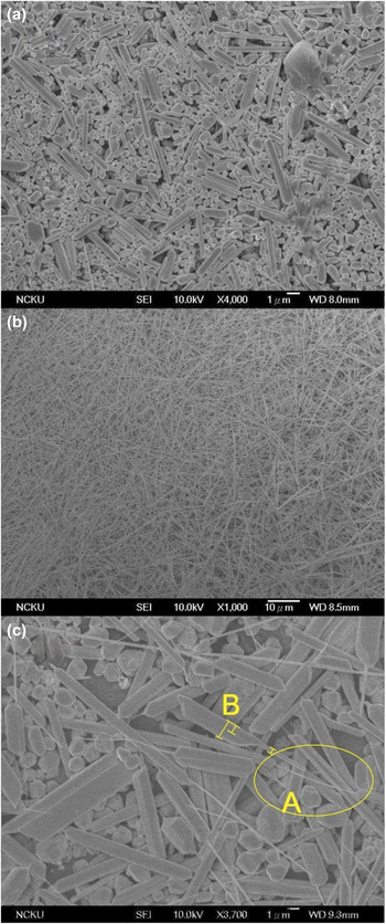
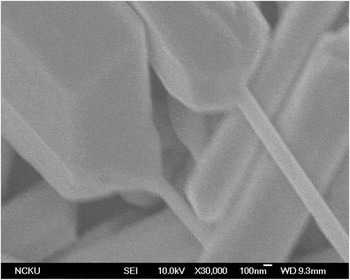
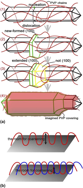
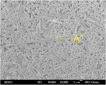
For the syringe rate study, the results of different syringe rates with the same concentrations of AgNO3 (0.05 M) and PVP (0.075 M) are shown in Fig. 1. In Fig. 1(a), many short rods and particles have been found when the syringe rate was 0.5 mL/min, which could be due to the syringe rate being too low to form the nuclei. When the concentration of AgNO3 increased with time, the tetrahedrons grew large enough and became particles. The appearance of rods is related to the mechanism of the growth of Ag NWs, and will be discussed later. In Fig. 1(b), when the syringe rate was 1 mL/min, the Ag NWs were synthesized successfully. The width of the Ag NWs was about 100–300 nm, and the length was in the range of 10–50 μm. Although the average aspect ratio of the Ag NWs is not higher than that from the polyol process with Cl−, Reference Li, Hu, Zhang and Oakes25,Reference Yang, Pan, Chen, Lin, Wu, Hung, Lin, Huang, Liu, Mao and Kuo26 the process without Cl− may find wide applications in the future, because it does not have the corrosion problem from Cl−. When the syringe rate is increased to 1.5 mL/min, an amazing phenomenon appeared in Fig. 1(c). The initial thin Ag NWs were covered by thick Ag. Their shape was like hotdogs, so we called them nano-hotdogs. This shape has not been discovered in the polyol process of Ag NWs synthesis before, but there is a similar shape for ZnO via the chemical vapor disposition. Reference Wang, Liu, Huang, Chen, Tseng and Kung27 We can see them more clearly in Fig. 2. It seems that the thin wires were coarsened instead of having the thin tail growing from the thick one. Because the cross-sections are unordered, the Ag atoms accumulated on the thin wires in different velocities. It is surprising that Ag atoms accumulated orderly over the thin wires when coarsening. In addition, if there were enough Ag atoms to accumulate, the thin wires might become thick wires with different widths. This indicates that coarsening might be part of the growth of Ag NWs. Because we only changed the syringe rate which affected the concentration of Ag+, we suggest that the concentration of Ag+ is the key to coarsening. We developed a model, which is shown in Fig. 3(a). In the presence of PVP, Ag+ was reduced and accumulated only on both ends for the lateral face (100) covered by PVP. The increase in Ag NWs length decreases the probability of Ag+ colliding to both ends. With the increase of syringe rate, more Ag+ needed to be reduced quickly. The driving force of reducing Ag+ became larger when more Ag+ existed. Therefore, Ag+ found places, such as nucleation sites on the (100) face where dislocations existed, that prevented PVP chains from covering well. Once the nucleation sites were formed, Ag atoms would accumulate on these sites until new (100) was formed. A study reported that there was a direct chemical bonding between PVP chains and Ag on the (100) face. Reference Saidi, Feng and Fichthorn28 Therefore, the (100) face with PVP chains would be stable, and no Ag atoms could penetrate between them and accumulate on the (100) face. The accumulation is like a step around the thin wire. The new-formed cross-section, which is not (100) of the step, would strongly attract Ag+ to be reduced and accumulated to release the driving force of reduction. Whenever Ag atoms are accumulated to the extended (100) face, it would be attached by PVP chains nearby soon and form the new-formed (100). As a result, the coarsening would create a consistent width, and the whole thin wires would coarsen gradually if there were enough Ag+. The appearance of nano-hotdogs was due to the coarsening becoming so fierce. This resulted in thick wires, but not enough Ag+ to accumulate on the whole thin wires. In the A part of Fig. 1(c), there was an Ag NW with the shape of nunchaku. According to the model, the coarsening would occur on the (100) face where PVP chains did not cover well, so there may be two of these types of sites on the same wire. However, the cross-section formed first would strongly attract Ag+ and suppress other nucleation site formed on the same wire. Therefore, it was rare to see the nunchaku-shaped wire. In the B part of Fig. 1(c), a wire with three different widths was observed, which may have coarsen twice. To understand the PVP protecting effect, we doubled the concentration of PVP, but other factors remained unchanged. The result is shown in Fig. 4. The widths of wires became smaller, because the coarsening is not easy to occur even if there is more PVP protecting. The reason is that the new-formed (100) face would be attached by PVP chains quickly, and there is less room to coarsen as shown in Fig. 3(b). It is hard to find a place that is not covered well for the second or third coarsening. A study mentioned that the Ag NWs became thinner when more PVP was added. Reference Coskun, Aksoy and Unalan29 In the A part of Fig. 4, the Ag NW with two widths was coarsening. Therefore, we suggest that the coarsening is one part of Ag NWs growth. The Ag NWs coarsen layer by layer gradually, and each layer has a consistent width.
Scanning electron microscope (SEM) images of Ag NWs with different syringe rates (a) 0.5 mL/min, (b) 1 mL/min, and (c) 1.5 mL/min.

SEM image of the nano-hotdogs.

(a) The model of coarsening of nano-hotdogs: (1) nucleation on the place where PVP chains do not cover well, (2) PVP chains attached to new-formed (100) and the transverse accumulation stopped, (3) extended (100) attached by PVP chains which induced a consistent width and (4) coarsening the whole wire with widening from the covering of PVP similar to blowing up a balloon. (b) Imaging PVP chains covered on the surface like a curve with the double PVP chains cover resulting in less room for coarsening.

SEM images of Ag NWs in the syringe rate of 1.5 mL/min with the concentrations of 0.05 M AgNO3 and 0.15 M PVP.

B. The concentration effect
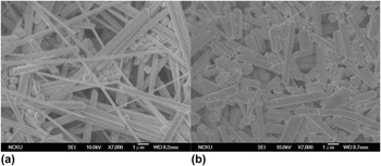
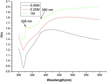
The effect of different concentrations of AgNO3 (0.25 or 1 M) and PVP (0.375 or 1.5 M) (the ratio of PVP to AgNO3 was fixed at 1.5) with the same syringe rate (1 mL/min) is shown in Fig. 5. The widths of Ag NWs increased with increasing AgNO3 concentration. The widths of Ag NWs were 200– 1 μm with 0.25 M AgNO3 and 0.375 M PVP, and 1–1.5 μm with 1 M AgNO3 and 1.5 M PVP. This is consistent with the previous observation. The coarsening would be fierce with the higher concentration of AgNO3 and stronger driving force for reduction. The UV–VIS absorption spectra of Ag NWs prepared from different concentrations of AgNO3 are shown in Fig. 6. All spectra had a shoulder peak at about 380 nm. Another shoulder peak at about 328 nm appeared when AgNO3 concentration increased. This has not been discussed in other literature. In Fig. 5, we can see that the biggest difference between the images with different concentrations was the aspect ratio, which is also related to the ratio of the surface area of (100) to that of (111). The surface area of (111) was larger, and the shoulder peak was also more obvious with the increasing concentration. As a result, we believe that the shoulder peak at 328 nm is attributed to the plasmon response of bulk Ag on (111). On the other hand, the lengths of Ag NWs were shorter with increasing concentration. There may be a mechanism of Ag NWs growth at the earlier stage that controls the lengths of Ag NWs.
SEM images of Ag NWs at the same syringe rate (1 mL/min) with (a) 0.25 M AgNO3 and 0.375 M PVP and (b) 1 M AgNO3 and 1.5 M PVP.

The UV–VIS absorption spectra of Ag NWs prepared from different concentrations of AgNO3.

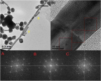
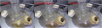
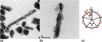
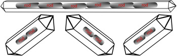
Hence, we repeated the experiment that synthesized Ag NWs whose shapes were as shown in Fig. 1(b), but we took the TEM image at 10 min right after syringing. The results are shown in Figs. 7(a) and 7(b). There were two things worth noticing. First, the width of the wires at the early stage was smaller than 40 nm. It was also smaller than the widths of wires of 70–100 nm length reported in many studies. This indicates that coarsening might exist. Second, the wires with many grain boundaries were observed. From the Fourier transforming diffraction patterns, we could find that A and C were different crystals with little lattice shift, so the diffraction points did not match well on the B grain boundary. This showed that a wire consisted of many rods. We also found a sign during the experiment as shown in Fig. 8. At the beginning of syringing AgNO3 and PVP, the solution was translucent and light yellow, meaning that some nanoparticles were synthesized. Gradually, the solution became opaque and gray within 40 s. The changing of color and transmittance represented the formation of wires. The long wires were synthesized in such a short period of time, just like the condensation reaction of polymers. When the Ag rods grew to some degree of length, the chance of the rods colliding with each other increased. The PVP chains attached to a rod might attach to another one when the rods are close by. Therefore, they could connect to each other in a short period of time, increasing the lengths of wires immediately. Some images of the rods connecting are shown in Figs. 9 and 10. The rods have not bonded well yet, and the PVP covering could be seen obviously at the connecting site. The PVP covering was extended from the edges of the rods, and made the wire a consistent width, as shown in Figs. 9(a) and 9(b), which could be a symbol of PVP chains attaching to different rods at the same time. Of course, the attachment between rods might not be end to end all the time. The rods could also be attached from the lateral edge to lateral edge, such as the X and Y parts in Fig. 7(a). This could explain the formation of a raft of Ag NWs that has been mentioned before in a study. Reference Jiang, Li, Xie, Gao and Song30 After the rods were connected, they continued to grow and then touch each other. For this reason, the crystals of rods are 5-fold symmetric. Both rods on the two ends of the connection just needed to rotate less than 20° as shown in Fig. 9(c). The lattices of both sides would almost match, so Ag atoms could accumulate around the connecting site to make the rod bonding together. In Fig. 9(b), we could see the rods bonding together with different widths. When the wider rod was connected to the thinner wire, a fault would be formed. The cross-section of the fault was not the (100) face, meaning that Ag atoms accumulate just like the process of coarsening, until the (100) face of the wider wire was extended and covered well by the PVP again. Then the width of the whole wire would become consistent as shown in Fig. 7(a). Therefore, Ag NWs became longer via comprising of the rods, and width-consistent via coarsening of the thinner rods. With these thoughts, we explain why the wires were shorter in the low syringe rate or with the high concentration. In the low syringe rate, the syringe rate was too low to form the nuclei, so the number of rods was small. This decreased the probability of rods connecting. Therefore, the wires were shorter, because they comprised fewer rods. In high concentrations, the driving force for reducing Ag+ was too big. The coarsening would be fierce and occur quite early. Hence, the wires would comprise fewer rods. It was difficult to connect two coarsened wires if they were too big and heavy. The PVP chains were not strong enough to fix them together until they were bonded together. The schematic diagram of several rods combined together to form a wire is shown in Fig. 10.
(a) The TEM image of wires at 10 min right after syringing and (b) the enlarged image of the grain boundary.

The change of color in the solution indicates the formation of Ag NWs.

(a) The SEM image of the rods do not bond well, (b) the SEM image of the rods connecting with different widths and (c) the situation of the most mismatching of two 5-fold symmetric rods. The angle A of a section of pentagon is 72°. The angle B of mismatching is 36°. The angle C that both rods need to rotate is about 18°.

Several rods are combined together to form a wire.

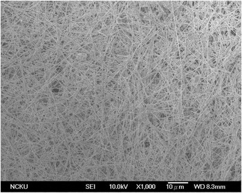
The rod connecting and coarsening are the major factors affecting the length and width of the wires. If we want to produce Ag NWs with a high aspect ratio, we have to prevent the occurrence of coarsening too early. For that purpose, the syringe rate must be low in order not to create a big driving force for reduction, and give more time for Ag atoms to accumulate on both ends. However, the syringe rate cannot be too low at the early stage, because it will reduce the probability of tetrahedrons colliding with each other to form nuclei as well as the probability of connecting rods. Therefore, in our experiment, we used a two-step synthesis with different syringe rates to control the rod connecting and coarsening. The first 10 mL of 0.25 M AgNO3 in EG and 10 mL of 0.75 M PVP in EG were syringed at 1.5 mL/min, and then the remaining 40 mL of the solutions were syringed at 0.5 mL/min. The solution was kept at 160 °C for 30 min after finishing syringing. The result was shown in Fig. 11. We could see that there was less coarsening of the Ag NWs. The widths of wires were less than 200 nm, and the longest wire was about 100 μm. This showed that we successfully synthesized Ag NWs with a high aspect ratio according to the mechanism.
The SEM image of high aspect ratio Ag NWs via a two-step synthesis with different syringe rates.

IV. CONCLUSIONS
In the past, the synthesis of Ag NWs by the polyol process was known in that the nuclei of Ag NWs were decahedrons and PVP chains would attach to the (100) face of the nuclei. This forces Ag+ to turn to the (111) face on both ends only, which resulted in anisotropic growth along 〈110〉. However, the growth mechanism and factors that control the width and length of Ag NWs were not understood. In our study, we found the phenomena of coarsening and formation of nano-hotdogs in the syringe rate testing. Coarsening created Ag NWs with consistent width. If coarsening occurred too early, the length would decrease as shown in the concentration effect testing. During the growth of Ag NWs, we have found that connecting of rods was just like the condensation reaction in polymer synthesis. We have developed a model of Ag NWs growth, and successfully synthesized Ag NWs with high aspect ratios according to the thoughts of the model.
ACKNOWLEDGMENT
The financial support provided by the Ministry of Science and Technology (Taiwan, ROC) through project MOST 103-2120-M-006 -004 -CC1 is greatly appreciated.