Introduction
The Leptopodomorpha are a group of true bugs with four extant and two extinct families, including approximately 400 extant and 64 fossil species. Known extant species are distributed mainly in semi-aquatic environments throughout temperate, subtropical, and tropical regions of the world (Moreira et al., Reference Moreira, Rodrigues, Sites, Cordeiro, Magalhães, Hamada, Thorp and Rogers2018). The extant families are Aepophilidae, Leptopodidae, Omaniidae, and Saldidae; the fossil families are Mesolygaeidae and Archegocimicidae.
Saldids are predators or scavengers of small invertebrates. These insects, known as shore bugs, usually inhabit the bases of waterfalls, mudflats, salt marshes, margins of lentic waters, and rocks in streams (Polhemus, Reference Polhemus1985; Cassis and Gross, Reference Cassis and Gross1995). The family Saldidae includes the extant subfamilies Saldinae (Amyot and Serville, Reference Amyot and Serville1843) and Chiloxanthinae (Cobben, Reference Cobben1959) plus the extinct Enicocorinae (Popov, Reference Popov1980). It comprises about 300 extant species (Larivière and Larochelle, Reference Larivière and Larochelle2019) and 20 fossil species, with the fossil record currently extending from the Lower Cretaceous to the Neogene.
The extinct subfamily Enicocorinae comprises the genera Mesolygaeus Ping, Reference Ping1928, Xishania Hong, Reference Hong1988, Ulanocoris Ryzhkova, Reference Ryzhkova2012, Propritergum Zhang et al., Reference Zhang, Engel, Yao, Ren and Shih2014, Baissotea Ryzhkova, Reference Ryzhkova2015, and Longianteclypea Popov, Reference Popov1986. The Chiloxanthinae comprises the extant genera Chiloxanthus Reuter, Reference Reuter1891, Enalosalda Polhemus and Evans, Reference Polhemus and Evans1969, Paralosalda Polhemus and Evans, Reference Polhemus and Evans1969, Pelachoris Drake, Reference Drake1962, and Pentacora Reuter, Reference Reuter1912 and the fossil genera Oligosaldina Statz and Wagner, Reference Statz and Wagner1950, Propentacora Polhemus, Reference Polhemus1985, Brevrimatus Zhang et al., Reference Zhang, Yao and Ren2011, Venustsalda Zhang et al., Reference Zhang, Song, Yao and Ren2012, and Luculentsalda Zhang et al., Reference Zhang, Yao and Ren2013. The Saldinae includes the extant genera Calacanthia Reuter, Reference Reuter1891, Lampracanthia Reuter, Reference Reuter1912, Teloleuca Reuter, Reference Reuter1912, Ioscytus Reuter, Reference Reuter1912, Micracanthia Reuter, Reference Reuter1912, Macrosaldula Leston and Southwood, Reference Leston and Southwood1964, Saldoida Osborn, Reference Osborn1901, and Saldula Van Duzee, Reference Van Duzee1914, as well as Salda Fabricius, Reference Fabricius1803, which comprises both extant and fossil forms.
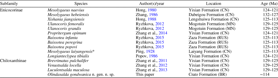
The fossil record of Saldidae in the Lower Cretaceous contains 14 species, 11 in the Enicocorinae and three in the Chiloxanthinae (Table 1). The oldest known saldid is Mesolygaeus hebeiensis (Zhang, Reference Zhang1986) from China, Dabeigou Formation, Lower Cretaceous (Hauterivian, ~132.9 Ma). From the Eocene Baltic amber (Priaborian, 37.8–33.9 Ma), Germar and Berendt (Reference Germar, Berendt and Berendt1856) described the first and so far only known fossil of Saldinae, Salda exigua. From the Oligocene of Germany, Rott Formation, three Chiloxanthinae are described by Statz and Wagner (Reference Statz and Wagner1950): Oligosaldina aquatilis, O. rhenana, and O. rottensis. The youngest fossil, described by Lewis (Reference Lewis1969), is a Chiloxanthinae, Propentacora froeschneri (= Oreokora froeschneri) from the Miocene, Latah Formation (Langhian, 16.0–13.7 Ma), of the United States of America.
Fossil record of Saldidae in the Lower Cretaceous of the world.

BR = Brazil; CN = China; MN = Mongolia; RUS = Russia.
* Junior synonyms of Mesolygaeus laiyangensis Ping, Reference Ping1928: M. rotundocephalus Ping, Reference Ping1928, Xishania fusiformis Hong, Reference Hong1981, Jiaodongia maershanensis Hong, Reference Hong1984, and Enicocoris manlaicus Popov, Reference Popov1980.
No fossil Saldidae has so far been described from the Gondwanan supercontinent. Here we describe, as a new genus and species, a fossil specimen of this family from the fossil Lagerstätte of Araripe Basin, Crato Formation (late Aptian). This is the first confirmed record of the family from Gondwana and one of the oldest of the world. Use of vacuum scanning electron microscopy allowed us to study three-dimensional features of the new taxon. Remarks comparing the new genus with other saldids and on the geology of the type locality are provided.
Materials and methods
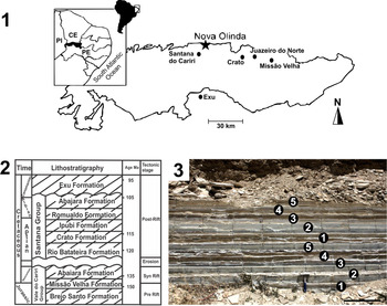
The specimen described here comes from Pedra Branca quarry (Crato Formation), located on a road connecting Nova Olinda and Santana do Cariri, Ceará state, northeastern Brazil (Fig. 1). The material studied belongs to the Museu de Ciências da Terra—Serviço Geológico do Brasil, Rio de Janeiro, Brazil (MCT/CPRM-RJ). A short characterization of the Crato Formation is provided.
(1) Araripe Basin map with the location of Nova Olinda indicated (modified from Batista et al., Reference Batista, da Costa Silva, Sales, Sá, Saraiva and Loiola2017); CE: Ceará state, PE: Pernambuco state, PI: Piauí state. (2) Simplified stratigraphical framework (modified from Valença et al., Reference Valença, Neumann and Mabesoone2003). (3) Fossiliferous layers at Pedra Branca quarry, type locality of Olindasalda gondwanica n. sp. 1 = laminated limestone deposition with high concentrations of algae and with few other fossils; 2 = laminated limestone of gray color with occurrence of insects; 3 = reddish-yellow oxidized laminated limestone with many Ephemeroptera nymphs and salt nodules (halite); this is the layer from which the holotype of O. gondwanica has been collected; 4 = yellow laminated limestone with low algal concentration; this layer is very fossiliferous, including many fishes (genus Dastilbe); 5 = deposition of laminated limestone with high algae concentration. (3) Scale bar = 50 cm.

Photographs were taken with a Zeiss Stemi 2000-C stereomicroscope with an attached Axiocam ERc5s camera. Scanning electron microscopy was conducted with a Hitachi TM3030Plus Tabletop Microscope (Centro de Tecnologia Mineral - CETEM, Rio de Janeiro).
Terminology for the head and thorax follows mainly Parsons (Reference Parsons1962, Reference Parsons1963), and that for other parts of the body and appendages follows Snodgrass (Reference Snodgrass1935). Measurements are given in mm.
Brief characterization of the Crato Formation
This formation is located in the Araripe Basin, which has areas in the states of Ceará, Pernambuco, and Piauí, comprising approximately 12,200 km2 (Fig. 1.1). It is the most important Konservat-Lagerstätte of the Lower Cretaceous from Gondwana. Fossil insects are usually found in the lower layers of the formation, in the Nova Olinda Member, mainly in Pedra Branca and Idemar quarries. Many of these fossils show three-dimensional preservation, which facilitates the study due to the presence of complete structures (see Barling et al., Reference Barling, Martill, Heads and Gallien2015), as is the case of the specimen herein described. During the Lower Cretaceous, the type locality of Olindasalda gondwanica n. gen. n. sp. was probably a freshwater lake, with occasional sea influence (Assine, Reference Assine2007). Our fossil has been found in a layer that also included a large number of mayfly nymphs (Fig. 1.3, layer 3). This proposed habitat is consistent with the biology of modern shore bugs. The lithostratigraphy of the Araripe Basin (Fig. 1.2) has been described and discussed in several works (Beurlen, Reference Beurlen1962, Reference Beurlen1963, Reference Beurlen1971; Assine, Reference Assine1992, Reference Assine2007; Neumann and Cabrera, Reference Neumann and Cabrera1999).
The holotype of O. gondwanica n. gen. n. sp. was collected from the Nova Olinda Member of the Crato Formation (Fig. 1.1, 1.2). In the twentieth century, since the mid-1960s, some authors have used biostratigraphy (Braun, Reference Braun1966) and palynology (Rios-Netto et al., Reference Rios-Netto, Regali, Carvalho and Freitas2012) to try to establish accurate ages for the Araripe Basin. When comparing the ostracod biotas from the Crato Formation with associations of the Lower Cretaceous of the Tucano-Jatobá and Mirandiba basins, they identified concomitant correlations. However, it is still not clear whether the Crato Formation is entirely Aptian or Aptian/Albian. This formation is considered to be correlated to the Inaperturopollenites microclavatus Superzone due to the presence of the gymnosperm species Galeacornea causea Stover, Reference Stover1963, which occurs in Albian-age layers from Morocco (Tarfaya Basin), Egypt (Kharita Formation), Nigeria (Bornu Basin), and Peru (Orient Formation) (Brenner, Reference Brenner1968; Abdel-Kireem et al., Reference Abdel-Kireem, Schrank, Samir and Ibrahim1996; Bettar and Méon, Reference Bettar and Méon2001; Ola-Buraimo and Boboye, Reference Ola-Buraimo and Boboye2011). Nevertheless, judging from previous lines of evidence, including faunal and palynological data within the overlying Nova Olinda Member, the age of the Crato Formation appears to be best constrained to the Aptian, approximately 114 Ma (see Rios-Netto et al., Reference Rios-Netto, Regali, Carvalho and Freitas2012). Regardless of the uncertainty over the Aptian/Albian status of the Crato Formation (see Rios-Netto et al., Reference Rios-Netto, Regali, Carvalho and Freitas2012; Kellner et al., Reference Kellner, Campos, Sayão, Saraiva and Rodrigues2013), we can safely conclude that O. gondwanica n. gen. n. sp. is a Lower Cretaceous taxon.
Repositories and institutional abbreviations
MCT/CPRM: Museu de Ciências da Terra, Serviço Geológico do Brasil, Rio de Janeiro, Brazil; CETEM: Centro de Tecnologia Mineral, Rio de Janeiro, Brazil.
Systematic paleontology
Order Hemiptera Linnaeus, Reference Linnaeus1758
Suborder Heteroptera Latreille, Reference Latreille1810
Infraorder Leptopodomorpha Popov, Reference Popov1971
Family Saldidae Amyot and Serville, Reference Amyot and Serville1843
Subfamily Chiloxanthinae Cobben, Reference Cobben1959
Genus Olindasalda new genus
Type species
Olindasalda gondwanica n. gen. n. sp., by present designation and monotypy.
Diagnosis
as for the type species by monotypy (see the following).
Etymology
The new genus name, Olindasalda (Olinda + salda), refers to the city of Nova Olinda (Ceará state, Brazil), where the specimen studied in this work has been collected. Salda is the type genus of the family.
Remarks
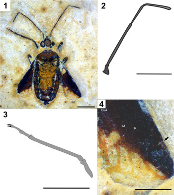
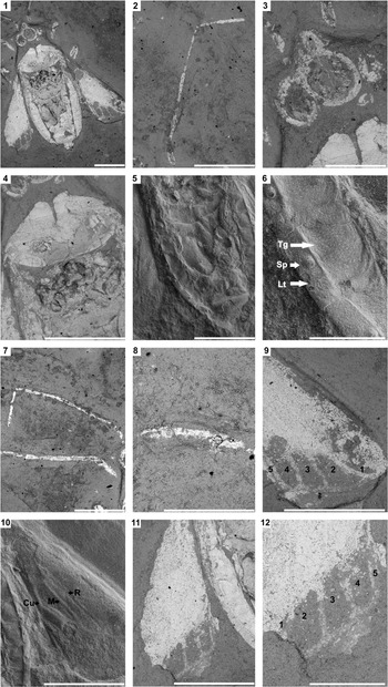
After carrying out a careful comparative study of the families included in the Leptopodomorpha, both fossil and extant, we have assigned our specimen to the Saldidae due to the remarkable similarity of the habitus to a modern representative of this family, including the posterior pronotal margin with indentation (Fig. 2.1), short and broad head with large compound eyes (Fig. 3.3), and hemelytron with five closed elongate cells in the membrane (Fig. 3.9). The body size in Aepophilidae and Omaniidae is usually less than 2 mm (Schuh and Slater, Reference Schuh and Slater1995), whereas in O. gondwanica n. gen. n. sp. it is distinctly longer, 4.49 mm. The Aepophilidae have reduced hemelytra and scutellum (Péricart, Reference Péricart1990), unlike O. gondwanica. In the Leptopodidae (subfamilies Leptopodinae and Leptosaldinae), at least the clavus, and sometimes most of the dorsum, is deeply and densely punctured (Schuh and Slater, Reference Schuh and Slater1995), a feature that is not observed in the new species (Fig. 3.4, 3.9, 3.10). The Leptopodinae have the two or three distalmost antennomeres setiform (Péricart, Reference Péricart1990), unlike O. gondwanica. The Leptosaldinae (= Palaeoleptidae) have quite short antennae (Cobben, Reference Cobben1971; Schuh and Polhemus, Reference Schuh and Polhemus1980; Poinar and Buckley, Reference Poinar and Buckley2009; Popov and Heiss, Reference Popov and Heiss2014); also, the hemelytron in this subfamily is nearly completely coriaceous and normally has eight apical cells (Poinar and Buckley, Reference Poinar and Buckley2009).
Olindasalda gondwanica n. gen. n. sp. (Crato Formation), MCT 6959 – I. (1) Body preserved in dorsal view. (2) Right antenna (four-segmented); the basal article is incomplete. (3) Foreleg: femur, tibia, tarsus (two-segmented), and pretarsus (with two claws). (4) Right hemelytron showing apical cells 1–5 (arrow indicates location of cuneal fracture). The fifth apical cell is very narrow (see also Fig. 3.9, 3.12). CuA = anterior cubital vein; M = median vein; R = radial vein. (1–3) Scale bars = 1 mm; (4) scale bar = 0.5 mm.

Olindasalda gondwanica n. gen. n. sp. (Crato Formation), MCT 6959 – I, MEV. (1) Body, dorsal view. (2) Right antenna. (3) Head, including compound eyes. (4) Pronotum. (5) Topography of abdomen. (6) Abdomen at higher magnification showing laterotergite (Lt), tergite (Tg), and spiracle (Sp). (7) Left antenna and foreleg. (8) Left foretarsus (two-segmented) and pretarsus (with two claws). (9) Apical cells (1–5) of right hemelytron. (10) topography of right hemelytron with radial (R), median (M), and cubital (Cu) veins marked. (11) Left hemelytron. (12) Apical cells (1–5) of left hemelytron. (1, 2, 4, 5, 7, 9–11) Scale bars = 1 mm; (3, 6, 8, 12) scale bars = 500 μm.

Olindasalda gondwanica cannot be included in any of the Mesozoic Leptopodomorphan families. The pronotum in Mesolygaeidae is divided into two parts and the terminalia are strongly curved (Zhang, Reference Zhang1991), two conditions that are not found in the new species. Nine apical cells occur in the hemelytron membrane of Archegocimicidae (Handlirsch, Reference Handlirsch1906–1908; Zhang et al., Reference Zhang, Engel, Yao, Ren and Shih2014), and they are distinct in form from those of O. gondwanica.
Olindasalda n. gen. can be assigned to the Chiloxanthinae rather than to the Saldinae because it has five cells in the hemelytron membrane, instead of four, and a distinct cuneal fracture (Fig. 2.4). The new genus is quite distinct from all other Lower Cretaceous Chiloxanthinae genera. Compared with Brevrimatus (Zhang et al., Reference Zhang, Yao and Ren2011), Olindasalda can be distinguished by the body size (approximately half as long as in Brevrimatus), corium without spots (Fig. 2.1), and absence of medial fracture (Fig. 3.10). Olindasalda differs from Venustsalda (Zhang et al., Reference Zhang, Yao and Ren2011) by the number of cells in the membrane, six in the latter and five in the former (Fig. 3.9). In Luculentsalda (Zhang et al., Reference Zhang, Yao and Ren2013), pale spots are present on the pronotum, mesonotum, and hemelytra. Olindasalda appears to be most similar to Oligosaldina (Statz and Wagner, Reference Statz and Wagner1950), from the Oligocene of Germany, from which Olindasalda can be distinguished by the more slender antennae (Fig. 3.2) and strongly reduced first cell of the membrane (Fig. 3.9); in Oligosaldina, the antennae are thicker and the first cell of the membrane is distinctly larger (see illustrations in Statz and Wagner, Reference Statz and Wagner1950).
Concerning the extant genera of Chiloxanthinae, Paralosalda (Polhemus and Evans, Reference Polhemus and Evans1969) has four well-developed cells on the membrane as in Olindasalda but shorter antennae, with the second antennomere nearly as long as the third. Enalosalda (Polhemus and Evans, Reference Polhemus and Evans1969) has the cells of the membrane more or less fused to one another or indistinct, and the compound eyes are smaller than in the new genus. The species of Pentacora (Reuter, Reference Reuter1912) have five well-developed cells on the membrane, which are longer and narrower than in the new genus, with the sublateral cell subequal in length to the lateral cell. The genera Chiloxanthus (Reuter, Reference Reuter1891) and Pelachoris (Drake, Reference Drake1962), as well as the Miocene genus Propentacora (Polhemus, Reference Polhemus1985), have also five well-developed cells on the membrane.
Holotype (MCT 6959 – I)
Pedra Branca quarry (7°7'35.2"S, 39°43'8.7"W), Nova Olinda (Ceará state, northeastern Brazil). The body is preserved in dorsal view; it shows taxonomically relevant structures such as completely preserved antennae, compound eyes, and left foreleg; middle legs are partially preserved, whereas hind legs are obscured; forewings (hemelytra) are well preserved. We have not been able to determine the sex of this specimen.
Diagnosis
A small Chiloxanthinae (4.49 mm) with oval-elongated body (Figs. 2.1, 3.1); macropterous, corium of forewing without spots, R long, first cell of membrane very reduced (Figs. 2.4, 3.9, 3.11, 3.12).
Occurrence
The holotype was collected from the Upper Aptian Nova Olinda Member, Crato Formation, Santana Group (see details in Materials and methods).
Description
Body (Figs. 2.1, 3.1) preserved in dorsal view; oval-elongated, small, 2.5 times as long as wide. Head (Fig. 3.3) with vertex somewhat circular, 1.4 times as long as wide, front pronounced. Compound eyes (Fig. 3.3) globular, occupying one-third of head area, interocular space 0.47 mm. Ocelli not preserved. Antennae (Figs. 2.2, 3.2, 3.7) long, four-segmented, ratio of antennomeres I:II:III:IV = 0.45:1.32:0.70:0.81; I short and stout compared with II; II slender and long, about 3 times longer than I; III shorter and slenderer compared with II; IV about same size as III but stouter. Pronotum (Fig. 3.4) ovoid, 1.8 times as wide as long, anterior margin narrower than head, lateral margins divergent posterad, posterior margin concave, posterior angles obtuse. Mesonotum (Fig. 3.4) longer than pronotum, triangular, 1.1 times as wide as long. Forelegs (Figs. 2.1, 2.3, 3.7, 3.8) with femur slender, same thickness as tibia; tibia 1.8 times as long as wide, 3.3 times as long as tarsus; tarsus two-segmented, tarsomere I longest, tarsomere II 0.8 times as long as I; pretarsus with two claws. Middle legs (Figs. 2.1, 3.1) partially preserved, femur 0.39 mm long, tibia 0.46 mm long. Hind legs not preserved. Hemelytron (Figs. 2.1, 2.4, 3.9–3.12) length 0.6 times body length, clavus length 0.6 times hemelytron length. Corium without spots; veins R, M, and Cu distinct; cuneal fracture distinct (indicated by an arrow in Fig. 2.4); membrane with five cells, first cell smallest, about 0.8 times length of second (sublateral) cell, third cell longest; R and M reaching cells 2 and 3, respectively; hypocostal ridge obscured. Abdomen (Figs. 2.1, 3.1, 3.5, 3.6) with seven to eight distinct tergites; spiracles located on laterotergites.
Measurements (mm): body length 4.49; body width 1.77; head length 0.66; head width 0.47; interocular space 0.47; antenna articles I–IV length 0.45, 1.32, 0.70, 0.81, respectively; scutellum length 0.69; scutellum width 0.73; foreleg: femur length 0.81; tibia length 1.44; tarsomere I length 0.24; tarsomere II length 0.19; hemelytron: length 2.66; width 1.07; clavus length 1.70; membrane cells I–V length 0.27, 0.32, 0.36, 0.35, 0.30, respectively.
Etymology
The name of the new species refers to the Gondwanan supercontinent.
Remarks
See detailed comparative remarks provided in the preceding for the new genus.
Conclusions
Some morphological features of O. gondwanica n. gen. n. sp., such as the indentation at the posterior pronotal margin, presence of the cuneal fracture, and five closed elongate cells in the hemelytron membrane, suggest a remarkable morphological stability of the Saldidae and Chiloxanthinae. These features occur in both fossil and extant forms of the family and subfamily. Perhaps O. gondwanica had a way of life similar to that of modern saldids (i.e., they were semiaquatic predators or scavengers of small invertebrates and were associated with freshwater habitats or salt marshes). The records of Chiloxanthinae fossils from the Lower Cretaceous of Brazil and China suggest that this subfamily arose before the Pangea breakup, probably at the end of the Lower Jurassic or Middle Jurassic (about 180–170 Ma). This suggestion implies that the Saldidae would be even older, perhaps 70 Myr older than the oldest known stratigraphic record of this family.
Acknowledgments
We are greatly indebted to R. Machado (MCT/DNPM), who loaned the specimen herein described. The first author thanks Conselho Nacional de Desenvolvimento Científico e Tecnológico (CNPq) and Fundação ‘Carlos Chagas Filho’ de Amparo à Pesquisa do Estado do Rio de Janeiro (FAPERJ) for stipends in connection with his doctoral studies in Museu Nacional, UFRJ (processes 140628/2019-3 and E-26/201.931/2017, respectively). We are grateful to L.D.R.S. Fernandes and B.S. Haiad (Departamento de Botânica, Museu Nacional), who kindly provided laboratory facilities after the tragic fire at our institution. We are also grateful to L.C. Bertolino and V.S. Brandão (CETEM), who made available the scanning electron microscope. G. Mejdalani is a research productivity fellow from CNPq (process 303229/2018-7). The latter author and S.M. Scheffler were supported by FAPERJ (processes E-26/200.082/2019 and E-26/200.110/2019; Apoio Emergencial ao Museu Nacional, UFRJ). F. Moreira benefited from grants provided by FAPERJ (processes E-26/203.207/2017 and E-26/200.149/2019). The manuscript benefited from very useful comments provided by T. Wappler, E. Heiss, and two anonymous reviewers.