Differentiation of the so-called sinus venosus from other defects permitting shunting between the atrial chambers remains problematic. The earliest account of the entity, provided by Peacock,Reference Peacock1 described how the hole permitting the interatrial communication was separated from the normal atrial septal structures. Hence, the lesions are not true septal defects. “Unroofing” of the right pulmonary veins was put forward to provide a mechanistic explanation for such holes, on the basis that a “party wall” normally separated these venous structures from the superior caval vein and the cavity of the right atrium.Reference Van Praagh, Carrera, Sanders, Mayer and Van Praagh2 This explanation, however, flounders underneath the fact that, in the normal heart, the so-called “septum secundum” is an interatrial groove, rather than a true “party wall.”Reference Anderson, Webb and Brown3 An alternative concept, therefore, is required to explain the existence of the sinus venosus defect. In a review of patients presenting with the inferior variant of the lesion, it was shown that the defining diagnostic feature is connection of a pulmonary vein to the inferior caval vein, but with the anomalous vein also retaining its connection with the left atrium.Reference Crystal, Al Najashi, Williams, Redington and Anderson4 In this respect, Edwards and HelmholzReference Edwards and Helmholz5 had postulated long since that the lesion could be explained on the basis of persistence of connections existing in the developing foetus of communications between the pulmonary and systemic veins. Such a veno-venous fistula had been discovered at autopsy in an infant with discordant ventriculo-arterial connections, albeit not diagnosed during life.Reference Devine and Anderson6 Others had also previously commented on the presence of extracardiac pulmonary-to-systemic venous channels that exist in the foetus.Reference Swan, Kirklin, Becu and Wood7 We have now encountered three adults with such systemic-to-pulmonary veno-venous communications that can explain the morphology of the superior sinus venosus defect. In this report, we discuss the diagnostic features of such communications, and show their relationship with the sinus venosus defects and with anomalous connection of the right upper pulmonary vein to the superior caval vein. We then provide supporting developmental and morphological evidence that the sinus venosus defect is a veno-venous malformation, rather than representing a problem with atrial septation.
We report three patients diagnosed at either Medical University of South Carolina or Toronto General Hospital as having venous bridges between the superior caval vein and the right pulmonary veins. We compare the diagnostic features of these patients with those known to have isolated connection of the right upper pulmonary vein to the superior caval vein, anomalous systemic connection of the upper pulmonary veins with retention of connection of the lower pulmonary veins to the left atrium, and the sinus venosus defect, respectively. We then review our existing knowledge concerning the development of the pulmonary veins and the morphology of the superior sinus venosus defect.
Clinical findings
Partially anomalous pulmonary venous connections
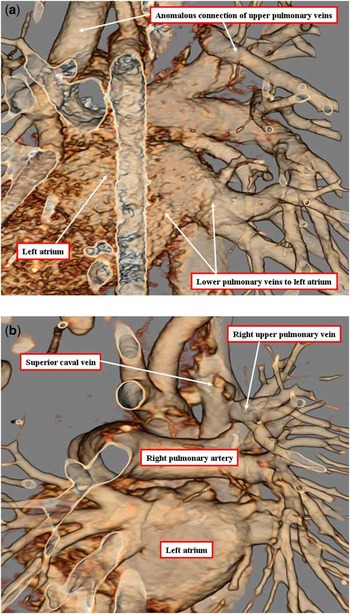
The first patient presented at 3 years of age with a mumur, but was otherwise asymptomatic. His medical history was unremarkable. Physical examination revealed a systolic ejection murmur, which was best heard at the right upper sternal border, and a diastolic murmur at the left lower sternal border. An echocardiogram was performed, showing an intact atrial septum with moderate right atrial enlargement and right ventricular dilation. As the pulmonary venous connections could not adequately be defined, computed tomographic angiography was performed to determine the pulmonary venous anatomy. The images revealed that the majority of the left lung drained through a vertical vein to the bracheocephalic vein. The right upper pulmonary vein connected to the superior caval vein. The right lower pulmonary vein and a small portion of the left lower lung field connected to the left atrium, albeit in a markedly atypical manner (see Fig 1a). The patient was referred for surgical repair.
The reconstruction of the computed tomogram (a), viewed from behind, shows anomalous connection of both upper pulmonary veins, but shows origin of the lower pulmonary veins from the inferior and rightward parts of the left atrium, reflecting the original location of the common pulmonary vein (see Fig 4b). Panel b, from a different patient, shows the arrangement in which the right upper pulmonary vein connects anomalously to the superior caval vein.

The second patient was 6 months old when she presented to the paediatric cardiologic clinic for evaluation of a mumur. She had no symptoms related to the cardiovascular system, and her growth was appropriate. Her medical history was unremarkable. Examination revealed a long, systolic ejection mumur, which was heard best at the left upper sternal border, with radiation to the back. Palpation of her pulses revealed brachiofemoral delay. On performing echocardiography, she was found to have a coarctation of the aorta, but was otherwise normal. The surgeon requested a computed tomographic angiogram to better evaluate the anatomy of the aortic arch. The angiogram revealed that the right upper pulmonary vein connected to the superior caval vein (Fig 1b). The patient then underwent an end–end repair of her coarctation. Given the size of the patient, and the distance between the anomalous pulmonary vein and the right atrium, the surgeon decided against altering the pulmonary venous connection. She is currently asymptomatic.
Veno-venous bridges
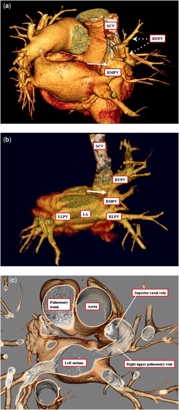
Mrs DW was a lady of 75 years who presented with left hemiparesis. Physical examination revealed a right ventricular heave, and a loud pulmonary component to the second heart sound. Her electrocardiogram showed sinus rhythm with incomplete right bundle branch block and right axis deviation. During an echocardiographic search for a source of embolus, she was shown to have a severely dilated right heart. Subsequent computerised tomography and magnetic resonance imaging revealed anomalous pulmonary venous return of the right upper pulmonary veins to the superior caval vein, along with a communication between the right upper pulmonary vein and the right middle pulmonary vein, the latter vein then connecting in a normal manner with the left atrium. The anomalous channel (Fig 2a) provided a venous bridge between the left atrium and the systemic venous circulation, producing the same physiological perturbation as would be experienced with a classic sinus venosus defect.
Three-dimensional reconstructions of computed tomograms viewed from above and behind of the three patients with veno-venous bridges (a–c). In each instance, the bridge (arrowed in panels a and b) runs from the right upper pulmonary vein (RUPV), which is connected anomalously to the superior caval vein (SCV), to the right middle vein (RMV), which retains its normal connection to the left atrium. The venous bridge, therefore, functions as an interatrial connection.

Mrs PM was aged 44 years. She presented with fatigue, a soft systolic murmur being heard at the left sternal border during the clinical examination, which was otherwise unremarkable. The electrocardiogram showed sinus rhythm, with a normal axis and QRS width. Transthoracic echocardiography showed moderate right ventricular enlargement, with normal systolic function. Magnetic resonance imaging confirmed the moderate enlargement of the right ventricle, and showed the right upper pulmonary vein to be connected anomalously to the superior caval vein, with a bridging vein joining the right middle pulmonary vein, which connected normally to the left atrium (Fig 2b). A transthoracic bubble contrast echocardiographic study was positive with early appearance of bubbles in the left atrium.
The third patient is a 72-year-old man, who presented to an outside institution with dyspnoea on exertion. He was otherwise in good health, with no history of use of tobacco or chronic illnesses. He had no risk factors for cardiac disease, and his examination was unremarkable. His echocardiogram revealed an intact atrial septum, with good biventricular function, and no atrial enlargement or ventricular dilation. On cardiac catheterisation, performed at the referring institution, he was noted to have an increase in oxygen saturation between the high superior caval vein and the right atrium. The catheterisation was otherwise normal. Computed tomographic angiography was performed to evaluate for the presence of partially anomalous pulmonary venous connection. The images revealed that one wall of the right upper pulmonary vein connected with the superior caval vein, with the pulmonary vein then continuing to connect normally with the left atrium (Fig 2c). He was subsequently taken to the catheterisation laboratory at our institution, where a covered stent was placed into the superior caval vein at the level of communication with the right upper pulmonary vein. This eliminated the anomalous drainage. His dyspnoea has improved since the procedure.
Sinus venosus defect
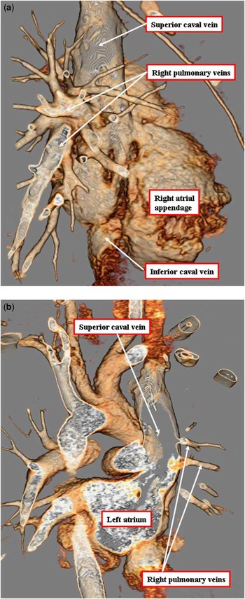
This patient initially presented to our institution at 9 years of age because of mild systemic desaturation. The medical history was significant for a cerebral arteriovenous malformation involving the vein of Galen and the Calcarine artery. She had undergone multiple occlusions of the arteriovenous malformations using coils, and it was noted that her systemic arterial oxygen saturations were in the mid-90s at each of her procedures. She was referred to our clinic for further evaluation. She admitted to shortness of breath with moderate exercise, and frequent palpitations. Initially, her shortness of breath was thought to be due to asthma, but did not improve with escalation of treatment. Her physical examination upon presentation did not reveal any abnormalities. Echocardiography revealed that the right upper pulmonary vein connected to the superior caval vein and the atrial septum appeared intact. There was no right atrial or ventricular enlargement. A decision was made to perform a computed tomographic angiogram to better assess the pulmonary venous connections, and to evaluate for additional venous malformations. The computed tomographic angiogram confirmed anomalous connection of the right upper and middle pulmonary veins to the superior caval vein (Fig 3a). The images also showed extreme overriding of the orifice of the superior caval vein. The orifice of the superior caval vein predominantly connects to the left atrium, and thus the majority of superior caval vein blood drains into the left atrium (Fig 3b). The resulting systemic venous return to the left atrium, albeit combined with some pulmonary venous return from the right lung, was deemed the likely explanation of her systemic desaturation. She was referred for surgical repair.
A three-dimensional reconstruction from a computed tomographic study of a typical patient with a superior sinus venosus defect (a and b). The images show how the right upper pulmonary vein connects anomalously to the superior caval vein, while retaining its connection to the left atrium.

Developmental considerations
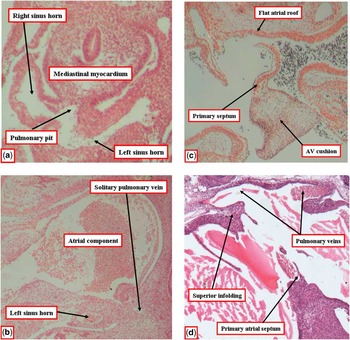
When first seen, the primordium of the pulmonary vein in the human heart is no more than a midline endocardial strand seen within the mediastinal mesenchyme. When traced towards the heart, the strand joins a pit at the dorsal wall flanked by persisting attachments of the myocardium to the pharyngeal mesenchyme, the so-called dorsal mesocardial connections. At this stage, the systemic venous tributaries are relatively symmetrical, opening dorsal to the mesocardial connections (Fig 4a). A crucial stage of normal development is reorientation of these venous tributaries so that they open to the right side of the developing atrial components. As the systemic veins come to open into the developing right atrium, the pulmonary vein canalises in the dorsal mesocardium. When first seen as a patent channel, the pulmonary vein enters the heart as a solitary structure, with its orifice directly adjacent to the developing left atrioventricular junction (Fig 4b). Although these changes have taken place in the venous connections to the heart, the primary septum has begun its growth from the roof of the atrial component, carrying on its leading edge a prominent mesenchymal cap. With time, the mesenchymal cap fuses with the atrioventricular cushions, dividing the atrioventricular canal into the prospective tricuspid and mitral valvar orifices. The dorsal area of fusion is itself reinforced by growth into the heart of tissue through the rightward migration of the dorsal mesocardial connections, this growth producing the so-called vestibular spine, which as it fuses with the primary atrial septum ensures that the pulmonary vein enters the cavity of the left atrium. By the time that the primary septum itself has fused with the atrioventricular cushions and the vestibular spine, its upper margin has broken down to form the secondary interatrial communication. At this stage, the atrial roof shows no evidence of infolding (Fig 4c). The infolding that produces the secondary atrial septum, or the “septum secundum”, does not become evident until after the 8th week of development, and appears as the solitary pulmonary vein migrates to adopt a position on the atrial roof, during which time there is formation of first two (Fig 4d), and then four, pulmonary venous orifices.
Stages in formation of the atrial septum. Initially in development, at Carnegie stage 12 in the human heart, the systemic venous tributaries drain in relatively symmetrical manner, flanking the site of a blind-ending pulmonary pit (a). A midline endocardial strand initially connects to this pit. By Carnegie stage 14, the strand has lumenised to form a solitary pulmonary vein, which connects to the developing left atrium adjacent to the atrioventricular junction (b). As the primary atrial septum, carrying a mesenchymal cap, fuses with the atrioventricular endocardial cushions to divide the right and left atrial chambers, the atrial roof remains flat, showing no evidence of the infolding that will eventually form the “septum secundum” (c). The secondary atrial septum is not formed until after the eighth week of development, by which time the pulmonary veins have migrated to adopt their postnatal position on the left atrial roof (d).

Morphological considerations
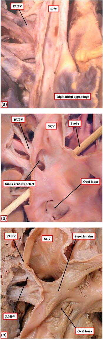
Remarkable similarities exist in the arrangement of the pulmonary veins in the setting of isolated connection of the right pulmonary vein to the superior caval vein (Fig 5a) and the superior sinus venosus defect (Fig 5b). The pulmonary veins join the superior caval vein at comparable distances from the atrial roof. However, in the setting of the sinus venosus defect, the anomalously connecting pulmonary veins retain their connection with the left atrium. It is this dual connection that creates a veno-venous channel through which blood is able to pass from right to left atrium. As is shown in Figure 5b, a probe can be placed through the gap between the superior rim of the oval fossa and the veno-venous communication. In the heart shown in Figure 5b, the superior caval vein itself retains its exclusive connection to the right atrium. More usually, in the setting of the superior sinus venosus defect, the superior caval vein overrides the superior rim of the oval fossa (Fig 5c). It is still possible, nevertheless, to pass a probe through the tube of atrial musculature that now forms the superior rim of the oval fossa. In most instances, the sinus venosus defect is associated with biatrial connection of the superior caval vein. As shown in Figure 5b, however, it is possible for the superior caval vein to connect exclusively to the morphologically right atrium, without any overriding.
The specimens illustrate the comparable location of the right upper pulmonary vein (RUPV) in the setting of isolated anomalous connection to the superior caval vein (SCV; a) and the sinus venosus defect (b). In the heart shown in panel b, only the right upper pulmonary vein is connected in an anomalous manner, and the defect is at a significant distance from the oval fossa, a probe having been placed through the tissue plane between the defect and the rim of the fossa. Note that the superior caval vein is connected exclusively to the right atrium. In the heart shown in panel c, in contrast, the superior caval vein overrides the rim of the oval fossa. In this heart, however, the right middle pulmonary vein (RMPV) is also anomalously connected to the superior caval vein, while retaining its connection with the left atrium.

Discussion
The advent of computed tomographic and magnetic resonance imaging has revolutionised the diagnosis of anomalous venous connections. Given the inherent complexity involved in understanding the three-dimensional relationships in these patients, these multiplanar imaging techniques allow one to understand the involved anatomy in a more straightforward manner. Pulmonary venous connections with a particularly tortuous course, as is seen in Figure 1a, can be difficult to follow by echocardiography. The new multiplanar modalities now permit even the most tortuous vessels to be followed in a relatively simple manner.
Since its initial description, although not then known as a sinus venosus defect,Reference Peacock1 it has been recognised that the lesion, despite its usual grouping as an atrial septal defect, had little to do with deficient atrial septation. It has been well established that the morphological criterion for diagnosis is the integrity of the rims of the oval fossa, albeit that true interatrial communications can coexist with the sinus venosus defect.Reference Crystal, Al Najashi, Williams, Redington and Anderson4, Reference Ettedgui, Siewers, Anderson, Park, Pahl and Zuberbuhler8, Reference Ettedgui, Siewers, Zuberbuhler and Anderson9 It has been difficult, however, to provide a rational explanation for the existence of such holes. As most patients diagnosed with the sinus venosus defect have also been known to have anomalous connection of the right pulmonary veins to the superior cavoatrial junction, a popular concept had been unroofing of the “party wall” that separates the pulmonary veins from the right atrium and the superior caval vein.Reference Van Praagh, Carrera, Sanders, Mayer and Van Praagh2 This concept is not dissimilar to the current concept of persistent veno-venous bridges as discussed below. It flounders, however, on the fact that no such “party wall” exists between the connection of the caval veins to the right atrium and the pulmonary veins to the left atrium.Reference Anderson, Webb and Brown3 Indeed, it has long been recognised that the so-called “septum secundum” is no more than a deep superior interatrial fold. Surgeons had recognised that dissection into this fold, known as Waterston's groove or Sondergaard's groove, provided access to the roof of the left atrium, or additional atrial walls to be used in atrial redirection procedures such as the Senning procedure. The aggregation of fat in the groove, producing so-called lipomas of the atrial septum, also pointed to the extracardiac nature of the tissues contained within the groove.
The formation of the groove is now well explained developmentally, as the pulmonary venous components, with separate orifices on both sides of the left atrial roof for the superior and inferior veins, do not achieve their definitive positions until after completion of atrial septation. As we have shown, the so-called “septum secundum” is only seen at the stage when four pulmonary venous orifices are present. This explains the presence of the Waterston's or Sondergaard's groove, enclosing a corridor of extracardiac fat, as described above. These developmental facts also rule out an alternative concept explaining anomalous connection of the pulmonary veins on the basis of malposition of the primary atrial septum.Reference Van Praagh, Carrera, Sanders, Meyer and Van Praagh10
The deep superior interatrial fold, also known as the septum secundum, the lack of connection of the pulmonary venous structure with the systemic venous tributaries, and the absorption and gradual incorporation of the common pulmonary vein and right and left-sided pulmonary vein orifices into the left atrial wall do not support the theory of “unroofing” of the right pulmonary veins to explain the nature of sinus venosus defects.Reference Van Praagh, Carrera, Sanders, Mayer and Van Praagh2 If unroofing of a “party wall” does not explain the morphology of the lesion, is there an alternative explanation? When reviewing the morphology of the lesion, Edwards and HelmholzReference Edwards and Helmholz5 had drawn attention to the description of collateral channels existing in the developing foetus between the systemic and pulmonary veins. Such a veno-venous collateral channel had been recognised at autopsy in a solitary case, with the suggestion made that this could be the “forme fruste” of the sinus venosus defect.Reference Devine and Anderson6 The case showed persistence of extracardiac connections between the pulmonary venous sinus and the primordium of the superior caval vein, as described previously in the foetus.Reference Swan, Kirklin, Becu and Wood7 Should such connections persist as the common pulmonary vein is moulded, and the four pulmonary venous orifices are gradually integrated into the left atrial wall, they would produce an interatrial communication just above the superior rim of the oval fossa, in other words, a sinus venosus defect.
This possibility is now confirmed by our findings of such veno-venous bridges in three adults. The collateral channels produce a shunt, to all intents and purposes, that is comparable to the interatrial shunting recognised in patients with sinus venosus defects. The anomalous channels may also produce a bridge between the superior caval vein, which retains its normal connection to the right atrium, and the pulmonary veins, which are normally connected to the left atrium. The sinus venosus defect is no more than an exaggerated version of this bridge, but with the anomalous pulmonary veins being directly connected to the caval vein, be it the superior or inferior caval vein, while retaining their normal left atrial connection. In many instances, the sinus venosus defect also results in a situation in which the orifice of the superior caval vein overrides the superior rim of the oval fossa, promoting the spurious impression that the lesion is truly a septal defect (Fig 5c). In other examples, however, the superior caval vein retains its exclusive connection to the right atrium; the extensive distance between the sinus venosus defect and the oval fossa then shows that the lesion has nothing to do with deficient atrial septation (Fig 5b).
The knowledge that has accrued over the past two decades concerning cardiac development then gives added weight to the explanation of the sinus venosus defect as a veno-venous malformation. Although it has long been suggested that, during its development, the pulmonary vein has connections to the tributaries of the systemic venous sinus, recent studies in chicken, man, and mouseReference Webb, Brown, Wessels and Anderson11–Reference Webb, Kanani, Anderson, Richardson and Brown13 show that, from the time it achieves its lumen, the pulmonary vein possesses its own discrete walls, which are always separate from the walls of the systemic venous channels. Furthermore, in humans, as shown in Figure 4, the pulmonary vein initially has but a solitary atrial orifice, which connects to the left atrium adjacent to the left atrioventricular junction. Only subsequent to the formation of the primary atrial septum does the pulmonary vein achieve its connection to the left atrial roof, and only at that stage is there development of the deep infolding still usually described as the “septum secundum”.
The lesions shown in our patients, therefore, represent the developmental history of the sinus venosus defect. They show the transition from veno-venous bridges (Fig 2) to partially anomalous pulmonary venous connections (Fig 1), and finally to the sinus venosus defect with anomalous connection of the right pulmonary vein or veins (Fig 3). Veno-venous bridges, therefore, are indeed a “form fruste”, or a forerunner, of the sinus venosus defect. In one of our patients, the lesion was repaired by placing a covered stent within the superior caval vein, closing off the entrance to the veno-venous collateral channel. In the second patient, it proved possible to ligate the channel, thus obliterating the effective interatrial shunt. The presence of pulmonary vascular disease in the third patient mandated medical treatment.
In summary, we support the notion that the essence of the sinus venosus defect is its existence outside the confines of the oval fossa, showing that it is not a defect of the true atrial septum. We have provided developmental and morphological evidence to support the concept that such defects are the result of persistent veno-venous bridges present in the foetus, rather than representing a problem with atrial septation. These persistent veno-venous connections present as a spectrum of malformation, with veno-venous malformations being at one end, and the sinus venosus defect with partially anomalous pulmonary venous connection at the other end.