Introduction
Most of the 42 territories that constitute Latin America and the Caribbean (LAC) are middle-income areas (PNUD, 2019) (fig. 1). Most countries in the region are heavily urbanized, with an estimate of 70% of LAC population living in cities. About 195 million people live in poverty (with a daily income of less than US$2), and 71 million people live in extreme poverty (with a daily income of less than US$1) (Schneider et al., Reference Schneider, Aguilera and da Silva Junior2011; PNUD, 2019). Under these conditions, the lack of appropriate sanitation and a close contact with infection reservoirs and vectors, along with inequality factors linked to ethnicity, age and gender, fosters the establishment of several pathologies (Franco-Paredes et al., Reference Franco-Paredes, Jones, Rodríguez-morales and Santos-Preciado2007; Hotez et al., Reference Hotez, Bottazzi, Franco-paredes, Ault and Periago2008).
Geographic distribution of states in Latin America and the Caribbean (areas filled in black).

Such is the case of neglected tropical diseases (NTDs), a group of infections (many of them parasitic) that are endemic in tropical and subtropical regions of LAC, Asia and Africa (Franco-Paredes et al., Reference Franco-Paredes, Jones, Rodríguez-morales and Santos-Preciado2007). About 1.7 billion people are estimated to require treatment and care because of an NTD (WHO, 2016).
Cysticercosis is regarded as an NTD. A mean prevalence of 4.08% and a mean seroprevalence of 13.03% have been estimated in LAC (Coral-Almeida et al., Reference Coral-Almeida, Gabriël, Abatih, Praet, Benitez and Dorny2015), making neurocysticercosis the most common neurological parasitic infection in humans (Atluri et al., Reference Atluri, Singhi, Khandelwal and Malla2009).
LAC has been recognized as one of the areas with the highest prevalence of infection by Echinococcus granulosus in humans and animals. The most common form of human echinococcosis is cystic echinococcosis (hydatidosis) by E. granulosus. The yearly incidence rates of echinococcosis are greater than 50 per 100,000 persons in endemic regions, and a prevalence near to 5–10% has been reported in some LAC zones (WHO, 2017a).
On the other hand, ascariasis is the most frequent intestinal parasitosis in humans; it affects about 800 million people, with children the most heavily affected group (Prieto-Perez et al., Reference Prieto-Perez, Perez-Tanoira, Cabello-Ubeda, Petkova-Saiz and Gorgolas-Hernandez-Mora2016). Ascariasis is one of the most prevalent parasitic diseases in the world (Beyhan et al., Reference Beyhan, Yilmaz and Hokelek2016; Melo et al., Reference Melo, Esteves, de Oliveira, Guerreiro, de Oliveira, de Oliveira Lima, Miné, Allegretti and Catharino2016; Shalaby & Shalaby, Reference Shalaby and Shalaby2016); it has been estimated that 514 million people in LAC live in a risk zone for ascariasis, and the disease shows a prevalence of 16% in the open population. In this region, 28 million cases have been reported in persons younger than 15 years, while 56 million people older than 15 years are infected with Ascaris lumbricoide s (De Silva et al., Reference De Silva, Brooker, Hotez, Montresor, Engels and Savioli2003).
Trichuriasis accounts for 35.5% of all estimated geohelminth infections in 18 countries (Saboyá et al., Reference Saboyá, Catalá, Nicholls and Ault2013), affecting about 100 million people in LAC (Hotez et al., Reference Hotez, Bottazzi, Franco-paredes, Ault and Periago2008); is one of the most prevalent NTDs in the region (19% in population older than 15 years) (De Silva et al., Reference De Silva, Brooker, Hotez, Montresor, Engels and Savioli2003).
Historically, about 500,000 people have been at risk of onchocerciasis infection in wide areas of Brazil, Colombia, Ecuador, Guatemala, México and Venezuela (Gustavsen et al., Reference Gustavsen, Hopkins and Sauerbrey2011). In recent years, some countries have made progress in reducing the transmission rates of the disease (WHO, 2017b). However, not all is good news, because some cases are still being reported in the Amazon area, and the parasite transmission in Brazil and Venezuela persists in 2018.
Helminths are the most common infective agents in humans. More than one-third of the world population is currently infected by one or more helminth species (Bourke et al., Reference Bourke, Maizels and Mutapi2011), mainly in developing countries (Lustigman et al., Reference Lustigman, Prichard, Gazzinelli, Grant, Boatin, McCarthy and Basáñez2012). The most prevalent helminthiases worldwide are caused by nematodes and soil-transmitted helminths (Bourke et al., Reference Bourke, Maizels and Mutapi2011). Usually, parasitoses establish as chronic infections, surviving in the host for long periods of time (Moreno et al., Reference Moreno, Gros, Tam, Segura, Valanparambil, Geary and Stevenson2011) and exhibiting complex life cycles (Bourke et al., Reference Bourke, Maizels and Mutapi2011). Supplementary fig. S1 shows the cycle life of the causative agents of ancylostomiasis, ascariasis, cysticercosis, echinococcosis, onchocerciasis and trichuriasis, the parasitoses discussed in this review.
Role of excretory/secretory proteins in parasitosis
Helminths have co-evolved with their hosts, which rely on their immune system to counter the infection; thus, helminths have resorted to several strategies to evade the host's immune response. One of such strategies is based on excreted/secreted (E/S) proteins (Moreno et al., Reference Moreno, Gros, Tam, Segura, Valanparambil, Geary and Stevenson2011). E/S proteins participate in various parasitic functions, including penetration, establishment and survival in host tissues, as well as incorporating host metabolites (Virginio et al., Reference Virginio, Monteiro, Drumond, de Carvalho, Vargas, Zaha and Ferreira2012). In addition, helminths secrete proteins with a significant homology to the host's own proteins that could explain their longevity in the host (Virginio et al., Reference Virginio, Monteiro, Drumond, de Carvalho, Vargas, Zaha and Ferreira2012; Gomez et al., Reference Gomez, Adalid-Peralta and Palafox-Fonseca2015). Furthermore, E/S proteins are capable of actively modulating the immune environment (Moreno et al., Reference Moreno, Gros, Tam, Segura, Valanparambil, Geary and Stevenson2011). Some of the most relevant E/S proteins from each parasite discussed in this review are described in the following sections.
Tissue invasion and parasite establishment
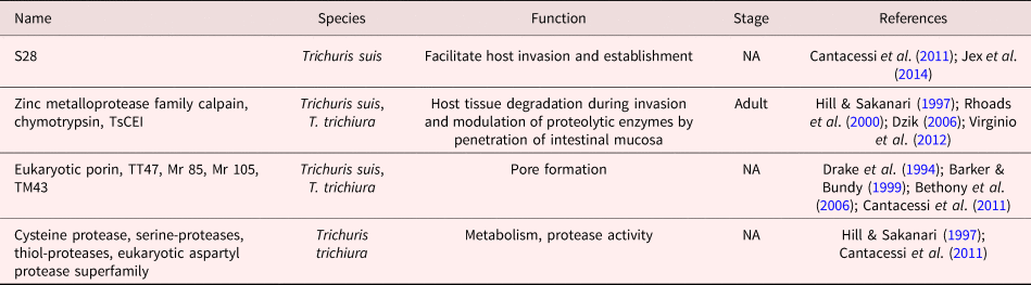
Echinococcosis is a zoonotic disease caused by the larval stages of Echinococcus spp. in humans. The life cycle of Echinococcus spp. involves several stages (supplementary fig. S1d), and in each stage the parasite releases E/S products which favour the invasion of host tissues, as well as its own survival and development. The proteins in hydatid fluid were characterized and reported recently (Moreno et al., Reference Moreno, Gros, Tam, Segura, Valanparambil, Geary and Stevenson2011; Virginio et al., Reference Virginio, Monteiro, Drumond, de Carvalho, Vargas, Zaha and Ferreira2012; Monteiro et al., Reference Monteiro, Lorenzatto, de Lima, dos Santos, Förster, Paludo, Carvalho, Brehm and Ferreira2017). Several of the proteins identified were proteolytic enzymes, protease inhibitors and chaperones probably associated with infection; some of these proteins could be E/S products. For instance, E. granulosus is known to secrete enzymes such as enolase, glyceraldehyde-3-phosphate dehydrogenase and aldolase during host–parasite interactions (table 1). In addition to their classical metabolic functions, these enzymes have been reported to bind host macromolecules and to have specific functions in invasion. As an example, plasminogen promotes the degradation of components of the extracellular matrix, favouring parasite propagation in host tissues (Lorenzatto et al., Reference Lorenzatto, Monteiro, Paredes, Paludo, Da Fonsêca, Galanti, Zaha and Ferreira2012; Virginio et al., Reference Virginio, Monteiro, Drumond, de Carvalho, Vargas, Zaha and Ferreira2012). Another relevant E/S molecule in helminths is calpain, found in Trichuris suis and E. granulosus, which apparently facilitates the digestion of host proteins and tissue invasion by modulating proteolytic enzymes that allow the parasite to penetrate the intestinal mucosa (tables 1 and 2) (Hill & Sakanari, Reference Hill and Sakanari1997; Dzik, Reference Dzik2006; Virginio et al., Reference Virginio, Monteiro, Drumond, de Carvalho, Vargas, Zaha and Ferreira2012).
Excretion/secretion proteins reported for Echinococcus granulosus.

P-29, hydatid disease diagnostic antigen; FBA, aldolase; GAPDH, glyceraldehyde-3-phosphate dehydrogenase; EgTeg, tegument protein; PS, protoscolex.
Excretion/secretion proteins reported for Trichuris spp.

TsCEI, elastase inhibitor; S28, serine carboxypeptidase; M24, peptidase; Mr 85 and Mr 105, proteases; calpain, family cysteine protease.
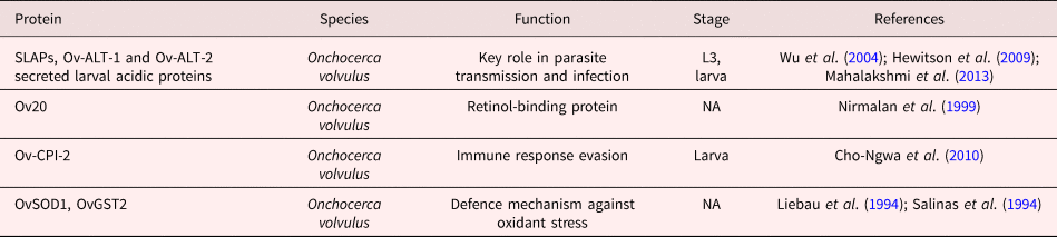
Several E/S molecules identified in the genome and transcriptome of the nematode T. suis could be involved in host–parasite interactions. A significant fraction of the predicted proteins are proteases that could participate in degrading epithelial cells in the host intestinal wall, probably to facilitate invasion and parasite establishment in the cecum, and to favour its evolution to adult stages (Cantacessi et al., Reference Cantacessi, Young, Nejsum, Jex, Campbell, Hall, Thamsborg, Scheerlinck and Gasser2011; Jex et al., Reference Jex, Nejsum and Schwarz2014). Trichuris suis resorts to E/S products like serine-proteases, metalloproteases, cysteine-proteases, aspartyl-proteases and thiol-proteases to penetrate the host intestinal mucosa (table 2) (Hill et al., Reference Hill, Gamble, Rhoads, Fetterer and Urban1993; Hill & Sakanari, Reference Hill and Sakanari1997; Cantacessi et al., Reference Cantacessi, Young, Nejsum, Jex, Campbell, Hall, Thamsborg, Scheerlinck and Gasser2011). In addition, homologues to pore-forming proteins play a role in the pathogenesis of infection by Trichuris spp. TM43 has been identified in T. muris and TT47 in T. trichiura, along with the eukaryotic porin; these proteins induce the formation of conducting pores in lipid bilayers and could be part of the syncytial structure or tegument that allows the parasite to be in direct contact with the host mucosal surfaces (table 2) (Drake et al., Reference Drake, Korchev, Bashford, Djamgoz, Wakelin, Ashall and Bundy1994; Barker & Bundy, Reference Barker and Bundy1999; Bethony et al., Reference Bethony, Brooker, Albonico, Geiger, Loukas, Diemert and Hotez2006). On the other hand, as shown in supplementary fig. S1e, Onchocerca volvulus has three larval stages (L1, L2 and L3). With respect to parasite transmission and infectivity, the secreted proteins Ov-ALT-1 and Ov-ALT-2 are expressed by the L2 and L3 larval stages in O. volvulus (table 3) (Wu et al., Reference Wu, Egerton, Pappin, Harrison, Wilkinson, Underwood and Bianco2004).
Excretion/secretion proteins reported for Onchocerca volvulus.

SLAPs, secreted larval acidic proteins; Ov20 20-kD, retinol-binding protein; Ov-CPI-2, onchocystatin; OvSOD1, superoxide dismutase 1; OvGST2, glutathione S-transferase 2.
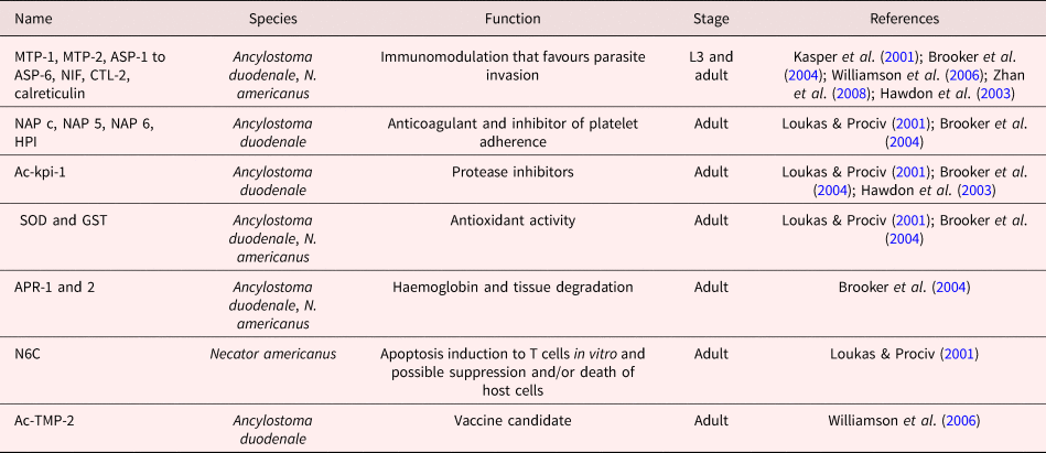
During hookworm infection, several molecules that facilitate mucosal invasion are secreted. Among them are various conjunctive tissue hydrolases such as metalloproteinases of the MTP-1 and MTP-2 astatine type, aspartic proteases like APR-1 and APR-2, and a Kunitz-type protease inhibitor (Ac-kpi-1) (Loukas & Prociv, Reference Loukas and Prociv2001; Brooker et al., Reference Brooker, Bethony and Hotez2004; Williamson et al., Reference Williamson, Lustigman and Oksov2006; Hawdon et al., Reference Hawdon, Datu and Crowell2003). On the other hand, Ancylostoma spp. and Necator americanus secrete products like activation-associated proteins and Ancylostoma-secreted proteins (ASPs); these abundant E/S proteins play an important role in the transition of the free-living to the parasitic stage during the invasion to a mammalian host (table 4) (Cantacessi et al., Reference Cantacessi, Young, Nejsum, Jex, Campbell, Hall, Thamsborg, Scheerlinck and Gasser2011). Additionally, Ancylostoma spp. secrete proteins that cleave eotaxin, retinol- and collagen-binding proteins, type-C lectins that compete with host lectins and acetylcholinesterases that could inhibit intestinal peristalsis (Loukas & Prociv, Reference Loukas and Prociv2001; Brooker et al., Reference Brooker, Bethony and Hotez2004).
Excretion/secretion proteins reported for Ancylostoma duodenale and Necator americanus.

MTP, astacin-like metalloproteinase; ASP, Ancylostoma-secreted protein; NAP, nematode anticoagulant protein; HPI, platelet inhibitor; KPI, Kunitz-type serine-protease inhibitors; NIF, neutrophil inhibitory factor; CTL-2, C-type lectins; TMP, tissue inhibitor metalloproteinases; SOD, Cu/Zn superoxide dismutase; GST, glutathione-S-transferase; ACHe, acetylcholinesterases; APR, aspartic protease; CP-1, cysteine-protease (cathepsin B); MEP-1, metallo-endopeptidase (neprilysin-like); N6C, six-cysteine domains.
While the functions of E/S products have been most widely described in those parasite stages involved in human infections, the role of E/S products in stages affecting other intermediate or definitive hosts should be considered as well to establish strategies aimed to prevent host invasion and parasite propagation.
Modulation of the host immune system
One of the most important functions of parasitic E/S proteins is to modulate the host's immune system to facilitate parasite survival. In hookworm infection, CTL-2, a C-type lectin, could interrupt the antiparasitic immune response or interfere with the coagulation mechanisms of the host (Loukas et al., Reference Loukas, Brown and Pritchard2002). Ac-TMP-2 is a highly immunogenic hookworm antigen; it is similar to the tissue inhibitor of metalloproteinase of vertebrates, except that it presents an unusual structure of C-terminal repeats, suggesting its possible role in the host–parasite relationship. Ac-TMP-2 could be acting to inhibit proinflammatory human metalloproteins as a form of modulation of the host's immune response (Zhan et al., Reference Zhan, Gupta, Wong, Bier, Jiang, Goud and Hotez2008). Another example of immunomodulating E/S products in hookworm infection is the neutrophil-inhibiting factor (NIF), an integrin that inhibits the function of CD11b/CD18-dependent leukocytes, binding the domain-I of CD11b, preventing adhesion and peroxide release (table 4) (Ali et al., Reference Ali, Brown, Stanssens, Timothy, Soule and Pritchard2001; Loukas & Prociv, Reference Loukas and Prociv2001).
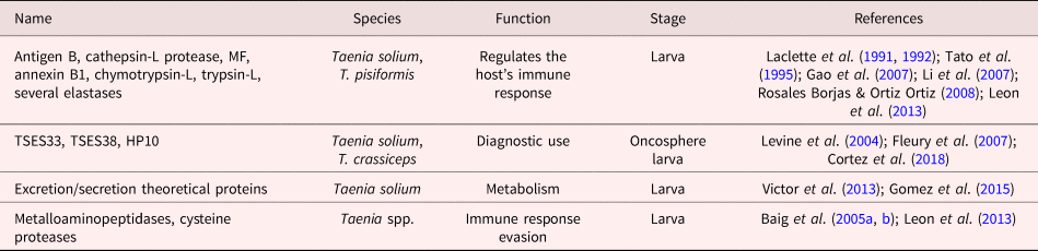
Other helminths such as Taenia spp. also secrete antigens into the environment that help them to modulate the host's immune system. For example, T. solium secretes antigen B (also known as paramyosin) (table 5), which can fixate the complement in soluble complexes, a property that could prevent damage to the parasite by complement fixation on its surface (Laclette et al., Reference Laclette, Landa, Arcos, Willms, Davis and Shoemaker1991, Reference Laclette, Shoemaker, Richter, Arcos, Pante, Cohen, Bing and Nicholson-Weller1992). An E/S protein also named as paramyosin has been reported in Echinococcus spp. (Virginio et al., Reference Virginio, Monteiro, Drumond, de Carvalho, Vargas, Zaha and Ferreira2012). On the other hand, calreticulin was identified as an allergen in infections by N. americanus. Upon secretion and contact with cells of the immune system, calreticulin is known to interact with the complement C1q fraction, inhibiting its hemolytic capacity (Kasper et al., Reference Kasper, Brown and Eberl2001); in addition, various six-cysteine domains (N6C) that could be inducing apoptosis in T cells (Loukas & Prociv, Reference Loukas and Prociv2001; Brooker et al., Reference Brooker, Bethony and Hotez2004) were identified (table 4). Dendritic cells differentiated in vitro have been reported to acquire a tolerogenic phenotype and induce regulatory T cells in the presence of T. crassiceps E/S products, apparently as a form of modulation of the host immune system (Adalid-Peralta et al., Reference Adalid-Peralta, Arce-Sillas and Fragoso2013).
Excretion/secretion proteins reported for Taenia spp.

TSES, Taenia solium excretion/secretion; Antigen B, paramyosin; MF, metacestode factor or RNA-peptide.
EgTeg is an E/S protein from E. granulosus that induces a higher intracellular expression of IL4, which promotes a Th2-type cell response, favouring the parasite survival in the host by limiting the inflammatory response (Ortona et al., Reference Ortona, Margutti and Delunardo2005). Similar products have been described in Ascaris spp., like the immunosuppressive PAS-1 protein, capable of suppressing inflammation and inducing IL-10 production by inhibiting leukocyte migration and the production of proinflammatory cytokines (table 6) (Oshiro et al., Reference Oshiro, Enobe, Araújo, Macedo and Macedo-Soares2006; De Araújo et al., Reference De Araújo, Perini, Martins, Macedo and Macedo-Soares2010; Antunes et al., Reference Antunes, Titz, Batista, Marques-Porto, Oliveira, Alves de Araujo and Macedo-Soares2015). An RNA-peptide conjugate known as the metacestode factor (MF) has been described in T. solium. This molecule inhibits cell proliferation and significantly reduces the inflammatory site in both size and composition. MF is believed to play a key role in down-regulating the host's immune response, which contributes to parasite survival (table 5) (Tato et al., Reference Tato, Castro, Rodríguez, Soto, Arechavaleta and Molinari1995, Reference Tato, White, Willms, Rodríguez, Solano, Sepúlveda and Molinari1996).
Excretion/secretion proteins reported for Ascaris spp.

PAS-1, protein 1 from A. suum; As, fatty-acid-binding protein.
Several studies have reported the presence of serpins in E/S products from nematodes; evidence indicates that many of them may have an immuno-evasive function. Serpins are serine protease inhibitors with a wide spectrum of functions in biological systems such as blood coagulation, complement activation and inflammation. A serpin-like protein was identified in E/S products from Ascaris suum. Apparently, serine protease inhibitors not only protect worms from degradation in the host's digestive environment, but also mask the surface of developing larvae, which allows them to evade the host's immune system by migrating from the intestine to the liver and lungs, although this has not yet been proven (table 6) (Wang et al., Reference Wang, Xiao and Shen2013). Serine proteases such as chymotrypsin-L, trypsin-L and several elastases that degrade the intestinal wall have been demonstrated to allow T. solium and T. saginata oncospheres to enter the bloodstream (table 5) (Leon et al., Reference Leon, Padilla, Pajuelo, Sheen and Zimic2013). Trichuris spp. also secrete a chymotrypsin and elastase inhibitor called TsCEI that the parasite uses as a defence, modulating the immune response of the host through a mechanism of evasion that involves mast cells of the intestinal mucosa (Rhoads et al., Reference Rhoads, Fetterer, Hill and Urban2000). Two proteases have been identified in T. muris, Mr-85 and Mr-105; both are capable of degrading collagen type-I in the epithelial structure of the intestine, thus allowing the parasite establishment (table 2) (Cantacessi et al., Reference Cantacessi, Young, Nejsum, Jex, Campbell, Hall, Thamsborg, Scheerlinck and Gasser2011).
Recently, 14-3-3 proteins have been isolated and characterized as secretory products in A. suum, Schistosoma mansoni and the genus Echinococcus (tables 1 and 6). This family of relatively conserved regulatory protein isoforms of 24–33 kDa can bind a range of functionally diverse signalling proteins and act essentially by scaffolding, sequestering or regulating the activity of target proteins. The interaction of 14-3-3 proteins with growth factor receptors (EGF, TGFβ, IGF) suggests a regulation of cell proliferation, survival and invasion in the context of the host–parasite relationship. In fact, they have been described as promising vaccine candidates against E. granulosus (Siles-Lucas et al., Reference Siles-Lucas, Nunes, Zaha and Breijo2000; Ridi & Tallima, Reference Ridi and Tallima2009; Wang et al., Reference Wang, Xiao and Shen2013).
Another form of host immunomodulation by helminths involves proteins that regulate the host's inflammatory response. Annexin, a multifunctional E/S protein from helminths, is associated with biological processes such as apoptosis, membrane fusion, exocytosis and signal transmission (Song et al., Reference Song, Hu, Zhong, Wang, Gu, Wang, Peng and Yang2016). Annexin B1 is found in the tegument of adult T. solium and is secreted by cysticerci; it causes apoptosis in host eosinophils and inhibits the protein PLA2 in vitro, which could decrease the host inflammatory response (table 5) (Gao et al., Reference Gao, Yan, Ding, Lu and Sun2007; Rosales Borjas & Ortiz Ortiz, Reference Rosales Borjas and Ortiz Ortiz2008). It is known that annexin II has anti-inflammatory and anticoagulant activities in Echinococcus spp. due to its ability to sequester phospholipids (table 1). It also plays an important role in cartilage calcification in the hydatid tumour by virtue of its Ca2+-binding properties (Díaz et al., Reference Díaz, Ibarguren, Breijo, Willis and Sim2000).
Evasion mechanism of the host's immune system
After installation, the survival of the parasite in an immunocompetent host depends on their capacity to evade immune response. During infection by E. granulosus, cytoskeletal proteins like actin and paramyosin have been found as E/S products. Although the presence of these proteins suggests that protein secretion could follow an alternate route in this parasite (Virginio et al., Reference Virginio, Monteiro, Drumond, de Carvalho, Vargas, Zaha and Ferreira2012), they could also be the product of tegument shedding, since it is known that the continuous moulting of this structure releases components that help the parasite to evade the host's immune response (Wang et al., Reference Wang, Van Steendam, Dhaenens, Vlaminck, Deforce, Jex, Gasser and Geldhof2015).
Another mechanism that seems to help helminths to evade damage is the secretion of antioxidant enzymes that allow the parasites to neutralize reactive oxygen species. Glutathione-S-transferases (GSTs) from helminths like E. granulosus, O. volvulus, Ancylostoma spp. and N. americanus participate in the detoxification of lipid hydroperoxides and carbonyl cytotoxics produced by oxygen-reactive intermediates (ORIs) (tables 1, 3 and 4). ORIs can come from the parasite metabolism or the host immune system. GSTs from helminths can conjugate glutathione with xenobiotic compounds or bind anthelmintic drugs (Brattig et al., Reference Brattig, Henkle-Dührsen, Hounkpatin, Liebau, Kruppa and Zipfel1997; Vibanco-Pérez & Landa-Piedra, Reference Vibanco-Pérez and Landa-Piedra1998). Another antioxidant enzyme described in in E/S products from O. volvulus, Ancylostoma sp. and N. americanus is superoxide dismutase (SOD) (Brophy et al., Reference Brophy, Patterson, Brown and Pritchard1995; Brattig et al., Reference Brattig, Henkle-Dührsen, Hounkpatin, Liebau, Kruppa and Zipfel1997; Brooker et al., Reference Brooker, Bethony and Hotez2004). SOD acts as a defence against host-generated oxygen free radicals but also plays an offensive role, damaging inflammatory cells (table 4) (Chiumiento & Bruschi, Reference Chiumiento and Bruschi2009). Another parasitic enzyme involved in redox homeostasis is thioredoxin peroxidase, which was found in E/S products from E. granulosus. This enzyme plays an important role protecting the adult worm from oxidative damage caused by host immune effector cells (Virginio et al., Reference Virginio, Monteiro, Drumond, de Carvalho, Vargas, Zaha and Ferreira2012; Wang et al., Reference Wang, Van Steendam, Dhaenens, Vlaminck, Deforce, Jex, Gasser and Geldhof2015). In O. volvulus, the cytosolic enzymes Cu/Zn SOD-1 (OvSOD1) and glutathione S-transferase-2 (OvGST2) represent two detoxifying molecules which may play an important role in parasite defence against host-induced oxidative stress (table 3) (Brattig et al., Reference Brattig, Henkle-Dührsen, Hounkpatin, Liebau, Kruppa and Zipfel1997).
Taenia spp. are also capable of surviving in the host by resorting to evasion strategies and defence mechanisms. It is known that Taenia spp. secrete proteolytic enzymes like metalloaminopeptidases and cysteine proteases to degrade cytokines and antibodies, which allows the parasite to evade the host's immune response (table 5) (Baig et al., Reference Baig, Damian, Molinari, Tato, Morales-Montor, Welch, Talhouk, Hashmey and White2005a, Reference Baig, Damian, Morales-Montor, Olecki, Talhouk, Hashmey and Whiteb; Leon et al., Reference Leon, Padilla, Pajuelo, Sheen and Zimic2013).
Parasite metabolism
Helminths have adapted their metabolism to survive within their host. For example, A. suum secretes a 293-kDa metalloaminopeptidase at the developmental stages L3 and L4. This enzyme could be associated with moulting, but some evidence suggests that it performs an essential digestive function for the parasite, providing amino acids for uptake and incorporation (Rhoads & Fetterer, Reference Rhoads and Fetterer1998).
The perivitelline fluid of developing A. suum larvae secretes proteins that could play a major role in parasite survival. As-p18, which shows some similarities with retinoid- and fatty acid-binding proteins (FABPs), has been proposed to regulate, store and/or transport fatty acids or other hydrophobic ligands in the fluid, being involved in the maintenance of the lipid layer of the egg, critical in the regulation and execution of hatching (table 6). The biology of the secretion of these compounds is poorly understood; however, it is known that FABPs of nematode origin contain putative secretion signals that are absent in any non-nematode FABPs. This suggests the existence of a new FABP class, apparently unique to nematodes (Mei et al., Reference Mei, Kennedy, Beauchamp, Komuniecki and Komuniecki1997; Plenefisch et al., Reference Plenefisch, Xiao, Mei, Geng, Komuniecki and Komuniecki2000; Ibáñez-Shimabukuro et al., Reference Ibáñez-Shimabukuro, Florencia Rey-Burusco, Cooper, Kennedy, Córsico and Smith2014). Another protein secreted in the perivitelline fluid of developing A. suum larvae is As-p50, a chitinase member of the glycosyl hydrolases family 19. Chitinases are involved in the first ascarid moults, and it is not clear whether p50 is synthesized and secreted specifically to digest the egg shell or it is a multifunctional protein released as a by-product when cuticles of L1 and L2 larvae are shed in the perivitelline fluid after moulting (table 6) (Geng et al., Reference Geng, Plenefisch, Komuniecki and Komuniecki2002).
ABA-1 from A. lumbricoides and A. suum is a secreted protein found in all stages of parasite life (table 6) (Xia et al., Reference Xia, Spence and Moore2000). Interestingly, it could bind a variety of fatty acids and retinol (Mc Gibbon & Lee, Reference Mc Gibbon and Lee1995; Xia et al., Reference Xia, Spence and Moore2000). It has been proposed that O. volvulus could be sequestering retinol from its host through the E/S protein Ov20, which would cause onset symptoms like retinol deficiency and blindness in the patient (table 3) (Nirmalan et al., Reference Nirmalan, Cordeiro, Kläger, Bradley and Allen1999).
Ancylostoma spp.-secreted products are molecules with anticoagulant activity, which facilitate blood consumption and cause poor coagulation to the host, leading to anaemia in severe parasitosis cases (Loukas & Prociv, Reference Loukas and Prociv2001). It is possible that Ac-TMP-1 and/or Ac-TMP-2 have a similar biological function to provide red blood cells from the host as a source of nutrients for the parasite (Zhan et al., Reference Zhan, Gupta, Wong, Bier, Jiang, Goud and Hotez2008).
Medical uses (diagnosis, vaccines, inflammation treatment) of E/S proteins
Various parasitic E/S proteins have been identified as possible vaccine candidates. The Ac-TMP-2 and Na-ASP-2 E/S proteins from N. americanus and Ancylostoma spp. have been proposed as vaccination antigens against hookworm infections (Loukas et al., Reference Loukas, Bethony, Brooker and Hotez2006). In N. americanus, ASP-2 (Na-ASP-2) was chosen as the most promising larval component of a vaccine targeting hookworms (Diemert et al., Reference Diemert, Bethony and Hotez2008), while Ac-TMP-2 was found to be an immunodominant antigen associated with the ir-L3 vaccine (Zhan et al., Reference Zhan, Gupta, Wong, Bier, Jiang, Goud and Hotez2008). Other vaccine antigens are Na-GST-1 and Na-APR-1, which are currently being developed as vaccines against human hookworm infection (table 4) (Hotez et al., Reference Hotez, Diemert and Bacon2013).
Secreted larval acidic proteins (SLAPs) are a family of E/S proteins from O. volvulus that are expressed specifically in the larval stage of the parasite and are of great interest as vaccine antigens (Wu et al., Reference Wu, Egerton, Pappin, Harrison, Wilkinson, Underwood and Bianco2004). OvSLAP1 is an immunogenic protein that induces an intense cell response; due to its protective capacity against filarial infections by Brugia malayi and Wuchereria bancrofti in endemic populations, it has been considered as an important vaccine candidate (Mahalakshmi et al., Reference Mahalakshmi, Aparnaa, Vishal and Kaliraj2013). Another interesting vaccine candidate is Ov-CPI-2 (Onchocystatin), a cysteine proteinase inhibitor from O. volvulus that has been associated with the development of a protective immunity in onchocerciasis (table 3) (Cho-Ngwa et al., Reference Cho-Ngwa, Liu and Lustigman2010).
Some E/S molecules have been used as antigens for immunodiagnosis. Taenia solium causes two diseases in humans, taeniasis and cysticercosis. Many serological tests have been developed to diagnose both conditions. An example of E/S proteins used in the immunodiagnosis of cysticercosis is the HP10 Ag-based ELISA, which detects a high-molecular-weight secreted protein from viable metacestodes; this serological test has already been used for the diagnosis of human and porcine cysticercosis (table 5) (Cortez et al., Reference Cortez, Rojas and Parkhouse2018). For taeniasis, a serological diagnostic test has been developed using the TSES33 and TSES38 proteins. Both recombinant proteins were recognized by a panel of serum samples from patients with taeniasis, but not with cysticercosis (table 5) (Levine et al., Reference Levine, Calderón and Wilkins2004). Other molecules have shown serodiagnostic potential, like the 17-kDa, E/S protein from T. trichiura that has been assayed in patients infected by the adult parasite (Lillywhite et al., Reference Lillywhite, Cooper, Needham, Venugopal, Bundy and Bianco1995). The antigen P-29 is a protein located in the tegument and rostellum of E. granulosus that has been used as a diagnostic antigen and as a potent serological marker for the post-treatment monitoring of echinococcosis (table 1) (González et al., Reference González, Spinelli, Lorenzo, Hellman, Nieto, Willis and Salinas2000; Boubaker et al., Reference Boubaker, Gottstein, Hemphill, Babba and Spiliotis2014). A protein secreted by O. volvulus that is important in the immunodiagnosis of onchocerciasis is Ov20, an immunodominant protein that exhibits a high specificity for onchocercal serum (Nirmalan et al., Reference Nirmalan, Cordeiro, Kläger, Bradley and Allen1999).
Finally, parasitic E/S proteins have also been used to treat inflammation-related pathologies. Trichuris suis E/S proteins were recently used as a therapy in Crohn's disease and ulcerative colitis, promoting a Th2 and T regulatory response that may buffer intestinal inflammation (Parthasarathy & Mansfield, Reference Parthasarathy and Mansfield2005; Summers et al., Reference Summers, Elliot, Urban, Thompson and Weinstock2005; Hiemstra et al., Reference Hiemstra, Klaver, Vrijland, Kringel, Andreasen, Bouma, Kraal, van Die and den Haan2014). Similarly, N. americanus larvae have been used against Crohn's disease in humans (Reddy & Fried, Reference Reddy and Fried2009); it is feasible that this effect is due to E/S proteins.
Conclusions
Numerous studies indicate that helminths have co-evolved with their hosts; this host–parasite adaptation is a very close relationship, where the parasite develops escape strategies through mimicry and manipulation of the host immune system, adapting itself to the host response to parasitism. Ultimately, this results in an environment that favours the survival and coexistence of both antagonist parties in a complex adaptive mechanism.
In this review, we have noted that some E/S proteins are shared by different parasite development stages, but others are unique for certain stages. This fact could be taken as an advantage for the possible development of biomarkers to indicate the parasite stage during infection, which would allow us to provide an appropriate therapeutic intervention. Additionally, stage-specific E/S proteins could be used as biomarkers of damage to the host, since some stages can target certain organs, directly affecting the health of the host.
The secretome of platyhelminths like Fasciola hepatica, T. solium and S. mansoni has been published. The bioinformatic analysis of these sequences could contribute to identify new E/S proteins related to parasite development, cell adhesion, proteolysis and extracellular matrix organization; in the context of host–parasite relationship, they could act as virulence factors or regulators of the host immune recognition.
Finally, this review allowed us to update our knowledge on E/S proteins from parasites endemic in LAC and offered an overview of the use of these proteins, which could facilitate the development of novel therapeutic, diagnostic and vaccine targets.
Author ORCIDs
L. Adalid-Peralta, 0000-0002-1010-698X
Supplementary material
To view supplementary material for this article, please visit https://doi.org/10.1017/S0022149X19000312
Conflicts of interest
None.