Introduction
Knowledge of nasal cavity circulation is essential for optimal treatment of epistaxis, particularly epistaxis that is intractable.Reference Babin, Moreau, Goullet, Rugy and Delmas1 Severe posterior epistaxis is a potentially life-threatening condition for the patient, and poses a therapeutic challenge to the otolaryngologist. Closure of the sphenopalatine artery by ligation or diathermy prevents direct retrograde and collateral blood supply from the contralateral and ipsilateral carotid systems.Reference von Buchwald and Tranum-Jensen2
Damage to the sphenopalatine artery during sinonasal surgery may cause severe bleeding. Therefore, sinonasal surgeons performing standard functional endoscopic sinus surgery or endonasal resection of vascular lesions (e.g. juvenile nasopharyngeal angiofibroma) should be aware of the treatment options available for this surgical complication.Reference von Buchwald and Tranum-Jensen2
Several studies have noted that the sphenopalatine artery frequently divides before its entrance into the nasal fossa.Reference Babin, Moreau, Goullet, Rugy and Delmas1, Reference Navarro and Filho3, Reference Legent, Boutet, Wesoluch, Viale, Galiba and Beauvillain4 The posterior superior nasal branches of the maxillary nerve also course through the sphenopalatine foramen, together with the sphenopalatine artery.Reference Navarro and Filho3
This study aimed to evaluate endoscopic cauterisation of the sphenopalatine neurovascular bundle as treatment for intractable posterior epistaxis, with regard to efficacy and safety. In addition, the study evaluated the technical difficulties and post-operative sequelae of this surgery.
Materials and methods
A prospective study studied 42 patients with severe posterior epistaxis who were treated with endoscopic cauterisation of the sphenopalatine artery, over a 17-month period (September 2008 to January 2010). Patients' ages ranged from 18 to 69 years, with a mean age of 51.1 years. The patients comprised 23 (54.7 per cent) men and 19 (45.3 per cent) women, seen at the otorhinolaryngology department of Mansoura University Hospital, Egypt.
Before commencement, the relevant institutional review board approved the research, and appropriate consent was obtained from all patients.
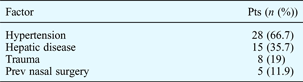
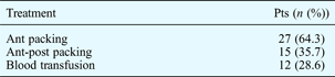
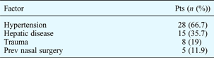
Our patients' predisposing factors for epistaxis are shown in Table I, and their pre-operative treatment in Table II.
Patients' Predisposing Factors
Pts = patients; prev = previous
Patients' Pre-Operative Treatment
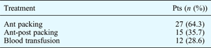
Pts = patients; ant = anterior; post = posterior
These patients' indications for endoscopic cauterisation of the sphenopalatine artery were: severe posterior epistaxis unresponsive to conservative treatment (i.e. refractory bleeding with a pack in place); severe, recurrent posterior epistaxis; bleeding after nasal surgery; and patient refusal or absolute intolerance of packing.
The basic principle of the surgical technique was to identify the branches of the sphenopalatine artery through an endoscopic endonasal approach, and then to perform endoscopic cauterisation of the sphenopalatine bundle under direct vision without separation of the posterior superior nasal nerve. In this study, access to the sphenopalatine foramen was limited in 26 cases, requiring resection of the crista ethmoidalis for complete skeletonisation of the artery, followed by cauterisation of the sphenopalatine artery with all its nasal branches.
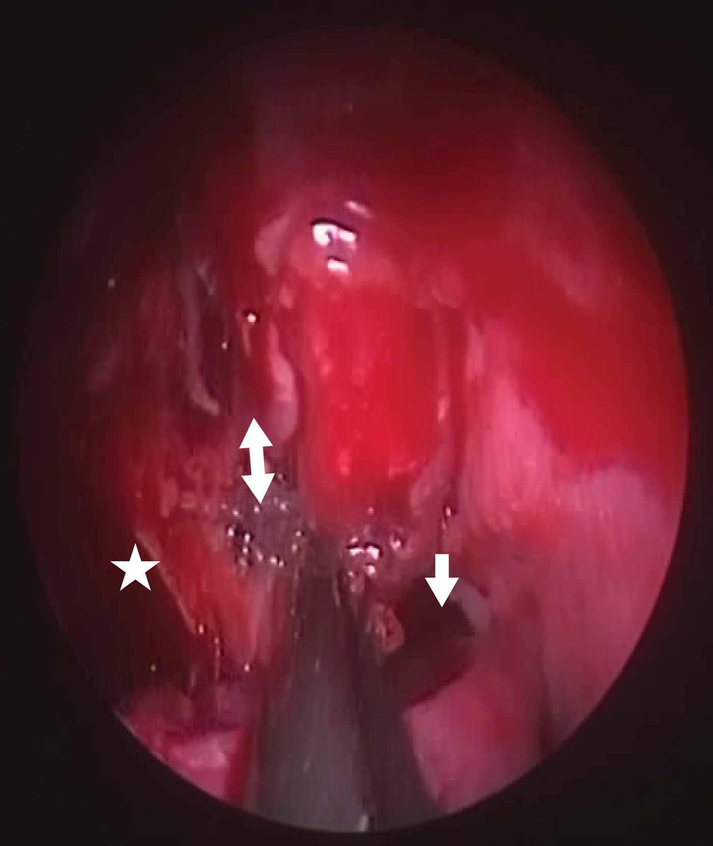
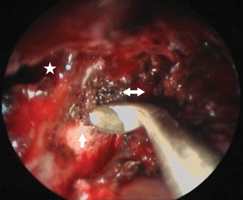
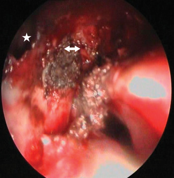
Surgery was carried out under general anaesthesia, using a 0° nasal endoscope (4 mm diameter). A vertical incision was made inferior to the posterior portion of the middle turbinate, 1 cm anterior to its posterior tip. A Freer elevator was used to raise a mucoperiosteal flap posteriorly and upwards. First, the sphenopalatine artery stump with its branches was identified (Figure 1). The neurovascular bundle was reached at its emergence from the sphenopalatine foramen. The ostium of the maxillary sinus was widened backwards to outline the posterior wall of the maxillary sinus, in order to confirm the anatomical topography. In addition, the bony ethmoidal crest was exposed just anterior to the sphenopalatine foramen, allowing the crista to be resected using a 2 mm up-biting Kerrison punch or curette, for proper exposure (Figure 2). This allowed identification of the artery as it emerged into the nose, and permitted proper cauterisation of all sphenopalatine vascular stumps.
Endoscopic view showing right posterior choanae (arrow), posterior maxillary wall (star) and right sphenopalatine bundle (double arrow).
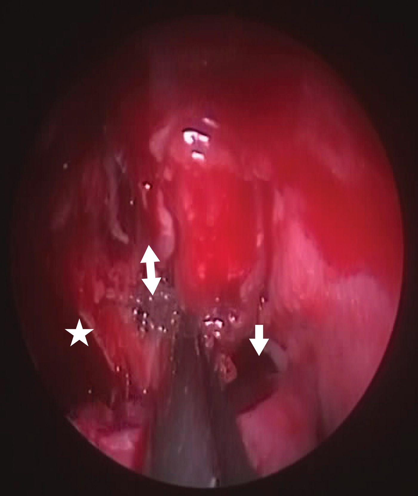
Endoscopic view showing curettage of the right crista ethmoidalis (arrow), the cauterised right sphenopalatine bundle (double arrow), and the posterior maxillary wall (star).

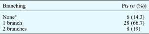
At this stage, the branching pattern of the sphenopalatine artery was noted (Table III). The artery was cauterised with bipolar diathermy just as it emerged from the sphenopalatine foramen. Bipolar cauterisation was applied laterally to the branching of the artery, to effectively treat the source of the bleeding (Figure 3). During this stage, one to two branches of the posterior superior nasal nerve, which runs along the artery, could be identified; however, in this study cauterisation of all sphenopalatine bundles was performed, without nerve dissection.
Endoscopic view showing the posterior maxillary wall (star) and the completely cauterised right sphenopalatine bundle (double arrow).

Sphenopalatine Artery Branching at Sphenopalatine Foramen
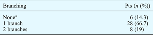
*Single trunk. Pts = patients
After cauterisation, the flap was replaced (as adequate flap coverage helps prevent crusting and inflammation). Blood loss was calculated intra-operatively. The operation was terminated when there were no points of bleeding, or when there was no accumulation of blood in the nasal cavities or the nasopharynx. However, it was important to avoid excessive cauterisation and injury to the nasal mucosa, in order to prevent scarring, adhesions, and unnecessary or unexpected complications.
In 13 cases, endoscopic septoplasty was performed, during the same operation, to facilitate surgery.
The nasal cavity was packed for the first 24 hours post-operatively, using a Merocel nasal tampon (Medtronic Xomed, FL, USA). Patients also received post-operative antibiotics.
Follow up in the second, eighth and 12th post-operative weeks included: history and nasal examination for recurrent epistaxis; evaluation of eye dryness, both subjective (rated as none, mild, moderate or severe) and objective (using Schirmer's test); evaluation of nasal dryness, both subjective (rated as none, mild, moderate or severe) and objective (i.e. nasal examination for dryness and crusting); evaluation of subjective nasal sensation and objective endoscopic nasal appearance (in the out-patient clinic); and evaluation of upper teeth and palate sensation.
Results and analysis
Pre-operative evaluation identified hypertension and hepatic disease as predisposing factors to epistaxis in 66.7 and 35.7 per cent of patients, respectively (Table I).
All patients were pre-operatively treated using anterior packing (alone in 64.3 per cent and combined with posterior packing in 35.7 per cent). Blood transfusion was performed in 28.6 per cent of patients (Table II).
No severe intra-operative bleeding was encountered in this study. However, the hypotensive technique was available in all cases. The sphenopalatine artery showed no branching in six patients (14.3 per cent), and one branch only in 28 patients (66.7 per cent).
Post-operative evaluation showed no recurrence of epistaxis (Table III).
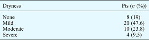
Post-operative subjective evaluation of eye dryness indicated no dryness in 90.5 per cent of patients (n = 38) and mild dryness in only 9.5 per cent (four patients, who complained of mild eye burning and congestion). Objective evaluation of eye dryness (using Schirmer's test) was negative in all cases.
Post-operative subjective evaluation of nasal dryness indicated that dryness was present in 81 per cent of patients (n = 34) but was severe in only 9.5 per cent (four), and resolved with medical treatment (i.e. nasal debridement and irrigation) (Table IV). These findings were similar to those from endoscopic examination for dryness and crusting.
Post-Operative Subjective Nasal Dryness
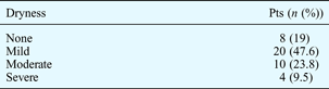
Pts = patients
Post-operative subjective evaluation for nasal sensation (i.e. paraesthesia or hypoaesthesia of the nose, upper teeth and palate) indicated numbness and paraesthesia of the upper teeth in three patients. Objective evaluation of nasal sensation (via endoscopy in the out-patient clinic) found hypoaesthesia of the nasal mucosa in eight patients, but without any patient complaint. No hypoaesthesia of the hard palate was detected.
No major post-operative complications or irreversible damage was encountered. The success rate was 100 per cent, with no episodes of recurrent epistaxis within the follow-up period.
Discussion
Bolger et al. have described the crista ethmoidalis, and its relatively constant position within 1 mm anterior or anteroinferior to the sphenopalatine foramen, in 21 of 22 specimens studied.Reference Bolger, Borgie and Melder5 The crest attachment is typically fibrous, and is thus easily removed during resection.Reference Orlandi6
Another study examined 61 cadavers (122 lateral nasal walls), and found the ethmoidal crest to be present in all of them.Reference Voegels and de Melo Padua7 The median distance from the sphenopalatine foramen to the anterior nasal spine was 6.6 mm. In 67.2 per cent of cases, only one artery emerged from the sphenopalatine foramen.
In our study, the crista ethmoidalis was present in all patients, and the sphenopalatine artery branched at its foramen in more than 85 per cent of cases. Other authors have noted that the sphenopalatine artery frequently divides before its entrance into the nasal cavity. In their study of anatomical variation of nasal cavity arteries, Babin et al. described two cases in which the sphenopalatine artery entered the nasal cavity with only one, unique trunk; in a further 18 cases, the artery divided within the infratemporal fossa and branched at the exit of the sphenopalatine foramen.Reference Babin, Moreau, Goullet, Rugy and Delmas1
The posterior superior nasal branches of the maxillary nerve course with the sphenopalatine artery through the sphenopalatine foramen. Neurological deficit from injury to these structures has not been described, but this risk should be kept in mind when performing sphenopalatine artery ligation.Reference Bolger, Borgie and Melder5 In the present study, cauterisation of the sphenopalatine neurovascular bundle did not cause any neurological deficits or major complications. Severe nasal dryness was present in only four patients (9.5 per cent), while objective nasal mucosa hypoaesthesia was detected in eight patients but without patient complaint. No hypoaesthesia of the hard palate was detected.
Surgical treatment for severe epistaxis is associated with several predisposing factors. Voegels et al. found hypertension in 36 per cent of cases, haematologic disorders in 9 per cent, previous nasal surgery in 45 per cent and trauma in 18 per cent.Reference Voegels, Curti, Vasquez and Butugan8 All their patients presented with posterior epistaxis, and 45 per cent needed a blood transfusion. No complications or difficulties were encountered in isolating the artery. Barlow et al. observed that surgical treatment for severe epistaxis was associated with posterior bleeding and blood transfusion.Reference Barlow, Deleyiannis and Pinczower9 In this study, predisposing factors for epistaxis comprised hypertension in 66.7 per cent of patients and hepatic disease in 35.7 per cent. Blood transfusion was performed pre-operatively in 12 patients (28.6 per cent).
Budrovich and Saetti were the first to report the endoscopic approach for ligation of the sphenopalatine artery.Reference Budrovich and Saetti10 This procedure interrupts the nasal vasculature at a point sufficiently distal to prevent direct, retrograde or anastomotic blood flow from the ipsilateral and contralateral carotid systems.Reference Sharp, Rowe-Jones, Biring and Mackay11 The risk of such carotid blood flow is one of the principal drawbacks of transantral ligation of the maxillary artery. Moreover, endoscopic sphenopalatine artery ligation avoids the risk of neurological injuries caused by inadvertent clipping of maxillary nerve branches.Reference Bolger, Borgie and Melder5 Endoscopic sphenopalatine artery ligation is facilitated by the constancy of the pterygopalatine fossa, the dimensions of which do not vary greatly (i.e. with respect to volume, width and depth).Reference Stojcev, Gacić, Popović and Stajcić12 In the present study, avoidance of excessive cauterisation and injury to the nasal mucosa and/or pterygopalatine fossa was important in order to prevent scarring, adhesions, and unnecessary or unexpected complications (e.g. ophthalmic artery spasm, injury to maxillary nerve branches within the pterygopalatine fossa, or necrosis of the inferior turbinate).
A number of studies have shown the effectiveness of endoscopic sphenopalatine artery ligation, with success rates ranging from 87 to 100 per cent.Reference Claes, Claes, Boudewyns, Rombaux and Daele13 Similar success has been described for endoscopic cauterisation of the sphenopalatine artery, although hypoaesthesia of the hard palate has been reported as a complication (presumably because of injury to the greater palatine nerve as it enters its canal just lateral to the sphenopalatine foramen).
No severe, irreversible sequelae of endoscopic sphenopalatine artery ligation have been reported to date.Reference Orlandi6 A retrospective review of 38 cases by Snyderman et al. revealed no major complications associated with such ligation.Reference Snyderman, Goldman, Carrau, Ferguson and Grandis14 Minor complications included: nasal crusting, which resolved with time; debridement; numbness of the teeth, palate and upper lip; acute sinusitis; decreased lacrimation; and septal perforation. Moorthy et al. described the previously unreported complication of necrosis of the inferior turbinate after endoscopic ligation of the sphenopalatine artery.Reference Moorthy, Anand, Prior and Scott15
• Endoscopic sphenopalatine neurovascular bundle cauterisation is a safe, effective treatment for refractory posterior epistaxis
• In the presented 42 patients, the sphenopalatine artery branched at its foramen in 36 (86 per cent)
• Excessive cauterisation must be avoided to prevent unnecessary nasal mucosa and pterygopalatine fossa trauma
In this study, the success rate was 100 per cent, with no recurrent epistaxis within the follow-up period. No major post-operative complications or irreversible damage was encountered. Minor complications were present, including nasal crusting and mild nasal dryness, which resolved with the correct medical treatment. The lack of serious complications could be attributed to avoidance of excessive cauterisation and nasal mucosa injury.
Conclusion
Endoscopic sphenopalatine neurovascular bundle cauterisation is a safe, easily performed, effective treatment for refractory posterior epistaxis. Cauterisation of the main sphenopalatine bundle before branching may require removal of the crista ethmoidalis for proper exposure. Branching of the sphenopalatine artery at its foramen was present in more than 85 per cent of the presented cases.
Cauterisation of the neurovascular bundle did not cause any neurological deficits or major complications. Severe nasal dryness was present in only four patients (9.5 per cent), nasal mucosa hypoaesthesia was found in eight patients and no hypoaesthesia of the hard palate was detected. There was no subjective eye dryness in 38 patients (90.5 per cent), whereas Schirmer's test was negative in all cases. No major post-operative complications were found. Avoidance of excessive cauterisation and injury to the nasal mucosa and/or pterygopalatine fossa was important in order to prevent scarring, adhesions, and unnecessary or unexpected complications.
The success rate was 100 per cent, and no recurrent epistaxis was encountered within the follow-up period.