

PerinatalFootnote 1 and infantFootnote 2 skeletal remains from archaeological sites have historically received minimal attention; sometimes discarded altogether,Footnote 3 many of those excavated are absent from excavation reports and published literature.Footnote 4 Today, interest in perinatal and infant studies from a range of disciplines is increasing, particularly within bioarchaeology, where much research has investigated factors related to childhood stress, social inclusion and care of children in the past.Footnote 5 Studies of perinatal and infant remains from Iron Age and Romano-British domestic sites have focused on the funerary and social contexts of these burials.Footnote 6 These have provided valuable insights into the social and cultural worlds into which these individuals were born, while the perceptions of infants within these societies have been widely discussed.Footnote 7 However, skeletal assessment of these young individuals has often concentrated on biological age and sex determination in support of theories of deliberate death and disposal.Footnote 8 Yet, skeletal analysis can reveal far more, both about the individuals and their mothers; detailed biological and pathologicalFootnote 9 assessment furthers our understanding of the complex ways morbidity, mortality and lifestyle factors within past communities affected these perinatal and infant individuals. Thus, this study examines the evidence for growth and health disruption through the analysis of 17 individuals from Piddington and considers the implications for perinatal/infant and maternal health in this late Iron Age to early Romano-British society. This research aims to highlight the value of palaeopathological evidence, in conjunction with funerary and other contextual evidence, when interpreting perinatal and infant remains from archaeological contexts.
RESEARCH CONTEXT
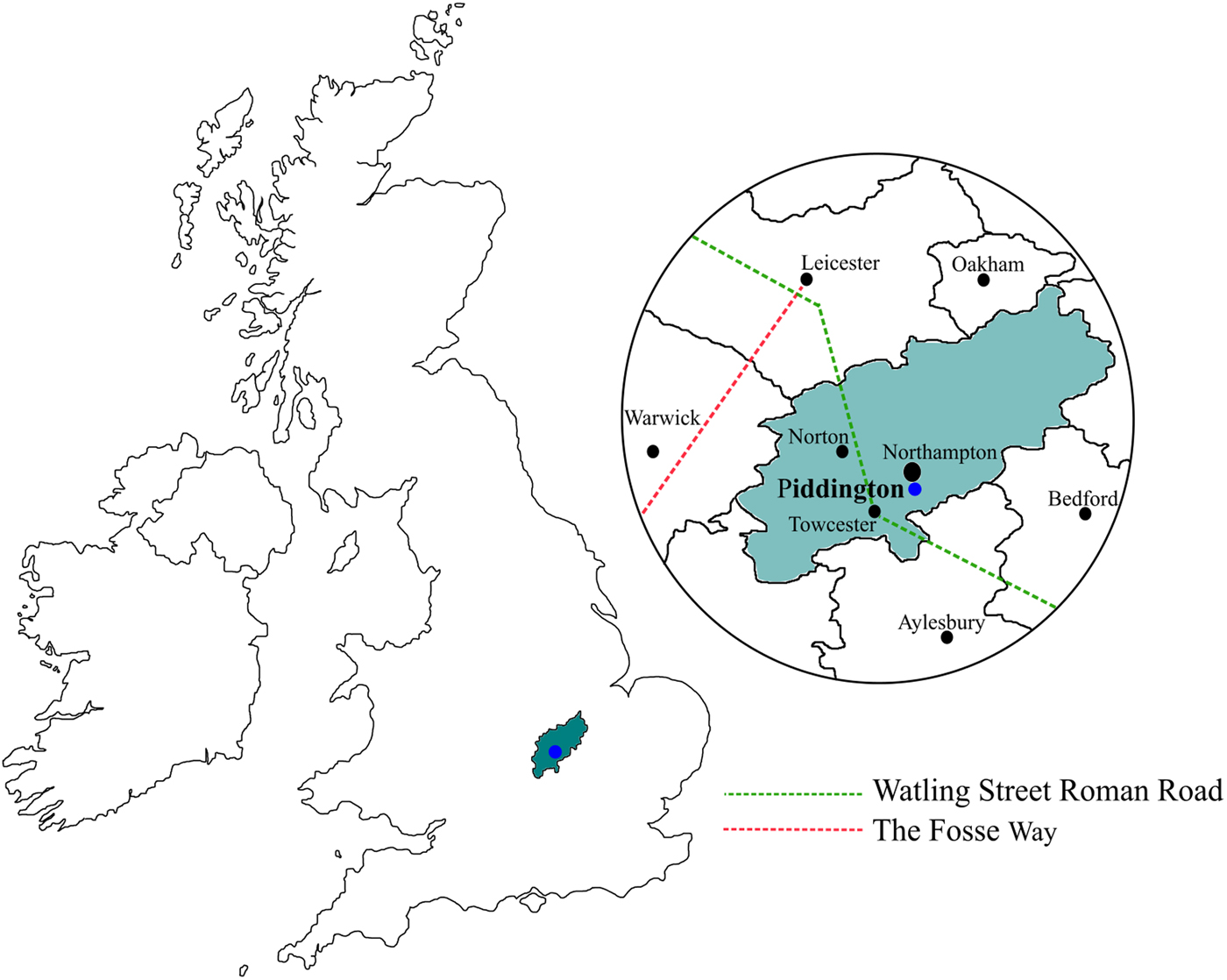
Since 1979 Piddington has undergone thorough and extensive excavation conducted by the Upper Nene Archaeological Society. These excavations have uncovered a large Roman villa and surrounding estate, with an underlying Iron Age settlement. At present the site is believed to have undergone seven main phases of occupation, with strong evidence for prehistoric activity on and within the immediate vicinity.Footnote 10 Piddington was situated among, and en route to, other much larger settlements: Towcester (Lactodorum), Norton (Bannaventa) and Leicester (Ratae) are all nearby, with the Watling Street Roman road and the Fosse Way accessible (fig. 1).
Map of the British Isles showing the location of Piddington, along with an enlarged map of Northamptonshire, which shows the position of the site and its proximity to other major Roman towns and two major Roman roads.

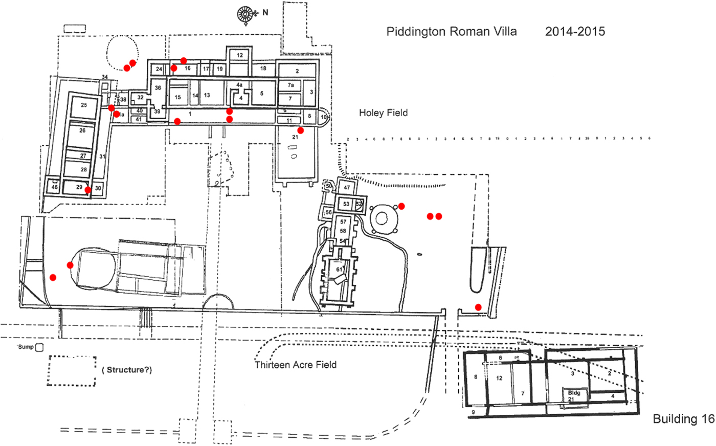
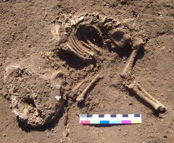
Assessment of the site plan shows that the burials all lie within the villa compound, with nine of the perinates/infants being within the main villa structure (fig. 2). The burials are likely to be contemporary with the main villa, dating them to between a.d. 90 and a.d. 190; however, stratigraphic records suggest that some perinates were interred earlier. Evidence of Iron Age features post-dating some of these perinatal burials has also been recorded, with many of the burials sealed by a layer of dark soil known to be of Iron Age date.Footnote 11 Miller suggests that although there is no patterning or zoning to the burials, it is notable that the majority are directly between the two larger Iron Age roundhouses, which underlie the main villa structure.Footnote 12 Thus, all burials from Piddington are considered to be first century a.d. in date, spanning the late Iron Age and early Roman occupation of the site. Six of the individuals were found to be buried in pairs, although no particular area appears to have been reserved for these infant burials. The undisturbed individuals were all found to be placed in a fetal positionFootnote 13 (fig. 3).
Site plan of Piddington. The red dots indicate the location of a single perinatal burial.

Photograph taken of Burial 24 in situ. The individual is in a fetal position, with upper and lower limbs flexed.

METHODS
This study examines whether growth disruption is identifiable in perinates and infants, and has used existing methods to explore this possibility. It is acknowledged that many of the methods are problematic in accurately determining age-at-death estimates. This is challenging as the article relies on the comparison of age estimates derived from metric and dental assessment of the individuals. However, this research highlights the potential of these methods to indicate perinatal and infant growth disruption, and hopes further study will help develop more accurate and appropriate methods for analysis.
Assessment of growth disruption was undertaken using two approaches: first to compare age estimates derived from dental development to those derived from metric assessment of skeletal elements and second by comparing measurements collected from metric assessment against a reference standard. The second of these approaches uses dental development to categorise the individuals by gestational weeks of age, thus enabling the measurements from those individuals to be compared to measurements of the reference individual of the same age.
Dental development is considered a more accurate method for estimating gestational age; tooth growth and development is less easily disrupted by external factors and more closely correlated with chronological age.Footnote 14 Methods of age-at-death estimation using teeth rely on assessing the stage of development for each tooth.Footnote 15 Teeth grow systematically from the tip of the crown down to the root, and this happens at varying, yet relatively precise ages for each tooth.Footnote 16 The tooth cusps, or developing teeth, recovered from perinatal and infant burials are therefore extremely important for calculating accurate age-at-death estimates. However, tooth cusps are notoriously small and easily lost in the archaeological record. At Piddington, tooth cusps were found to be present in eleven of the burials, although only ten individuals have been included within dental analysis as Burials 12A and 12B were commingled. This means that 66 per cent of the individuals analysed (excluding 12A/B) have tooth cusps present. Tooth-cusp development was recorded in accordance with Moorrees et al.Footnote 17 and age estimates attributed using the tooth-development atlas developed by AlQahtani et al.Footnote 18 This is the most recent revision of the Moorrees et al. methodFootnote 19 and is based on 704 clinical and archaeological individuals of known age-at-death.Footnote 20 The major limitation of this method is that it assumes tooth development of the archaeological child and of the modern reference children are identical.Footnote 21 However, this limitation is a feature of all skeletal ageing techniques and Hillson confirms that tooth development varies less between populations than skeletal development.Footnote 22 Where tooth-cusp development was considered to fall between two developmental stages a midpoint between those two stages was provided. All age estimates were assigned in gestational weeks as is standard practice with perinatal skeletal remains.
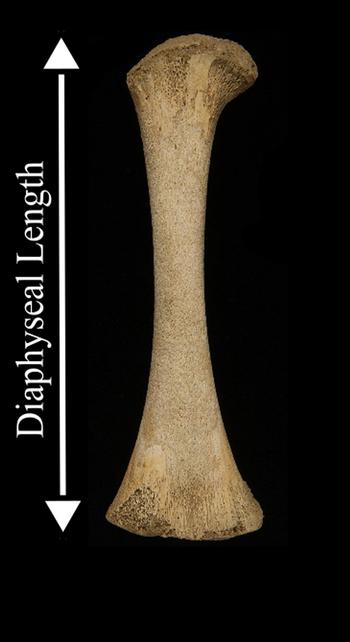
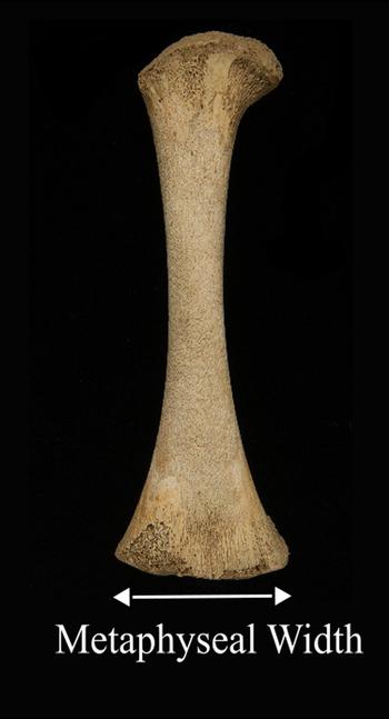
To obtain skeletal age estimates, selected long bones and cranial elements were measured in accordance with existing methods.Footnote 23 All measurements were taken using digital sliding callipers and the majority were of the long-bone diaphyseal lengths to calculate gestational ageFootnote 24 (fig. 4). MetaphysealFootnote 25 widths of long bones were also recorded. This was to observe whether differences in diaphyseal length and metaphyseal width age estimates could be identified. Growth is widely known to be disrupted when subjected to episodes of stressFootnote 26 and research has also suggested that prioritisation of growth in certain dimensions can be suspected.Footnote 27 Thus, metric assessment of these two dimensions was analysed in order to examine whether inconsistencies and differences between age estimates — suggestive of growth disruption — could be identified. The metaphyseal width measurements were taken at the distalFootnote 28 ends of the long bones (fig. 5). Measurements were also taken of cranial elements, in particular those of the pars basilaris,Footnote 29 a bone at the base of the cranium forming part of the foramen magnum. Due to the changes in morphology, and thus the measurements obtained, the pars basilaris is considered to be indicative of certain age thresholds.Footnote 30
Photograph of a right perinatal femur from Piddington. The white arrow indicates the dimensions of a diaphyseal length measurement. Diaphyseal length measurements are taken from the proximal to distal ends of all long bones to obtain the maximum length.

Photograph of a right perinatal femur from Piddington. The white arrow indicates the dimensions of a metaphyseal width measurement. Metaphyseal width measurements are taken at the distal end of the femora and humeri, measuring from the furthest point of the lateral edge to the medial edge to obtain the maximum width.

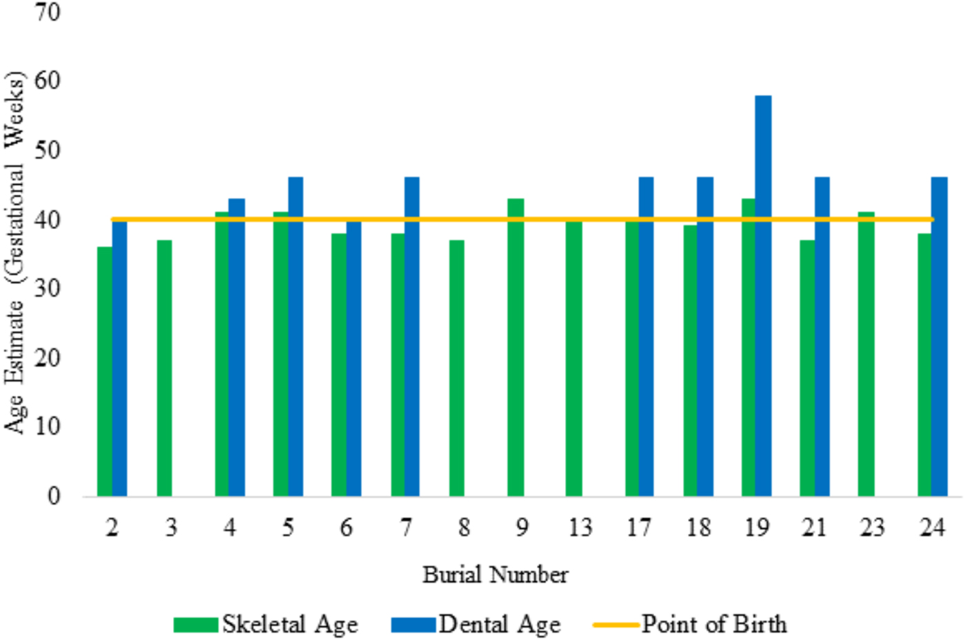
Linear regression equations, developed by Scheuer et al.,Footnote 31 were used to calculate age estimates based on long-bone diaphyseal length measurements. This method was employed as it is based on clinical data of known age-at-death individuals.Footnote 32 In addition, some of the Piddington individuals had long-bone diaphyseal length measurements that were over the upper bounds of those given by Fazekas and Kósa,Footnote 33 but considerably under the measurements recorded by Maresh.Footnote 34 Thus, this method proved useful in determining more specific age estimates for those which fell between 40 and 46 gestational weeks.Footnote 35 The age estimates derived from this method of assessment have been used in fig. 6 to compare differences in age estimates derived from dental and skeletal assessments, and in figs 7, 8 and 10 where measurements derived from this method have been used as reference points against which data from the Piddington individuals have been plotted.
Graph displaying age estimations for all individuals from Piddington. Both age estimations derived from metric assessment and tooth development have been presented where possible. The yellow line indicates what may typically be considered the ‘standard point of birth’ at 40 weeks and demonstrates the mid-point of the perinatal period (36–44 weeks).

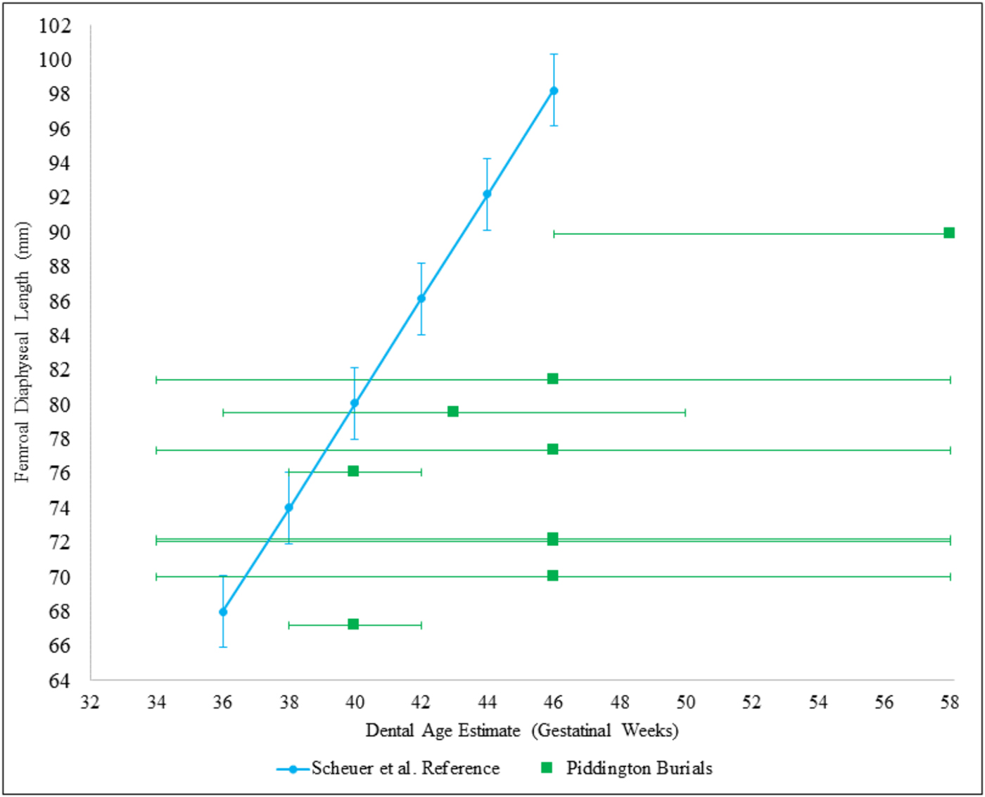
Femoral length plotted against dental age for individuals with both data points available. The Piddington individuals, represented by a square symbol, can all be seen to plot to the right of the reference data (Scheuer et al. Reference Scheuer, Musgrave and Evans1980). This suggests that given their dental age estimates, their femoral growth is less than expected. However, error bars given suggest that most individuals have dental/skeletal data points which are within an accepted range. Two individuals, one aged 40 weeks, the other 58 weeks, are suggested to show definitive evidence of growth disruption as their error bars do not intercept.

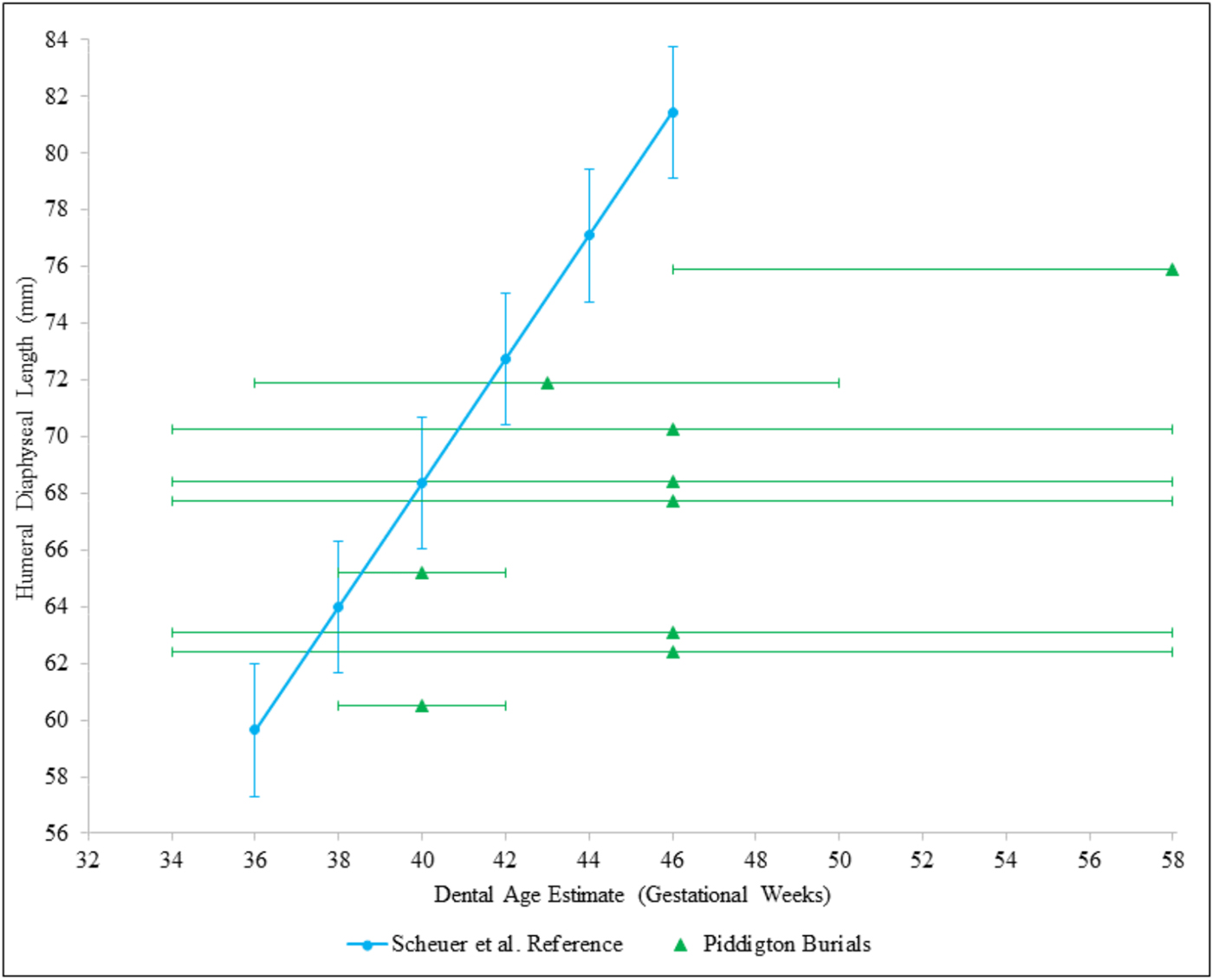
Humeral length plotted against dental age for individuals with both data points available. Once again, the Piddington individuals can be seen to plot to the right of the reference data (Scheuer et al. Reference Scheuer, Musgrave and Evans1980), suggesting humeral length is less than expected given their dental ages. Similarly, all individuals, except the same two identified in fig. 7, have data points which fall within an accepted range. Thus, femoral and humeral lengths appear to be growing in a similar pattern, with the same individuals suggesting evidence of growth disruption is present.

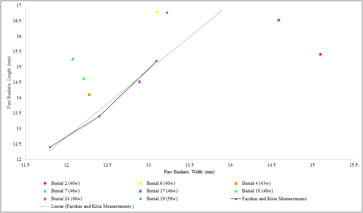
Reference data published by Fazekas and KósaFootnote 36 have been used in consideration of the pars basilaris (fig. 11) and when comparing long-bone diaphyseal lengths and metaphyseal widths (figs 9i and 9ii). This is because these are still the only published data which detail averaged measurements, for individuals aged between 30 and 40 gestational weeks at two-week intervals, for these skeletal dimensions. This method has not been used to estimate age-at-death from skeletal measurements, but is included as a reference sample.
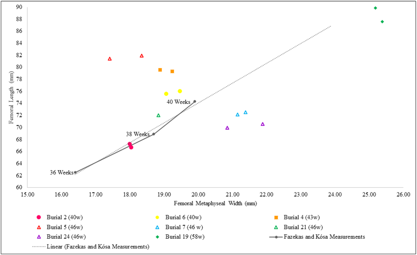
Femoral diaphyseal length plotted against femoral metaphyseal width for those individuals with both measurements and tooth age estimations available. The solid grey line indicates the reference measurements given for individuals between 36 and 40 gestational weeks by Fazekas and Kósa (Reference Fazekas and Kósa1978), with the dashed grey line indicating a linear trend line for these measurements. By plotting measurements of those with tooth age estimates it can be discerned whether there is growth disruption of the femora and in which dimension. All of the eight individuals with measurements plotted are suggested to display disruption to growth.

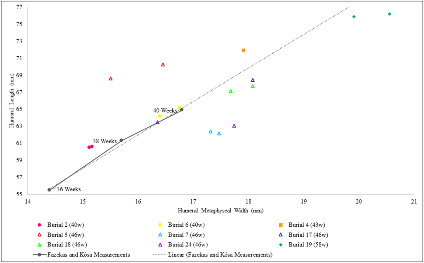
Humeral diaphyseal length plotted against humeral metaphyseal width for those individuals with both measurements and tooth age estimations available. The solid grey line indicates the reference measurements given for individuals between 36 and 40 gestational weeks by Fazekas and Kósa (Reference Fazekas and Kósa1978), with the dashed grey line indicating a linear trend line for these measurements. Of the nine individuals plotted on the graph only Burial 6 appears to have humeral measurements similar to those of the reference measurements for the corresponding age.

The primary limitation of using these various methods is that their results rely on different study populations. This means that the Piddington individuals have been aged dentally in comparison with European nineteenth- and twentieth-century individuals and aged skeletally in comparison to European twentieth-century individuals. This range of comparative material means that differences between age estimates and variation from the reference data may be expected. However, evidence from living populations suggests that early development and growth is relatively similar between populations, as long as the environment is optimal.Footnote 37 Consequently, differences between growth and age estimates identified in such young individuals are considered to be indicative of health stress.Footnote 38
Assessment of the presence and characteristics of pathological lesions was undertaken macroscopically. The location of the pathological change, its extent, severity and nature (bone formation/destruction) and the type of new bone formed (woven/lamellar; pitted/striated/spiculated)Footnote 39 were all documented descriptively and photographically. Pathological changes to long bones, and the ectocraniumFootnote 40 and endocranium,Footnote 41 were recorded in detail. The identification and assessment of pathological indicators in these very young individuals provides further challenges and limitations; distinguishing between normal growth and pathological new bone formation has been, and remains, an ongoing area of discussion.Footnote 42 Bone has a limited response to health stress: it can either form new bone or resorb bone. When bone forms in response to infection or dietary deficiency, it is sometimes difficult to distinguish it from normal bone growth, which is constant and rapid in perinatal and infant individuals.Footnote 43 While caution must be exercised when recording pathology in such young individuals, the findings of this study suggest that severe pathological bone growth can be differentiated from normal growth processes on the remains from Piddington. The author has, to date, analysed the perinatal remains of 434 individuals and thus the pathologies observed at Piddington are contextualised against an extensive knowledge of the appearance of normal perinatal growth. The pathological lesions from Piddington were also examined by another experienced bioarchaeologist.Footnote 44
RESULTS
fig. 6 indicates that all age estimations derived from tooth development are older than those estimated from metric assessment. The degree to which the age estimates differ is variable; eight of the individuals have at least a four-week difference between their tooth and metric age estimations, with a difference of 15 weeks for Burial 19. Even when taking into consideration a margin of error for these methods, there is still a considerable discrepancy between some of the age estimations given. These results suggest that over half of the Piddington individuals may have experienced growth disruption. Burials 12A and 12B have been excluded from analysis due to commingling of the dental and skeletal remains.
Analysis of femoral and humeral diaphyseal length measurements against reference data, calculated using Scheuer et al.,Footnote 45 supports the suggestion that growth was disrupted. Using the linear equations given for femoral and humeral diaphyseal lengths, reference measurements have been plotted to indicate the expected length of these long bones for individuals aged between 36 and 46 gestational weeks. Error bars have been given for these measurements, +/− 2.08 mm for the femora and +/− 2.33 mm for the humeri. Diaphyseal length measurements recorded from the Piddington individuals have been plotted in accordance with their dental age. Error bars have been included for the dental age estimations of the Piddington individuals in accordance with those stated in AlQahtani et al.Footnote 46
figs 7 and 8 demonstrate that all the individuals from Piddington with dental age estimates available fall considerably below the expected attainment of long-bone diaphyseal length growth, if the dental age is presumed accurate. However, when the error levels of both the dental age estimates and the diaphyseal length measurements are considered, only two individuals, for both the femoral and humeral analysis, can be interpreted as showing significant differences between dental and skeletal estimates. Although these may be the only two individuals that can be definitively identified as showing growth disruption, a couple of other individuals, estimated dentally to be 46 weeks of age, intercept the reference measurements at the tail end of their error bars, also suggesting growth disruption.
figs 9i and 9ii compare diaphyseal length and metaphyseal width for the femora and humeri. These measurements have been plotted against reference measurements for an individual of 36, 38 and 40 gestational weeks.Footnote 47 The symbol used to plot each of the individuals is representative of their tooth-age estimate.Footnote 48 This enables consideration of where the individuals would be expected to be approximately plotted; if dental and skeletal age estimates were the same, the individuals should align closely with the reference data plotted. Two points have been plotted for some of the burials, this is due to both left and right measurements being available for recording.
Both figs 9i and 9ii display similar patterns of growth disruption for the femora and humeri. Burials 2 and 21 align closely with the reference measurements for the femora. By contrast the femoral measurements of Burials 4, 5, 7 and 24 indicate that growth has been disrupted. Burials 5, 7 and 24 are all claimed to be 46 weeks of gestational age, yet there is around a 12 mm difference between minimum and maximum femoral length of these individuals and 4.5 mm difference between minimum and maximum metaphyseal width. Both Burial 7 and Burial 24 have femoral lengths well below what is expected, whereas Burial 5 has femoral length measurements more consistent with a 46-week-old individual, yet has metaphyseal measurements which fall on and below the 38-week reference point.
For the humeral measurements there are similar patterns of growth disruption, with Burials 7, 17, 18 and the left humerus of Burial 24 again failing to attain their expected diaphyseal lengths. The right humerus of Burial 24, however, aligns with the reference measurements, despite falling below what would be expected of a 46-week-old individual. This bilateral difference is also mirrored in the femoral measurements. A greater discrepancy between the bilateral measurements can be seen in Burial 24, although Burial 5 also appears to show bilateral differences in measurements. This may indicate that not only is growth being disrupted between dimensions but also between sides of the body.
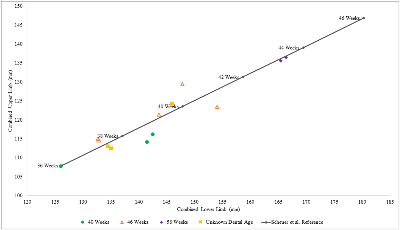
Overall, there is a greater range of growth disruption present in the femora than in the humeri. Interestingly, the femora and humeri of the same individual are not always disrupted correspondingly or to the same degree, suggesting that growth disruption is not equal throughout the skeleton and that individual elements can be affected in isolation. To consider differences between upper and lower limbs further, total lower limb length (femoral + tibial diaphyseal length) was plotted against total upper limb length (humeral + radial diaphyseal length). Reference measurements were also plotted for comparison. Eight individuals from Piddington had all four skeletal elements from one side available for assessment, with five individuals having both left and right elements present. Results (fig. 10) suggest that growth to the lower limb is being disrupted more with the average difference between Piddington and reference measurements being greater in the lower limb. However, statistical analysis revealed no significant difference for the upper or lower limb combined measurements for those aged 40 gestational weeks when compared to the reference measurements. For those aged 46 weeks, however, a significant difference was found for both the upper and lower limb combined totals when compared to the 46-week reference measurement. No statistical analysis could be undertaken of Burial 19 (58 gestational weeks based on dental development) as no reference standard exists for this age.
Graph displaying total upper limb length plotted against total lower limb length, with reference measurements plotted for comparison (Scheuer et al. Reference Scheuer, Musgrave and Evans1980). Many of the combined measurements for the Piddington individuals plot close to the reference measurements, indicating that femoral and humeral growth is typically consistent. However, when taking dental age estimates into consideration, many of these individuals have measurements which fall significantly below those expected. This suggests that growth is disrupted but equivalently between upper and lower limbs.

Metric assessment of cranial bones indicated that growth disruption was not limited to the limb bones. fig. 11 shows that there is extensive growth disruption to the pars basilaris in the majority of the individuals, despite this typically being the most reliable cranial element for estimating age-at-death.Footnote 49 The pars basilaris of Burial 24 aligns closely with the reference measurements of Fazekas and Kósa,Footnote 50 but suggests an age estimate over six weeks younger than that generated from the tooth development. Burial 2 displays the most anomalous measurement, with the length of this element substantially larger than what is typical of a 40-week-old individual. Burial 6, has a width measurement far beyond that expected, yet almost exactly aligns with the reference measurement in terms of length. In contrast, Burials 4, 7 and 18 all have measurements which fall short, both in terms of length and width, for their age estimates; these burials have length measurements suggestive of 38 weeks or less meaning there is a five-week difference between age estimates for Burial 4 and at least an eight-week difference for Burials 7 and 18.
The pars basilaris sagittal length plotted against pars basilaris width for those individuals with both measurements and tooth age estimations available. The solid grey line indicates the reference measurements for individuals between 36 and 40 gestational weeks based on those provided in Fazekas and Kósa (Reference Fazekas and Kósa1978), with the dashed grey line indicating a linear trend line for these measurements. It can be seen that seven out of eight of the individuals plotted on the graph display growth disruption with only measurements from Burial 24 appearing to align with those of the reference measurements.

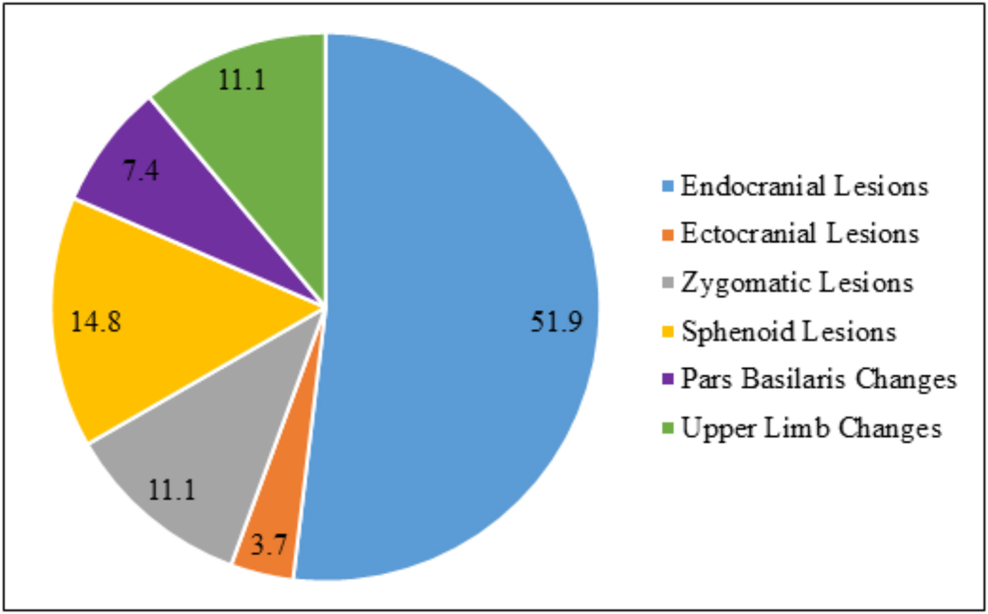
Pathological lesions were identified on 14 of the perinates. While these lesions varied in location (fig. 12), severity and prevalence, most lesions were found to be cranial. Overall, 14 out of 17 perinates demonstrated new bone formation on the endocranial surface of the cranial bones. The three remaining burials had no cranial elements to assess. The most commonly affected cranial element was found to be the endocranial surface of the frontal bones (11 individuals), followed by the occipital squama (9 individuals) and then the parietal bones (8 individuals).
The percentage of pathological lesions identified on the perinatal individuals. The chart clearly shows that endocranial lesions were the most commonly found lesions.

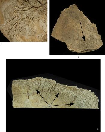
Both wovenFootnote 51 (active) and lamellar (healed) new bone formation was identified on the cranial bones of all 14 perinates with endocranial lesions. In those individuals where lamellar bone is present the bone often appeared as a plateau, raised above the endocranial surface and with a ‘web-like’ appearance (fig. 13i). Many of these lesions, found particularly on the frontal and parietal bones, also showed vascular impressions running through the new bone deposits. On the occipital squama woven bone was more typically present. These deposits were more porous in nature and often appeared to be in association with vascular activity; all the lesions were found within the sagittal and transverse sulci of this element (fig. 13ii). A further type of bone deposit was also identified at the sutural edges of the cranial vault elements. These bone deposits were irregular in form, with lattice-like organisation and were identified primarily as lamellar bone (fig. 13iii).
(i). Photograph of frontal bone fragment displaying the raised surface, multiple layers, as well as the grey hue, of the endocranial bone deposits. The ‘grooves’ are considered to be a result of meningeal vessels. (ii). Photograph of occipital bone, where the deposit (seen centre) appears more porous, and is located around and radiating from a foramen, which would have contained a vascular structure. (iii). Photograph of a frontal bone fragment showing endocranial deposits of bone at the sutural edge, which appear more irregular.

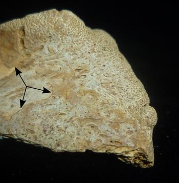
Ectocranial surface changes were only identified in Burial 6. The characteristics of the lesions were similar to those found on the endocranial surface, appearing raised from the original cortical surface and ‘web-like’ in character (fig. 14). More than one layer of bone was deposited, suggesting a recurrent bone response and healing.
Photograph of a frontal bone where the ectocranial deposits can be seen as the ‘web-like’ covering of grey-coloured bone towards the lateral and orbital edge of the fragment (indicated by arrows). The original endocranial surface, seen partially on the far left-hand side, is differentiated by its colour and surface level.

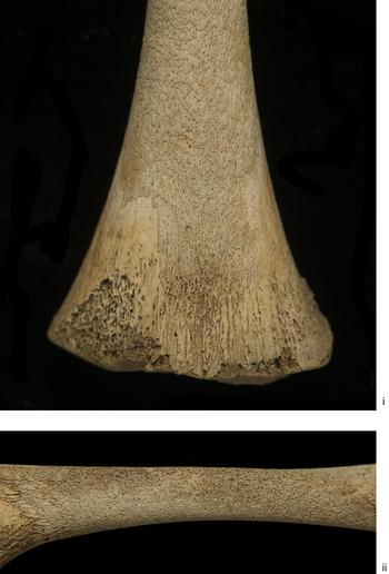
Pathological changes were also identified on the limb bones, particularly those of the lower limb where new bone formation was identified on eight of the perinates. The tibiae were the most commonly affected of the long bones and displayed additional layers of porous, woven new bone formation (figs 15i and 15ii). These were distinct additional layers of bone on top of the original cortical surface which gave them an overall ‘thickened’ morphology.
(i). Photograph of the right femur of Burial 24. This image supports evidence of pathological lesions identified on the long bones from Piddington, showing new bone formation to the anterior (front) of the femur laying over the cortical surface. (ii). Photograph of the left tibia of Burial 24 displaying evidence of pathological new bone formation. The new bone can be seen to once again overlie the original cortical surface, forming multiple layers and giving a very porous appearance. There is also discolouration to this area, with the new bone formation appearing more grey in nature.

DISCUSSION
Pathological and metric assessment indicates that the individuals from Piddington experienced severe episodes of poor health that resulted in growth disruption. Despite the limitations of existing methods of assessment, over 80 per cent of the individuals showed evidence of pathology and many have measurements that are inconsistent with growth standards. A correlation between growth and health disruption was identified; health stress, affecting overall well-being, is likely the major factor affecting normal growth.
Metabolic stress (malnutrition) is the most probable cause of the growth and health disruption identified. The lack or limited intake of various nutrients and vitamins can cause both acute and chronic changes in the human skeleton. Vitamin C and D deficiencies can affect growth and development of the skeleton; lack of vitamin C results in defective osteoid formation (development of new bone cells),Footnote 52 while vitamin D deficiency affects the calcification of bone.Footnote 53 Enlarged or widened and porous metaphyses, identified in some of the individuals (figs 9i and 9ii), are also an indication of vitamin D deficiency, in which cartilage at the growth plates does not mineralise properly.Footnote 54 Metaphyseal changes can also indicate the presence of vitamin C deficiency as widening at the metaphyses could suggest chronic bleeding at the joints.Footnote 55 Lack of vitamin C can result in blood vessels becoming fragile and easily ruptured.Footnote 56 This can lead to haemorrhaging, including in the vessels supplying the metaphyses, but particularly within the cranium.Footnote 57 The Piddington perinates exhibited evidence of endocranial lesions, suggestive of cranial haemorrhagingFootnote 58 and chronic bleeding.Footnote 59 Lesions associated with cranial haemorrhaging are typically found around meningeal grooves, on the frontal bones and at the cruciate eminence of the occipital bone, where they present a web-like appearance.Footnote 60 Although their exact aetiology is debated,Footnote 61 other possible causes are meningitis, anaemia and venous drainage disorders.Footnote 62
The presence of these pathological lesions in such young individuals is suggestive of a poor intrauterine environment. Maternal malnutrition, whether as a result of limited access to food sources or infection, has been identified as causing and exacerbating conditions such as anaemia and maternal haemorrhage and is known to restrict fetal growth and development.Footnote 63 Critically, these conditions can often lead to more unfortunate outcomes such as low birth weight, preterm birth and even birth defects.Footnote 64 Although at Piddington no adult remains have been found within the Iron Age or Roman contexts, disruption to perinatal/infant growth and health indicates that the mother was facing adverse conditions. There is a constant interaction between the fetus, placenta and mother,Footnote 65 with the mother and placenta typically acting as barriers and regulators, ensuring that the fetus is receiving the highest level of nutrition that the mother can provide.Footnote 66 When this nutrient transfer is disrupted, in particular during periods of nutritional deficiency experienced by the mother, these deficiencies become expressed in the fetus. The fetus will be unable to accumulate any stores of nutrients such as vitamin C, vitamin D, calcium and iron, which are vital in the initial stages of life outside the womb.Footnote 67 Therefore, the indication of metabolic stress presented on the skeletal remains of perinatal/infant individuals suggests that maternal health and nutrition was reduced for a sustained period of time so that the mother could no longer act as an effective barrier to protect them. This is corroborated by the fact that vitamin C deficiency is considered to be skeletally identifiable only after at least six months of the vitamin being absent or reduced in the diet.Footnote 68 Therefore, evidence from the Piddington remains suggests either chronic under-nutrition, lack of fresh fruits and vegetables or vitamin C absorption was being prevented due to maternal illness and infection. Parasitic infections are known to cause chronic disease but can often manifest as a nutritional deficiency,Footnote 69 causing severe changes in fetal development, as the parasite can prevent nutrients and vitamins being absorbed by the mother. Furthermore, nutritional deficiency is also known to cause intrauterine growth restriction (IUGR).Footnote 70
The predisposition of fetuses to such deficiencies can have long-term implications for health and disease.Footnote 71 Many significant studies into the long-term health consequences of a poor in-utero environment have revealed that major health problems in older adulthood are a result of reduced circumstances during growth and development, in both the pre-natal and post-natal period.Footnote 72 From the Developmental Origins of Health and Disease hypothesis (DOHaD)Footnote 73 it is known that gene expression can alter in response to a variety of stressors.Footnote 74 Were the mothers from Piddington predisposed to a reduced health status as a result of their own in-utero development and the life course of their own mothers? Epidemiological studies have suggested a link between IUGR and chronic metabolic disease in both children and adults.Footnote 75 As a result, the chronic metabolic stress inferred from the perinatal remains may be a result of long-term, multi-generational exposure to stress, in particular under-nutrition. Furthermore, studies have demonstrated that there was a marked reduction in health during the transition from Iron Age to Roman life, particularly within rural settlements.Footnote 76 Consequently, the health stress identified at Piddington must be considered within the changing landscape of Britain and might in fact be representative of a population transitioning from an Iron Age to a Romanised lifestyle. In particular, an increase in metabolic stress, vitamin C and D deficiency, during the Iron Age to Roman transition was identified in Dorset.Footnote 77 This again may suggest that the pathological lesions identified on the Piddington remains, thought to show evidence of metabolic stress, may demonstrate a similar pattern, and may even be expected for such a population.
Metric assessment of the Piddington burials also shows clear disruption to growth in some individuals, with the suggestion that others were also likely affected. The lower limbs were found to have a bigger difference to the reference population, in comparison with the upper limbs, suggesting growth was more disrupted in the femora and tibiae. However, the tibiae are known to be the most variable and susceptible to growth disruption when exposed to stress.Footnote 78 Therefore, these greater differences in the lower limbs may be as a result of greater variation within the tibial measurements. However, it must also be considered that during fetal and infant development, 87 per cent of growth is prioritised to the cranium.Footnote 79 Consequently, the fact that all limbs individually and combined show disruption, may be a result of a general prioritisation of resources towards cranial, rather than infracranial growth. In addition, if the pathological lesions and the illness/conditions these individuals may have been facing are also considered, general disruption to growth is expected. Very little difference in bilateral measurements was observed. However, Burial 24 does show the most extreme differences in measurements and, interestingly, this individual does have some of the more severe pathology. This asymmetry could be considered to be a further indicator of growth disruption.Footnote 80
Multiple possibilities for the cause of endemic and long-term health stress must be considered. There is a complicated relationship between the health stress and the pathological lesions identified, and the social and physical environment that these perinates, infants and their mothers experienced.Footnote 81 As Gowland states,Footnote 82 cultural practice can affect the exposure and response to health stress within and between populations, meaning that depending on who you are within a society, you may be differentially exposed to both health risks and treatment. Therefore, understanding the pathological lesions and growth changes identified on the perinates and infants from Piddington pivots around a discussion of who these mothers were, their social status and their position within Iron Age and Roman society. With no adult remains having yet been excavated, it is impossible to understand the true dynamic of the community at Piddington, or why maternal health appears to have been so compromised. However, consideration as to whether these mothers were young or old, high or low status, native Iron Age peoples or ‘Roman’ enables reflection on some of the various factors that may have affected perinatal, infant and maternal life at Piddington.
In both Iron Age and Roman communities in Britain, many people would have been present and working in the settlement in addition to the inhabitants and owners. Servants and slaves were an integral part of the social organisation of Iron Age and Romano-British communities, particularly on agricultural and villa estates.Footnote 83 WebsterFootnote 84 suggests that positions of servitude and slavery were common among these communities and, even though little archaeological evidence has been found to substantiate this, it is commonly accepted that there was a large network in the trading of people and slaves within Britain before the Roman conquest. Therefore, it is plausible that these individuals were present at Piddington; if these perinatal and infant remains are those of the lesser, lower ranks of women living and working on site, this may explain the high levels of pathological conditions being identified. The extensive evidence of metabolic stress may indicate limited access to certain food types, or even that these women were engaged in work that involved limited exposure to sunlight (vitamin D). Social inequality is known to increase susceptibility to poor health and disease.Footnote 85 Thus, a reduced social status would mean a limited access to high-quality food, harsher working and living environments and limited access to medical care. However, these perinatal and infant remains may equally be those of the high-status women at the site. If this is the case, it suggests that maternal health was reduced regardless of social position. This may indicate a more general limited availability of food, some form of endemic infection, or even cultural practices of pregnant mothers such as restrictive diets and avoidance of sunlight.
Furthermore, it must also be considered whether these women and mothers were native Iron Age people or ‘Romans’. As these burials are from the first century a.d. it must be remembered that this is a period of great transition, especially at Piddington, where the influx of the Roman army would potentially have led to wide-scale social and cultural changes.Footnote 86 From modern clinical studies, it has been identified that any form of stress, be that physical, mental, social or cultural, can lead to growth disruption.Footnote 87 Thus, the pathological lesions identified might be a result of the mother experiencing a multitude of stressors simultaneously.
Regardless of social status, working or living conditions, it is vital to consider the precarious nature of pregnancy for any mother, even today. Today, stressors experienced by the mother and developing child alike during the in-utero period are known to cause a multitude of both short-term and long-term consequences to the health of the individual.Footnote 88 Lifestyle factors are primarily the cause of growth disruption, changes to gene expression and overall well-being. However, the fragility of fetal life and growth remains even today.Footnote 89 The evidence indicates that these 17 individuals from Piddington represent ‘natural’ deaths. It is likely that there were high mortality rates among young infants in Iron Age and Romano-British contexts;Footnote 90 estimates suggest between 20 and 40 per cent of infants would not survive to one year of age.Footnote 91 With a prolonged reduction in health status during in-utero life, the likelihood of survival after birth is decreased further, especially if the mother was weakened from the birth process, unable to feed the child or potentially died herself in childbirth. Paradoxically, the individuals from Piddington, although showing extensive pathological lesions, also show signs in many cases of healing, or a recurrent response by the bone, indicating multiple episodes of poor health. Although this indicates greatly reduced maternal health status, it also suggests that the perinatal and infant individuals, as well as the mothers, were able to endure health stresses.
The mortality of these perinatal individuals from Piddington must ultimately be considered in terms of poor maternal health. Such severe pathological lesions and irregular growth patterns indicate that maternal health greatly affected the lives of these perinatal individuals and ultimately led to their premature deaths. With such high rates of infant mortality, death was a common experience within such societies; that is not to say that it was expected or attitudes towards death were indifferent, but simply death was a part of life and consequently evidence of infant death is not surprising. This likelihood of death combined with evidence of such chronic and endemic health reduction suggests that interpretations of infanticide at Piddington are unlikely. The burial of perinates and infants within a domestic setting, and not within a formal cemetery, is a common practice within Iron Age and Roman contexts.Footnote 92 The burial of infants close to, within and under settlements and dwellings has been argued by some to show deviant, hidden burial, suggestive of ‘foul play’ and the deliberate killing of individuals based on sex or disability.Footnote 93 However, many scholars have rejected these notions, instead considering the ‘spiritual’ infant and its place within the community.Footnote 94 Burial of infants within the domestic context may be nothing more than simply pragmatic. It is known that infants were afforded different burial ‘rites’ to those of older children and adults; perinates and young infants were often not considered to be a part of the community until a certain age threshold had been attained.Footnote 95 The burial rites of these perinates at Piddington suggest careful placement of the individuals in many instances (seen in fig. 3).
CONCLUSION
Detailed analysis of the perinatal burials from Piddington reveals a wealth of biological and pathological information. This evidence is for understanding these young deaths, not simply in terms of casual or careless disposal, but as providing insights into the unpredictable and tough lives of expectant mothers 2,000 years ago. Metric and pathological assessment suggests evidence of disruption to growth and health in these individuals. The implications of these findings indicate intrauterine life and survival was precarious, suggesting that the health status of the mothers was greatly reduced and ultimately led to the shortened lives of these individuals. Therefore, it can be suggested that there is no indication of deliberate killing, thoughtless disposal or lack of care surrounding these burials. These individuals instead died weakened and ill, nothing more than the unlucky victims of chronic suffering.
ACKNOWLEDGEMENTS
I wish to thank Dr Becky Gowland and Professor Charlotte Roberts for their guidance and constructive comments on an earlier version of this article. I also wish to thank Professor Christopher Knüsel who inspired and developed my interest in infant burials and who supervised the initial data collection for this project. I would like to warmly thank Roy and Liz Friendship-Taylor, site directors at Piddington Excavation, for their constant support, interest and help with this research and for allowing me to work with the Piddington perinates. I also wish to thank the AHRC for funding this research (Grant Number: AH/K502996/1).