Introduction
Anania hortulata (Linnaeus, Reference Linnaeus1758) (Lepidoptera: Crambidae: Pyraustinae), commonly called the magpie moth with its black-and-white patterned wings and contrasting yellowish-orange body, is one of the most common and widespread species of the subfamily Pyraustinae. It is superficially distinct enough that it has long been placed in its own genus, Eurrhypara Hübner, 1825, which is now regarded as a subgenus of Anania Hübner, 1823 (Leraut Reference Leraut2005; Tränker et al. Reference Tränker, Li and Nuss2009). It is widespread from Europe to far-east Russia (Ganev Reference Ganev, Karsholt and Razowski1996; Sinev Reference Sinev and Sinev2008; Slamka Reference Slamka2013; Nuss et al. Reference Nuss, Landry, Mally, Vegliante, Tränkner and Bauer2017). It has been introduced in North America where it occurs in the East (it was first discovered in Nova Scotia in 1907) and on the west coast (with a more recent introduction in British Columbia in 1977) (Gillespie and Gillespie Reference Gillespie and Gillespie1982). Munroe (Reference Munroe, Dominick, Ferguson, Franclemont, Hodges, Munroe and Classey1976) has stated that this species was confined to the mainland of the northern parts of temperate East Asia and not represented in Japan. However, it has been reported from China, Japan, and Korea since the 1980s (Wang Reference Wang1980; Inoue Reference Inoue, Inoue, Sugi, Kuroko, Moriuti and Kawabe1982; Bae et al. Reference Bae, Byun and Paek2008). Atay and Can (Reference Atay and Can2007) have redescribed the external and genital morphology of the male based on material from Turkey. Larvae feed on several different host plants on which they gregariously spin or roll leaves. Although nettle (Urtica Linnaeus; Urticaceae) seems to be the main host, it has also been recorded from Lamiaceae (Mentha Linnaeus, Stachys Linnaeus), Convolvulus Linnaeus (Convolvulaceae), Ribes Linnaeus (Grossulariaceae), and others (Slamka Reference Slamka2013).
Taxon delineation in species complexes in Lepidoptera may underestimate the number of species when based solely on morphological features (Hebert et al. Reference Hebert, Penton, Burns, Janzen and Hallwachs2004; Mutanen et al. Reference Mutanen, Aarvik, Landry, Segerer and Karsholt2012, Reference Mutanen, Kekkonen, Prosser, Hebert and Kaila2015; Kozlov et al. Reference Kozlov, Mutanen, Lee and Huemer2017). Two common problems may occur when using morphological characters alone as proxies to define biological species. First, the process of speciation is not always accompanied with character change at all levels in morphology, and the rates of character change can vary. Second, biological species are not easily tested and clarified because of the possibility of incongruence among lines of evidence (Padial and De La Riva Reference Padial and De La Riva2010). Integrative taxonomy is becoming a widely accepted and reliable approach of species delineation to extract convincing evidence and diagnoses of the differences among the closely related and cryptic species. As part of integrative taxonomy, many recent studies in Lepidoptera have discussed the power of DNA-based approach as an effective way to assist in discovering previously unrecognised cryptic species (Dincă et al. Reference Dincă, Lukhtanov, Talavera and Vila2011; Huemer and Mutanen Reference Huemer and Mutanen2012; Mutanen et al. Reference Mutanen, Aarvik, Landry, Segerer and Karsholt2012, Reference Mutanen, Kekkonen, Prosser, Hebert and Kaila2015; van Nieukerken et al. Reference van Nieukerken, Doorenweerd, Stokvis and Groenenberg2012; Yang et al. Reference Yang, Landry, Handfield, Zhang, Solis and Handfield2012; Huemer et al. Reference Huemer, Elsner and Karsholt2013, Reference Huemer, Karsholt and Mutanen2014; Landry and Hebert Reference Landry and Hebert2013; Kozlov et al. Reference Kozlov, Mutanen, Lee and Huemer2017).
During the assemblage of the DNA barcode reference library of the subfamily Pyraustinae from North America and China, we found that the specimens of A. hortulata were divided into two deeply divergent haplotype clusters corresponding to two different geographic groups, Europe and North America versus China. Initially, no obvious differences in external morphology were detectable between the two haplotype groups. However, a detailed examination of genitalia revealed subtle differences in both sexes that corresponded to the barcode–geographic split and suggested that the specimens from China represented an overlooked, undescribed species.
In this paper we describe this new species, provide comparative illustrations to differentiate it from A. hortulata, present strong evidence from both morphometric and genetic analyses that support the taxonomic recognition of two species, and provide an estimate of possible divergence time.
Materials and methods
Sampling
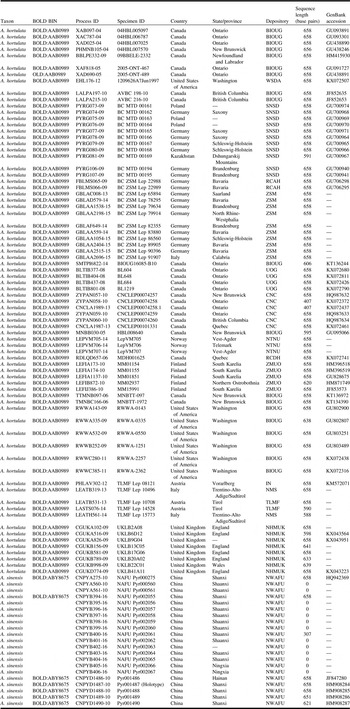
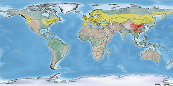
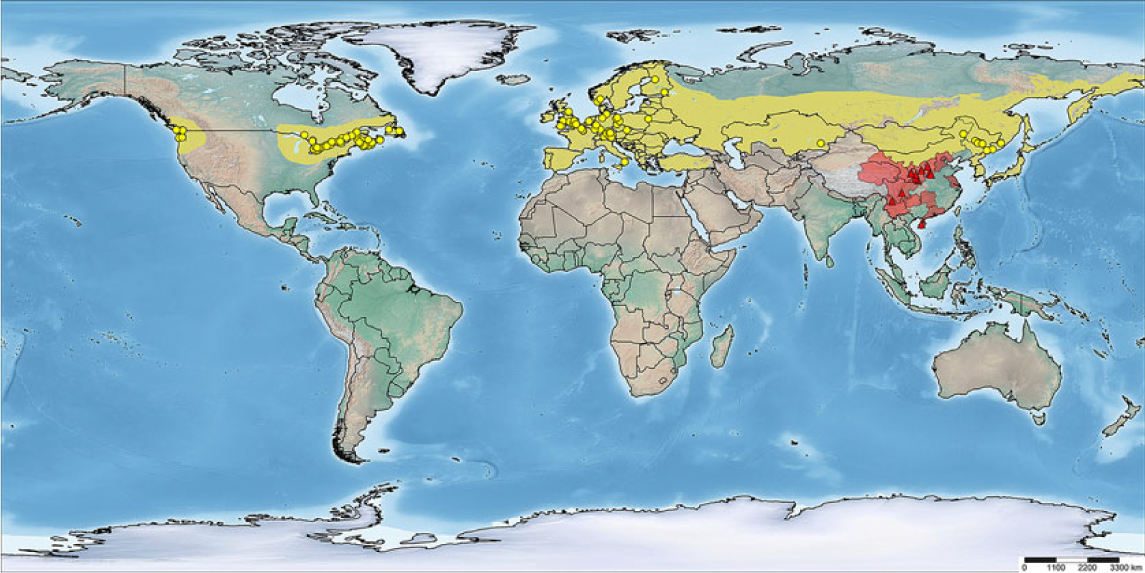
For molecular analysis, Sikes sequences were obtained from 96 specimens sourced from various depositories (Table 1): 43 specimens from Europe (Austria, Finland, Germany, Italy, Norway, Poland, United Kingdom), 31 from North America (Canada, United States of America), a single specimen from Central Asia (Kazakhstan), and 21 from China. In addition, we also included 21 specimens from Canada, The Netherlands, and the United Kingdom (deposited in the Canadian National Collection of Insects, Arachnids, and Nematodes, Ottawa, Ontario, Canada) and two specimens from China (deposited in the National Museum of Natural History, Smithsonian Institution, Washington, District of Columbia, United States of America) to complement the morphological analysis. Specimens of A. hortulata were identified by expert curators from where the material was obtained and verified by us for accuracy using the taxonomic accounts of Leraut (Reference Leraut2005) and Tränker et al. (Reference Tränker, Li and Nuss2009). No other known species of Anania, other than the newly discovered cryptic one described below, resembles A. hortulata, so the possibility of confusion with some other taxa was ruled out. To illustrate the global distributional range of these two allopatric species (Fig. 1), geographical coordinates were extracted from more than 337 specimens from Asia, Europe, and North America (mainly deposited in the Canadian National Collection of Insects, Arachnids, and Nematodes; Northwest A&F University, Yangling, China; and National Museum of Natural History, Smithsonian Institution, including those barcoded voucher depositories). Distributional data were also extracted from Fauna Europaea (Karsholt and van Nieukerken Reference Karsholt and van Nieukerken2013) and previous studies (Wang Reference Wang1980; Inoue Reference Inoue, Inoue, Sugi, Kuroko, Moriuti and Kawabe1982; Xu et al. Reference Xu, Lu, Yuan and Kang1999; Atay and Can Reference Atay and Can2007; Bae et al. Reference Bae, Byun and Paek2008; Li et al. Reference Li, Ren, Zhang, Du, Li and You2009, Reference Li, Wang, Zhen, Zhang, Hu and Jing2012; Chen et al. Reference Chen, Wu, Xing and Wu2013; Slamka Reference Slamka2013). A distribution map was generated using the online SimpleMappr software (Shorthouse Reference Shorthouse2010).
Sample information for the sequenced specimens of Anania hortulata species complex.

Distribution of collection localities of Anania hortulata species complex in this study: yellow circles = A. hortulata; red triangles = A. sinensis.
Morphometric analysis
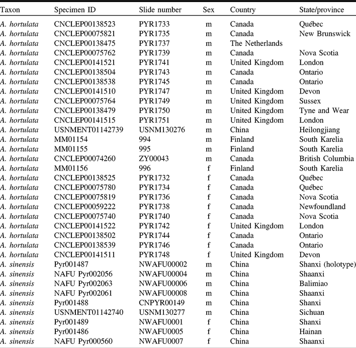
A total of 13 females and 21 males were dissected for morphological analysis (Table 2). Protocols for the preparation and photography of genitalic slides followed Landry (Reference Landry2007), whereas genital terminology followed Munroe (Reference Munroe, Dominick, Ferguson, Franclemont, Hodges, Munroe and Classey1976) and Kristensen (Reference Kristensen and Kristensen2003). Variation in genital features was assessed with quantitative multivariate analyses following Yang et al. (Reference Yang, Landry, Handfield, Zhang, Solis and Handfield2012). Principal components analysis was used to study the patterns of covariation among the features of female and male genitalia. Discriminant function analysis and canonical analysis were used to determine the degree to which samples could be maximally distinguished. Statistical comparisons were performed with Statistica version 13.0 (www.statsoft.com/Products/STATISTICA-Features). The following measurements were taken (schematics of measurements shown in Yang et al. Reference Yang, Landry, Handfield, Zhang, Solis and Handfield2012, fig. 2): for male genitalia: DUTV, distance from uncus to vinculum; LUNC, length of uncus; LPHA, length of phallus; LSAC, length of sacculus; LVAL, length of valva; PSEL, perimeter of sella; WPHA, width of phallus; WSAC, width of sacculus; WUNC, width of uncus; WVAL, width of valva. For female genitalia: DBUR, diameter of corpus bursae; LAAP, length of anterior apophyses; LLDB, length of lamina ducti bursae; LPAP, length of posterior apophyses; LSIG, length of signum; WSIG, width of signum.
Anania genitalia preparations measured in this study.

DNA extraction, polymerase chain reaction amplification, and sequencing
Genomic DNA was extracted from a single leg from dried specimens using a standard protocol (Ivanova et al. Reference Ivanova, deWaard and Hebert2006). We sequenced one mitochondrial gene (cytochrome c oxidase subunit I, COI) from all specimens and one additional protein-coding nuclear gene (carbamoylphosphate synthase domain protein, CAD) for two paratypes of A. sinensis, new species (GenBank accessions: MH446375, MH446376). CAD sequence from one specimen (MM01851) of A. hortulata was obtained from a previously published study (Mutanen et al. Reference Mutanen, Wahlberg and Kaila2010) and downloaded from GenBank (accession: GU828170.1). Polymerase chain reaction amplification and DNA sequencing followed standard protocols (Wahlberg and Wheat Reference Wahlberg and Wheat2008; deWaard et al. Reference deWaard, Ivanova, Hajibabaei, Hebert and Cristofre2008; Hebert et al. Reference Hebert, deWaard, Zakharov, Prosser, Sones and McKeown2013; Yang et al. Reference Yang, Landry and Hebert2016). Target regions were amplified using the primer sequences and annealing temperatures following the literature (Hebert et al. Reference Hebert, Penton, Burns, Janzen and Hallwachs2004; Hajibabaei et al. Reference Hajibabaei, Janzen, Burns, Hallwachs and Hebert2006; Haines and Rubinoff Reference Haines and Rubinoff2012). Complete specimen information, including images, coordinates, voucher depositories, sequences, trace files, and GenBank accession numbers, is available in the Barcode of Life Data systems (BOLD; www.boldsystems.org) public dataset “DS-ANAN, Anania hortulata complex taxonomy” at https://doi.org/10.5883/DS-ANAN.
Phylogenetic and haplotype analysis
Sequences of mtDNA and nDNA gene were aligned separately with MUSCLE (Edgar Reference Edgar2004), and uncorrected pairwise genetic distances within and among lineages were estimated based on Kimura two-parameter model for base substitution in MEGA 7.0 (Kimura Reference Kimura1980; Kumar et al. Reference Kumar, Stecher and Tamura2016). Bootstrap values were calculated with 1000 replicates (Kimura Reference Kimura1980; Kumar et al. Reference Kumar, Stecher and Tamura2016), and neighbour-joining trees and minimum evolution trees based on distance were built in MEGA software. Anania lancealis (Denis and Schiffermüller Reference Denis and Schiffermüller1775) and Loxostege sticticalis (Linnaeus, Reference Linnaeus1761) were selected as outgroups to root the trees based on COI and CAD sequences, respectively, because the former species is morphologically and genetically the most similar species in the genus, and the latter species was previously proposed to be closely related to Anania by Leraut (Reference Leraut2005). Due to the low number of sequences available for CAD gene, all the following analyses were carried out on the COI sequences only.
The number of haplotypes was calculated with DnaSP 5.10 (Rozas et al. Reference Rozas, Sanchez-Delbarrio, Messeguer and Rozas2003). Phylogenetic analyses were implemented using maximum likelihood and Bayesian inference, using RAXML version 8.2 (Stamatakis Reference Stamatakis2014) and MrBayes 3.1.2 (Ronquist and Huelsenbeck Reference Ronquist and Huelsenbeck2003), respectively. MrModeltest 2.3 (Nylander et al. Reference Nylander, Ronquist, Huelsenbeck and Nieves-Aldrey2004) was used to identify the Akaike information criterion best-fit nucleotide substitution model. For RAxML, the best maximum likelihood tree was calculated from 200 RAxML runs, followed by 1000 bootstrap replicates. In Bayesian analysis, the parameters were set as follows: generations = 1 000 000, samplefreq = 100, nchains = 4, nst = 6. The first 2500 trees were discarded as burn-in, and those remaining were used to estimate posterior parameters and probability distributions (Ronquist and Huelsenbeck Reference Ronquist and Huelsenbeck2003). The convergence among the runs was assessed by checking the potential scale reduction factor values of each parameter in MrBayes and the effective sample size values of each parameter in Tracer version 1.7 (Rambaut et al. Reference Rambaut, Drummond, Xie, Baele and Suchard2018). Median-joining haplotype networks were constructed in Network 4.6 (Bandelt et al. Reference Bandelt, Forster and Röhl1999) to visualise the relationships among haplotypes.
Estimation of divergence time
The analysis of divergence time was estimated using BEAST 1.8.0 (Drummond and Rambaut Reference Drummond and Rambaut2007) based on sequences longer than 600 base pairs. We selected the widely accepted molecular clock of the mtDNA gene (0.0115 per site per million years) (Brower Reference Brower1994) because there is no fossil record or geological evidence available for calibration. We used strict model and substitution mode (TN93; Tamura and Nei Reference Tamura and Nei1993) based on the results of jModelTest 0.1.1 (Guindon and Gascuel Reference Guindon and Gascuel2003; Posada Reference Posada2008) in the analysis. Default priors were used, with the chains run for 20 million generations sampling every 1000 generations. Tracer 1.7 was used to verify the posterior distribution and effective sample sizes from the MCMC output. TreeAnnotator 1.8.0 in the BEAST package was used to summarise trees with “mean height” after we discarded the first 25% as burn-in.
Abbreviations for specimen depositories
BIOUG – Biodiversity Institute of Ontario (now Centre for Biodiversity Genomics), University of Guelph, Guelph, Ontario, Canada
CNC – Canadian National Collection of Insects, Arachnids, and Nematodes, Ottawa, Ontario, Canada
IN – inatura, Dornbirn, Austria
NHMUK – Natural History Museum, London, United Kingdom
NMNH – National Museum of Natural History, Smithsonian Institution, Washington, District of Columbia, United States of America
NMS – Naturmuseum Suedtirol, Bolzano, Italy
NTNU – University Museum, Department of Natural History, Trondheim, Norway
NWAFU – Northwest A&F University, Yangling, China
RCAH – Research collection of Alfred Haslberger, Teisendorf, Germany
RCDH – Research collection of Daniel Handfield, Saint-Mathieu-de-Beloeil, Québec, Canada
SNSD – Senckenberg Natural History Collections Dresden, Museum of Zoology, Dresden, Germany
TLMF – Tiroler Landesmuseum Ferdinandeum, Innsbruck, Austria
WSDA – Washington State Department of Agriculture, Olympia, Washington, United States of America
ZMUO – University of Oulu, Oulu, Finland
ZSM – Zoologische Staatssammlung Muenchen, Munich, Germany
Results
Genetic distance analysis
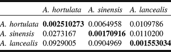
In total, 83 COI sequences were obtained (range 307–658 base pairs; mean 641 base pairs). The pairwise genetic distances within and among the three species of Anania are shown in Table 3. Intraspecific genetic distances ranged from 0.00 to 0.92% (mean 0.25%), whereas interspecific divergences among congeners varied from 2.19 to 12.04% (mean 5.25%). On average, the mean genetic distance among the species was 10 times higher than that within species. The interspecific distance between A. hortulata and A. sinensis was 2.73%, whereas the intraspecific divergence was 0.25 and 0.17%, respectively, and the distance to the outgroup A. lancealis was over 9%. For nDNA gene, the alignment of the three recovered CAD sequences of two Anania species was 615 base pairs long. The mean interspecific distance between A. hortulata and A. sinensis was 17.4%, indicating that nuclear CAD gene would be helpful in resolving the status and relationships of closely related species due to having much greater genetic distance than COI distances between species and faster rate of substitution (Wahlberg and Wheat Reference Wahlberg and Wheat2008; Dincă et al. Reference Dincă, Lukhtanov, Talavera and Vila2011).
Kimura two-parameter genetic distances calculated within (in bold) and between species of Anania.

Upper triangle indicates standard error estimates obtained by bootstrap procedure (1000 replicates) as implemented in MEGA 7.0.
Phylogenetic and haplotype analysis
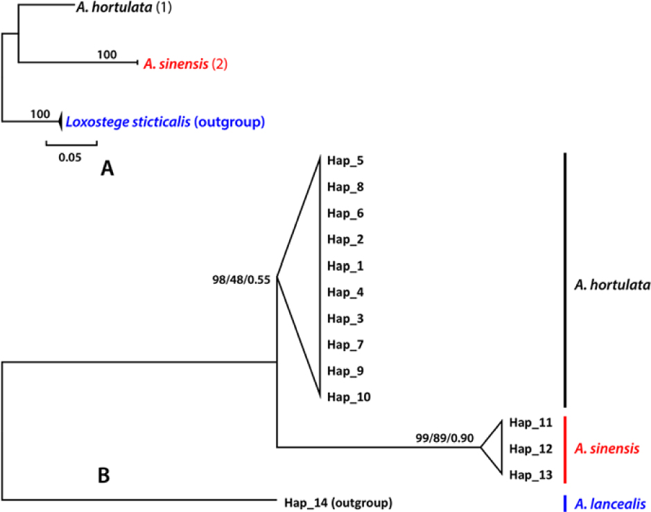
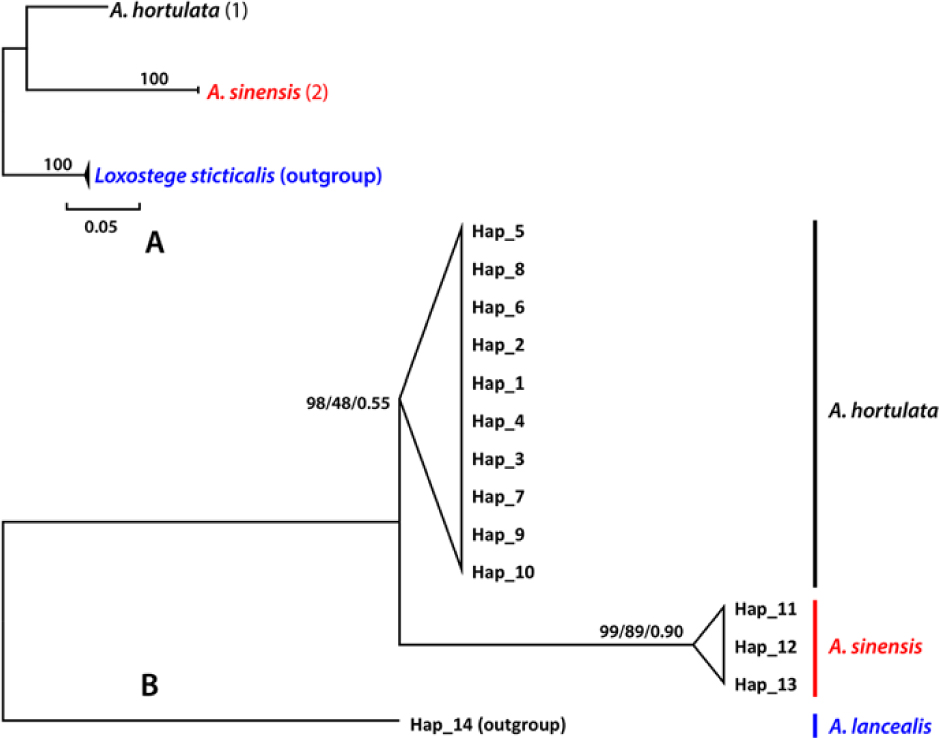
In both minimum evolution and neighbour-joining analyses with CAD (Fig. 2A) and COI based on Kimura two-parameter model, each species within the A. hortulata species complex was recovered as a well-supported monophyletic group. Thirteen different haplotypes were detected among 83 COI sequences, with nine haplotypes from Europe, three haplotypes shared with North America, and one haplotype from Kazakhstan. There were three haplotypes from China. The best-fit model of nucleotide substitution for all haplotype sequences selected was GTR with a relative Akaike information criterion weight of 0.5321. Tracer plots indicated that MrBayes runs reached convergence before 1 000 000 generations (results not shown). Convergence of the analyses was also supported by the average standard deviation of split frequencies (< 0.01), all potential scale reduction factor values close to 1.0 (range between 1.0 and 1.008), and effective sample size values above 200 (with many > 1000) for the post burn-in trees. The results obtained from neighbour-joining, maximum likelihood, and Bayesian inference came to the same topology, and all records were assigned to the same major clades (Fig. 2B).
Anania hortulata species complex molecular analyses. A, Neighbour-joining tree (Kimura two-parameter model) based on three CAD sequences of the two Anania species, rooted with Loxostege sticticalis as the outgroup; B, Majority rule consensus trees based on a Bayesian phylogenetic analyses of 13 COI haplotypes, rooted with Anania lancealis. Node support values: bootstrap neighbour joining/maximum likelihood/Bayesian posterior probabilities.
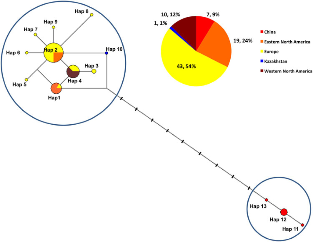
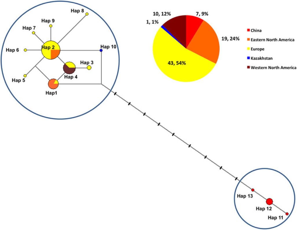
Network analysis results (Fig. 3) showed that Europe (frequency 43; 54%), western North America (frequency 10; 12%), eastern North America (frequency 19; 24%), and Kazakhstan (frequency 1; 1%) clustered together, whereas all haplotypes from China (frequency 7; 9%) were distinctly differentiated in nine variable sites. The topology of the network showed a clear differentiation between the two species, congruent with the phenetic results below. Most notably, there was no haplotype sharing between the two species.
Haplotype networks of Anania hortulata species complex.
Estimation of divergence time
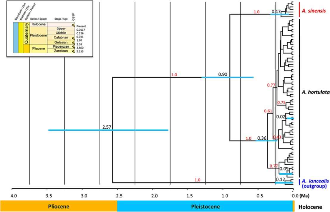
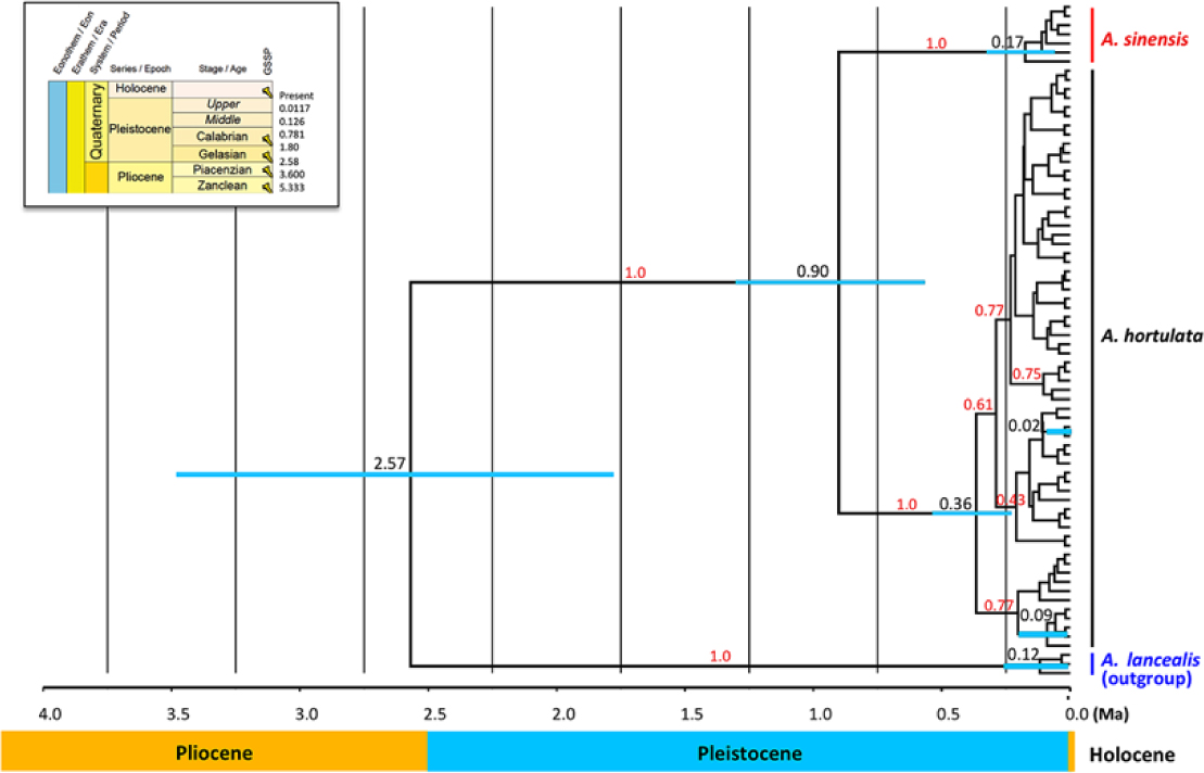
The divergence time (Fig. 4) between A. hortulata and A. sinensis was estimated to be 0.90 million years ago (95% highest posterior density: 0.57–1.31 million years ago), and the divergence time between A. lancealis and A. hortulata plus A. sinensis occurred 2.57 million years ago (95% highest posterior density: 1.78–3.48 million years ago), all during the transition from the early Pleistocene to the mid-Pleistocene.
Divergence times of Anania hortulata species complex estimated using a Bayesian inference analysis of COI sequences with a strict clock model. Thick bars indicate 95% highest probability density intervals of node ages. Red numerals to the left of nodes are posterior probabilities (> 75 shown). Black numerals above blue bars are divergence times. Ma, million years ago; GSSP, global boundary stratotype section and point.
Morphological characters and morphometric analysis
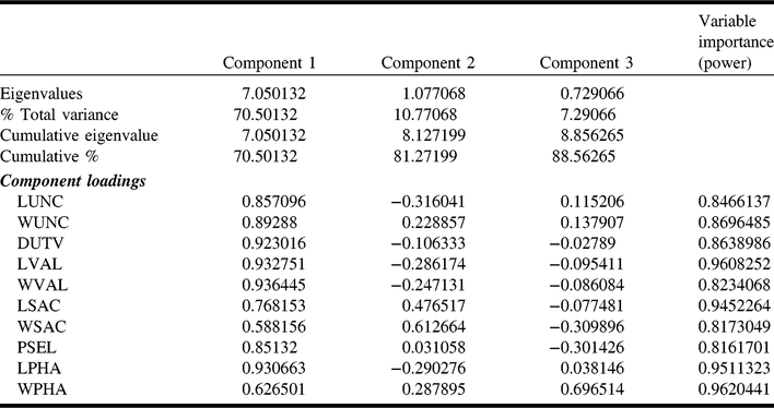
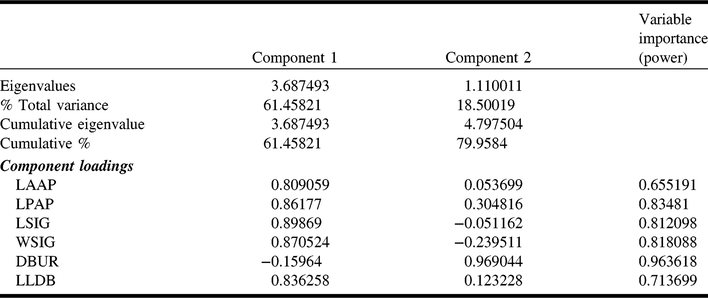
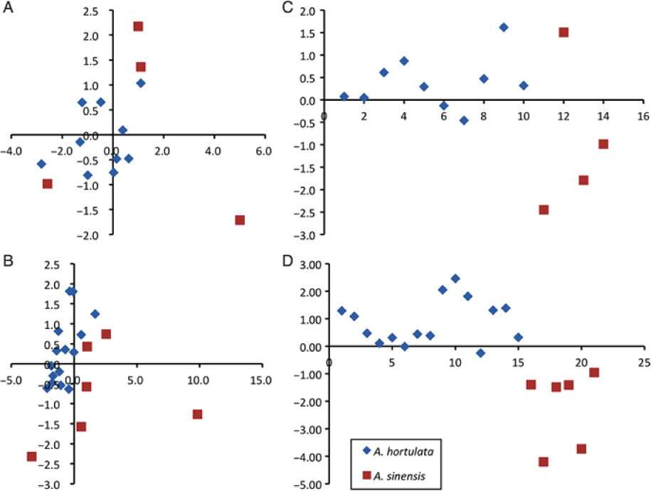
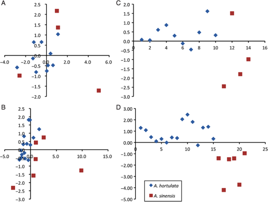
The principle components analysis of female and male genitalia variables is summarised in Tables 4–5. The results show that the first two components of the principle components analysis together explain 79.96 and 81.27% of the total variance for the females and males, respectively. Four female genitalia measures (LAAP, WSIG, LSIG, DBUR) (P > 0.80) and seven male genitalia measures (WUNC, DUTV, LVAL, WVAL, LSAC, LPHA, WPHA) (P > 0.85) were the most important variables. Principle components analyses separated A. hortulata and A. sinensis from each other (Fig. 5A–B). In discriminant function analysis, the most significant diagnostic variables for adult males were WPHA (F = 2.46; P < 0.14) and LSAC (F = 1.53; P < 0.24). For females, the most significant diagnostic variable was WSIG (F = 1.03; P < 0.34). Moreover, the first roots of canonical analysis yielded similar results, separating all specimens into two distinct groups (Fig. 5C–D).
Results of principal components analyses of 10 male genitalia measurements from 21 specimens of the Anania hortulata species complex.

Abbreviations: DUTV, distance from uncus to vinculum; LPHA, length of phallus; LSAC, length of sacculus; LUNC, length of uncus; LVAL, length of valva; PSEL, perimeter of sella; WSAC, width of sacculus; WPHA, width of phallus; WUNC, width of uncus; WVAL, width of valva.
Results of principal components analyses of six female genitalia measurements from 13 specimens of the Anania hortulata species complex.

Abbreviations: DBUR, diameter of corpus bursae; LAAP, length of anterior apophyses; LLDB, length of lamina ducti bursae; LPAP, length of posterior apophyses; LSIG, length of signum; WSIG, width of signum.
Principal components analysis scatter plot comparing variation of the first two principal components for all morphological characters analysed. A, Females, B, males. C–D, Scatter plots of the canonical measures calculated after the discriminant function analysis of morphometric data along the first roots; C, females. D, males.
Taxonomy of Anania hortulata species complex
Anania hortulata (Linnaeus, Reference Linnaeus1758)
(Figs. 6A, C; 7A, C; 8A, C, E, G, I; 9A; 10A, C, E, G, I)
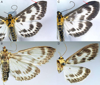
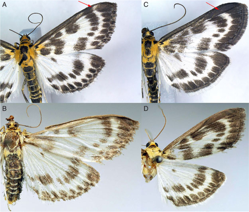
Adults of Anania hortulata species complexes. A, Female of A. hortulata (United Kingdom: London, specimen CNCLEP00141523, forewing length = 13.2 mm). B, Female of A. sinensis (China: Shaanxi, specimen NAFU Pyr002058, forewing length = 15.7 mm). C, Male of A. hortulata (Canada: Québec, specimen CNCLEP00101331, forewing length = 11.3 mm). D, Male of A. sinensis (China: Shanxi, specimen NAFU Pyr002055, forewing length = 14.0 mm). Red arrows indicate female/male difference in forewing tip.
Phalaena (Geometra) hortulata Linnaeus, Reference Linnaeus1758: 529; type locality: Sweden; type locality restricted by Munroe (Reference Munroe, Dominick, Ferguson, Franclemont, Hodges, Munroe and Classey1976: 29).
Geometra urticata Linnaeus, Reference Linnaeus1761: 340; type locality: Sweden.
Eurrhypara hortulata: Pierce and Metcalfe Reference Pierce and Metcalfe1938: 29; Bae et al. Reference Bae, Byun and Paek2008: 101; Li et al. Reference Li, Ren, Zhang, Du, Li and You2009: 211; Li et al. Reference Li, Wang, Zhen, Zhang, Hu and Jing2012: 513; Chen et al. Reference Chen, Wu, Xing and Wu2013: 22.
Eurrhypara (Eurrhypara) hortulata: Hannemann Reference Hannemann and Dahl1964: 340.
Eurrhyparodes hortulata: Wang Reference Wang1980, 21: 123; Inoue Reference Inoue, Inoue, Sugi, Kuroko, Moriuti and Kawabe1982: 359; Xu et al. Reference Xu, Lu, Yuan and Kang1999: 25.
Anania (Eurrhypara) hortulata: Tränker et al. Reference Tränker, Li and Nuss2009; Slamka Reference Slamka2013: 69; Nuss et al. Reference Nuss, Landry, Mally, Vegliante, Tränkner and Bauer2017.
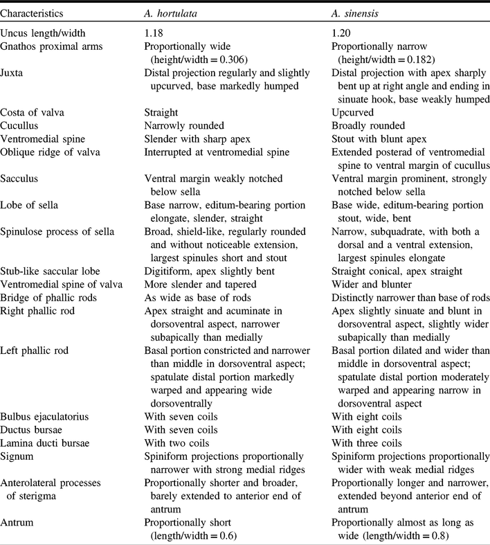
Diagnosis. The combination of orange-yellow head and thorax, yellow-banded black abdomen, and white wings with black spots and bands is highly distinctive. The dark spots and bands of the forewings are darker and more contrasting than in A. sinensis. The diagnostic characters of female and male genitalia that distinguish it from A. sinensis are detailed in Table 6.
Diagnostic characteristics of Anania hortulata species complex.

Length of forewing. Female, 13.20 ± 0.18 mm (n = 24); male, 13.39 ± 0.18 mm (n = 27).
Distribution. Widespread in Europe, Russia, Central Asia, Korea, North Korea, Japan (Inoue Reference Inoue, Inoue, Sugi, Kuroko, Moriuti and Kawabe1982; Slamka Reference Slamka2013), and northeastern China (Heilongjiang, Inner Mongolia, Jilin) (Wang Reference Wang1980; Xu et al. Reference Xu, Lu, Yuan and Kang1999; Chen et al. Reference Chen, Wu, Xing and Wu2013), also in North America where it has an disjunct pattern reflecting separate introductions: in the east from the Canadian Maritime Provinces west to Ontario in Canada and Minnesota, south to Maryland in the United States of America; in the west in southern British Columbia in Canada and Washington, and northwestern Oregon in the United States of America (Moth Photographers Group 2017).
Host plants. Urtica dioica Linnaeus (Urticaceae); Mentha Linnaeus, Marrubium vulgare Linnaeus, Stachys Linnaeus (Lamiaceae), Calystegia sepium (Linnaeus) Brown, Convolvulus Linnaeus (Convolvulaceae), Ribes Linnaeus (Grossulariaceae) (Munroe Reference Munroe, Dominick, Ferguson, Franclemont, Hodges, Munroe and Classey1976; Slamka Reference Slamka2013) and others.
Remarks. Male and female differ in forewing markings: in male the apical area between R2 and R4 is uniformly greyish black to black (Fig. 6C) from a thick overlay of densely set scales, resulting in the subterminal band not being extended to costa; in female the apical area lacks the thick overlay of black scales, is transversely divided by a white band continuous from the subapical band, and the portion distal to the white subterminal band is vaguely suffused with white scales (Fig. 6A). This sexual dimorphism was not noted by other authors (e.g., Atay and Can Reference Atay and Can2007, who studied the morphology).
Anania sinensis Yang and Landry, new species
(Fig. 6B, D; 7B, D; 8B, D, F, H, J; 9B; 10B, D, F, H, J)
http://zoobank.org/urn:lsid:zoobank.org:act:0CB3CD6B-AA17-4A45-916B-AA175180703F
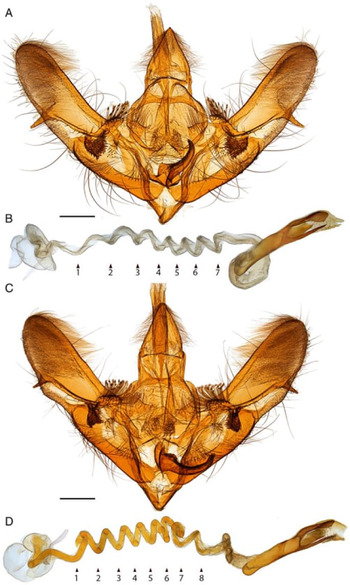
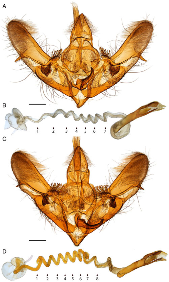
Male genitalia. A, C, Anania hortulata (genitalia slide PYR1741, CNC). B, D, Anania sinensis (genitalia slide NWAFU00006, NWAFU). A–B, Whole genitalia, ventral aspect. C–D, Phallus complex in ventral view; small triangles with numbers indicate individual loops (coils) of the bulbus ejaculatorius. Scale bar = 0.5 mm.
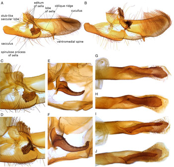
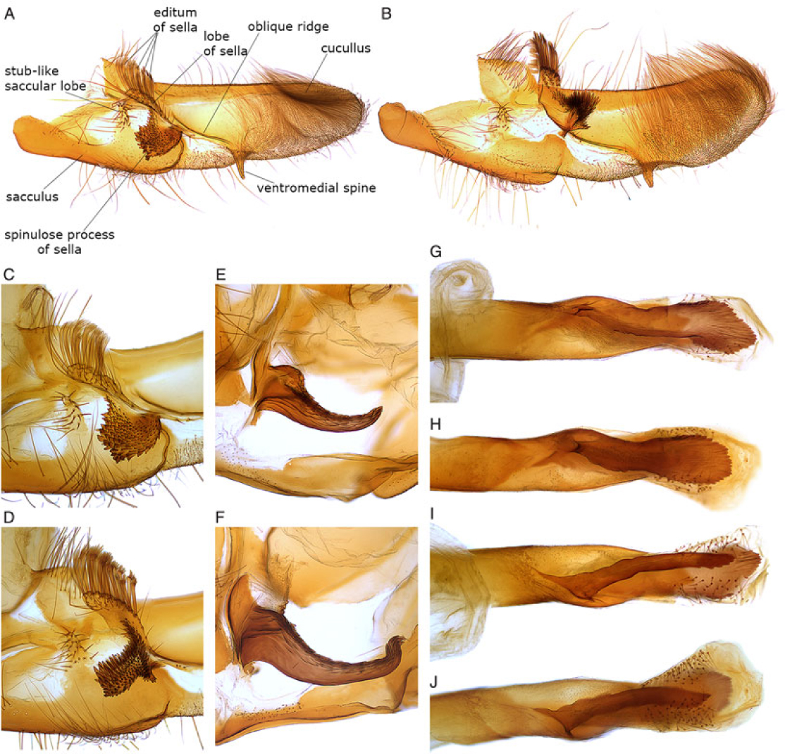
Male genitalia details. A, C, E, G, I, Anania hortulata (genitalia slide PYR1741, CNC). B, D, F, H, J, Anania sinensis (genitalia slide NWAFU00006, NWAFU). A–B, Valva. C–D, Details of valva showing editum and spinulose process of sella. E–F, Juxta lobe. G–H, Left spatulate lobe of phallus in lateral aspect. I–J, Right digitiform lobe of phallus in lateral aspect.
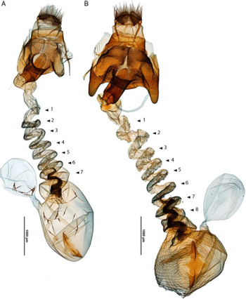
Female genitalia. A, Anania hortulata (genitalia slide PYR1738, CNC); B, Anania sinensis (genitalia slide NWAFU00005, NWAFU). Small triangles with numbers indicate individual loops (coils) of the ductus bursae. Scattered male deciduous cornuti visible inside the corpus bursae of A. Wrinkled membrane of corpus bursae of B due to virgin condition of female. Scale bar = 1.0 mm.

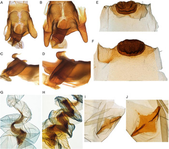
Female genitalia. A, C, E, G, I, Anania hortulata (genitalia slide PYR1738, CNC); B, D, F, H, J, Anania sinensis (genitalia slide NWAFU 00005, NWAFU, except J from slide NWAFU00007). A–B, Sterigma and antrum, ventral aspect. C–D, Antrum, lateral aspect. E–F, Abdominal sternum 7 and lamella antevaginalis, ventral aspect. G–H, Lamina ducti bursae. I–J, Signum. All figures at same scale.

Type material examined. Holotype, ♂, “CHINA: Shanxi, Jiaocheng County, Pangquangou, 29.vii.2006, Yang Zhaofu” (NWAFU); BOLD sample ID and process ID: Pyr001487, CNPYD1487-10; genitalia slide number: NWAFU 00002.
Paratypes: 13 ♂♂, 7 ♀♀ as follows: 1 ♂, CHINA: Shanxi, Jiaocheng County, Pangquangou, 29.vii.2006, Yang Zhaofu, BOLD sample ID and process ID: Pyr001488, CNPYD1488-10; 1 ♀, CHINA: Shanxi, Jiaocheng County, Pangquangou, 28.vii.2006, Yang Zhaofu, BOLD sample ID and process ID: NAFU Pyr000275, CNPYA275-10, genitalia slide number: NWAFU 00003; 1 ♀, CHINA: Shanxi, Yuanqu, Li Mountain, 17.vii.2006, Yang Zhaofu (NWAFU), BOLD sample ID and process ID: Pyr001489, CNPYD1489-10, genitalia slide number: NWAFU 00001; 1 ♂, CHINA: Shanxi, Yuanqu, Li Mountain, 17.vii.2006, Yang Zhaofu (NWAFU), BOLD sample ID and process ID: NAFU Pyr002055, CNPYB394-16; 1 ♀, CHINA: Shanxi, Yangcheng, Manghe, 21.vii.2006, Yang Zhaofu (NWAFU), BOLD sample ID and process ID: Pyr001490, CNPYD1490-10; 1 ♂, CHINA: Shaanxi, Taibai Mountain, Haopingshi, 1200 m, 13.vi.1983 (NWAFU), BOLD sample ID and process ID: NAFU Pyr002061, CNPYB400-16, genitalia slide number: NWAFU 00008; 1 ♂, CHINA: Shaanxi, Taibai Mountain, Haopingshi, 1200 m, 13.vi.1983 (NWAFU), BOLD sample ID and process ID: NAFU Pyr002062, CNPYB401-16; 1 ♂, CHINA: Shaanxi, Taibai Mountain, Haopingsi, 2.vi.1982 (NWAFU), BOLD sample ID and process ID: NAFU Pyr002057, CNPYB396-16; 1 ♀, CHINA: Shaanxi, Taibai Mountain, Haopingsi, 7.vi.1982 (NWAFU), BOLD sample ID and process ID: NAFU Pyr002058, CNPYB397-16; 1 ♂, CHINA: Shaanxi, Taibai Mountain, Haopingsi, 4.vi.1982 (NWAFU), BOLD sample ID and process ID: NAFU Pyr002059, CNPYB398-16; 1 ♀, CHINA: Shaanxi, Taibai Mountain, Luotuoshu, 27.v.1983. 2100 m, Chai Yonghui (NWAFU), BOLD sample ID and process ID: NAFU Pyr000560, CNPYA560-10, genitalia slide number: NWAFU 00007; 1 ♂, CHINA: Shaanxi, Taibai Mountain, Luotuoshu, 9.vi.1983. 2100 m (NWAFU), BOLD sample ID and process ID: NAFU Pyr002060, CNPYB399-16; 1 ♂, CHINA: Shaanxi, Taibai Mountain, Luotuoshu, 2.vi.1982 (NWAFU), BOLD sample ID and process ID: NAFU Pyr002056, CNPYB395-16, genitalia slide number: NWAFU 00004; 1 ♂, CHINA: Shaanxi, Ningshan, Huoditang, 19.vi.1985. Liu Lan (NWAFU), BOLD sample ID and process ID: NAFU Pyr000561, CNPYA561-10; 1 ♂, CHINA: Shaanxi, Yanan, 4.vii.1973. Luojinghui (NWAFU), BOLD sample ID and process ID: NAFU Pyr002064, CNPYB403-16; 1 ♀, CHINA: Shaanxi, Changwu, 1976 (NWAFU), BOLD sample ID and process ID: NAFU Pyr002065, CNPYB404-16; 1 ♂, CHINA: Ningxia, Liupanshan, Fengtai Forest Farm, 5.vii.2008. Li Tao (NWAFU), BOLD sample ID and process ID: NAFU Pyr002066, CNPYB405-16; 1 ♂, CHINA: Ningxia, Liupanshan, Qiuqianjia Forest Farm, 25.vi.2008. Men Qiulei and Li Tao (NWAFU), BOLD sample ID and process ID: NAFU Pyr002067, CNPYB406-16; 1 ♀, CHINA: Hainan, Nada, 23.viii.1974, Zhou Yao and Lu Zheng (NWAFU), BOLD sample ID and process ID: Pyr001486, CNPYD1486-10, genitalia slide number: NWAFU 00005; 1 ♂, CHINA: Balimiao (collecting locality data undecipherable), 30.vii.1976 (NWAFU), BOLD sample ID and process ID: NAFU Pyr002063, CNPYB402-16, genitalia slide number: NWAFU 00006.
Additional specimen examined. 1 ♂, CHINA: Sichuan, Mt Omei (no date but evidently old), D.C. Graham; specimen ID: USNMENT01142740, genitalia slide number: USNM 130277 (NMNH). This specimen was excluded from the type material because it was not barcoded and lacks a collecting date.
Diagnosis. Similar to A. hortulata in colouration and wing pattern but appearing paler overall. However, the type material was trap-collected and it is possible that a certain amount of wear or fading is responsible for the paler appearance of the specimens. The species is slightly larger than A. hortulata (mean forewing length 15.5 versus 13 mm). The diagnostic characters of female and male genitalia that distinguish it from A. hortulata are presented in Table 6.
Description. Head with frons flat, smoothly scaled, and fuscous; laterally edged with yellowish white. Vertex pale yellow. Labial palpus pale yellow ventrally, dark brown dorsally; first and second palpomeres oblique, third palpomere exposed and porrect, distal part fuscous. Maxillary palpus rather short and inconspicuous. Chaetosemata absent. Haustellum base covered with yellowish-white scales. Antenna slightly thickened in male, filiform in female. Thorax dark brown dorsally, posterior part pale yellow. Tegula bright orange yellow, suffused with fuscous scales basally. Legs dark fuscous. Abdomen elongated, black, each segment yellow banded posteriorly. Forewing slender; apex rather angulate, termen evenly curved to tornus. Ground colour above white, suffused with fulvous or dark brown at costa. Basal area with yellow. Antemedial spot and discocellular spot dark brown; postmedial fascia and subterminal band a series of dark brown spots. White subterminal band sexually dimorphic: in female anteriorly extended to costa, but in male interrupted at R4 by black patch between R2 and R4. Fringe fuscous. Discal cell less than half as long as wing. R1 arising from the cell at about four-fifths. R2 stalked with R3+4. R3 and R4 stalked more than halfway from cell to apex. R5 from just behind apex of cell. M1 arising from discal cell a short distance behind R5. M2, M3, and CuA1 arising close together at posterior angle of cell, weakly curved and approximate basally. CuA2 from cell at about two-thirds. Hindwing broadly triangular. Ground colour and postmedial and subterminal bands as on forewing but without evident sexual dimorphism, fringe white. Sc+R1 and Rs anastomosed for a considerable distance. Rs and M1 short-stalked. M2, M3, and CuA1 arising close together at posterior angle of cell, basally curved and approximate. CuA2 from cell at three-fourths.
Forewing length. Female, 15.43 ± 0.69 mm (n = 7); male, 15.57 ± 0.43 mm (n = 14).
Male genitalia (five preparations examined). Tegumen broadly trapezoid. Uncus triangular, densely setose dorsally. Gnathos with thin, flat lateral arms joined mesially with subscaphium. Juxta with small base and markedly expanded and sclerotised, anchor-shaped distal projection, with a basal hump and strong longitudinal keel with sinuate, upcurved apex and fish scale-like dorsal microsculpture. Vinculum trapezoid. Valva slender; costa narrowly margined; cucullus broadly rounded, inner surface densely setose, mesially delineated by oblique ridge ending in ventrally directed spine protruded beyond ventral edge of cucullus; sacculus with four processes: a stub-like membranous saccular lobe stemming from mediodorsal edge of sacculus, situated mesad of spinulose process, bearing several short, normal-looking setae; lobe of sella pad-like, situated at anterodorsal end of oblique cucullar ridge, bearing a series of dorsally projected, flat setae with comb-like apices (editum); spinulose process of sella extended from oblique cucullar ridge and ventrad of pad-like lobe, oriented mesad, heavily sclerotised, covered with a dense set of sharp spinules of various sizes; spinulose process and pad-like process with common base on oblique mesial ridge. Phallus more or less straight, distal third deeply and unequally cleft into two rods; right rod narrow, attenuate with pointed, crenate apex; left rod distally dilated, spatulate with serrate apex; apex of phallic tube with spatulate, free sclerotised apical projection; ductus ejaculatorius about two times longer than phallus, with eight coils, anterior end expanded into oval chamber; vesica with bundle of deciduous cornuti.
Female genitalia (four preparations examined). Ovipositor and apophyses short, shorter than sterigma. Sterigma surface concave, covered with dense microtrichia, with a mesial groove, anterolateral angles expanded into deep, ear-like, thickly sclerotised smooth pockets. Antrum cylindrical, approximately 0.8 as long as wide, melanised, dorsal wall with tongue-like plate protruding out of ostium bursae. Inception of ductus seminalis at anterior (cephalad) end of antrum. Ductus bursae long, membranous, with eight coils, anterior-most two coils with an inner, spinulose sclerotised lamina. Corpus bursae globular, with an elongate-rhomboid signum and a thin-walled globular appendix bursae about half smaller than corpus bursae.
Distribution. China (Beijing, Gansu, Guangdong, Guizhou, Hainan*, Hebei, Henan, Jiangsu, Ningxia*, Qinghai, Shaanxi*, Shanxi*, Sichuan*, Yunnan Provinces). Distribution data were obtained from the samples examined in this study (distinguished with an asterisk *), and from literature sources where genitalia illustrations clearly allowed the recognition of the species (Li et al. Reference Li, Ren, Zhang, Du, Li and You2009, Reference Li, Wang, Zhen, Zhang, Hu and Jing2012).
Host plant. The larvae most likely feed on Marrubium vulgare Linnaeus (Lamiaceae) (Li et al. Reference Li, Ren, Zhang, Du, Li and You2009, Reference Li, Wang, Zhen, Zhang, Hu and Jing2012 as reported under A. hortulata).
Etymology. Named after the country where the type series was collected.
Remarks. Overall the genitalia of A. sinensis appear larger and more robust than those of A. hortulata. In males, the most striking differences are in the spinulose process of the sella, which is smaller with longer spines in A. sinensis versus larger with shorter spines in A. hortulata; the bulbus ejaculatorius has one more coil in A. sinensis than in A. hortulata (eight versus seven), with the same number of coils in the ductus bursae of the respective females. In female A. sinensis, the antrum is proportionally longer (length/width = 0.8); the anterolateral lobes of the sterigma are very large, prominent, and longer than the antrum, and the lamina ducti bursae has three coils; in A. hortulata, the antrum is proportionally shorter (length/width = 0.6); the sterigma lobes are shorter than the antrum and wider; and the lamina ducti bursae has two coils. Other more subtle differences are described in Table 6. This new species was formerly overlooked and misidentified as A. hortulata throughout China. The diagnostic characteristics of male and female genitalia in the illustrations of A. hortulata in Li et al. (Reference Li, Ren, Zhang, Du, Li and You2009, Reference Li, Wang, Zhen, Zhang, Hu and Jing2012) match those of A. sinensis. The wrinkled membrane of the corpus bursae of A. sinensis visible in Fig. 9B, which contrasts with the smooth membranes of A. hortulata in Fig. 9A, is believed to be due to the virgin condition of the female, and not to represent a specific difference. The sexual difference in the subapical forewing spot noted for A. hortulata (Fig. 6) is also present in A. sinensis.
Discussion
Anania is a large genus currently with 117 described species (Nuss et al. Reference Nuss, Landry, Mally, Vegliante, Tränkner and Bauer2017). Several species groups present heterogeneous, strikingly different aspects, especially in colouration: wing patterns range from brown wavy lines and spots over an off-white ground colour (e.g., A. coronata (Hufnagel, 1767) species complex, A. murcialis (Ragonot, 1895)), to black with white spots (e.g., A. funebris (Ström, 1768)), to nearly uniformly rusty brown with faint markings (e.g., A. terrealis (Treitschke, Reference Treitschke1829)), to white with radiating black markings (e.g., A. hortulata species complex) (Tränker et al. Reference Tränker, Li and Nuss2009; Slamka Reference Slamka2013). The generic concept of Anania was recently expanded by synonymising several generic names and reclassifying several species from other genera into the genus (Leraut Reference Leraut2005; Tränker et al. Reference Tränker, Li and Nuss2009). Despite superficial heterogeneity, two putative synapomorphies unite members of Anania: that is, the apex of phallic tube has a free sclerotised apical projection, and the female antrum possesses an inner digitiform strip with a free apical ledge (Leraut Reference Leraut2005). Tränker et al. (Reference Tränker, Li and Nuss2009) have added that the sclerotised projection of the phallus has a serrated edge; however, the serrate condition is not shared by all Anania, for example, it is lacking in A. plectilis (Grote and Robinson, 1867) (Yang et al. Reference Yang, Landry, Handfield, Zhang, Solis and Handfield2012), although this can represent a secondary loss. The apical projection is double in A. hortulata and several other species. These hypotheses need further testing through more comprehensive phylogenetic analyses.
Anania hortulata was historically treated as Eurrhypara, which was reduced to a subgenus of Anania by Leraut (Reference Leraut2005). It possesses some unique genital traits: for example, the large, protruding hook-like or anchor-like juxta and the enlarged spinulose process of the sella in males, and the heavily sclerotised, shield-like lamella antevaginalis that covers the ostium bursae in females – all traits shared by A. sinensis as well.
In this study, both morphological and molecular analyses revealed that what has long been known as A. hortulata actually comprises two superficially indistinguishable species, A. hortulata and A. sinensis. Several other studies have used DNA barcodes effectively as an initial means to detect potential cryptic species (Huemer and Mutanen Reference Huemer and Mutanen2012; Mutanen et al. Reference Mutanen, Aarvik, Landry, Segerer and Karsholt2012; Yang et al. Reference Yang, Landry, Handfield, Zhang, Solis and Handfield2012; Huemer et al. Reference Huemer, Elsner and Karsholt2013; Landry and Hebert Reference Landry and Hebert2013). This in turn led to a closer morphological scrutiny to ascertain whether subtle differences previously unnoted or construed to represent intraspecific variation are actually indicative of separate taxonomic entities.
The COI genetic distance between A. hortulata and A. sinensis at 2.73% is of the same magnitude as that observed between other pairs of sibling species of Anania coronata species complex at 2.1–3.3% (Yang et al. Reference Yang, Landry, Handfield, Zhang, Solis and Handfield2012). Other Lepidoptera studies have revealed much greater genetic distances between sibling species with weakly differentiated morphology: for example, 8.6% between the Plutellidae Plutella australiana Landry and Hebert, Reference Landry and Hebert2013 and P. xylostella (Linnaeus, 1767) (Landry and Hebert Reference Landry and Hebert2013); 6.1% for the Tortricidae Epinotia cinereana (Haworth, 1811) and E. nisella (Clerck, 1759) (Mutanen et al. Reference Mutanen, Aarvik, Landry, Segerer and Karsholt2012); 6.14–13.86% among Eulamprotes Bradley, 1971 (Gelechiidae) (Huemer et al. Reference Huemer, Elsner and Karsholt2013). In the Teleiopsis albifemorella Hofmann, 1867 species complex of gelechiids, Huemer and Mutanen (Reference Huemer and Mutanen2012) have found weak (1.54–2.42%) barcode differentiation overlapping with significant intraspecific variation, but nuclear markers corroborated interspecific distinctness. In all these instances, morphological differences were slight or subtle but congruent/consistent with barcode divergences (and nuclear gene differences were tested), and supported that both entities are valid taxa.
Despite the short sequences recovered due to the age of specimens, the CAD genetic distance between A. hortulata and A. sinensis at 17.4% was similar in magnitude to those observed in other Lepidoptera species (Wahlberg and Wheat Reference Wahlberg and Wheat2008; Dincă et al. Reference Dincă, Lukhtanov, Talavera and Vila2011; Haines and Rubinoff Reference Haines and Rubinoff2012) and much greater than COI distances differentiating species (see above), indicating that the CAD gene provides substantial evidence for the resolution of species-level taxa.
As presently known, A. hortulata and A. sinensis are allopatric: the former has a wide range across the Palaearctic region, in addition to being introduced into the Nearctic region where it is adventive. In contrast, A. sinensis has been found only in all major geographical regions of China except the northeastern part. Our findings suggest that the evolutionary process within the A. hortulata species pair perhaps involved geographical separation leading to its divergence in China. The estimated divergence time between A. sinensis and A. hortulata occurred from early to mid-Pleistocene (approximately 0.9 million years ago), suggesting that Pleistocene glacial cycles may have led to the partitioning of their common ancestor, which would have led to genetic and morphological divergence and subsequent speciation. Further analysis of specimens from the east Palaearctic region, such as far-east Russia and Japan, would likely provide further insights into the phylogeographic patterns of these two sibling species.
Acknowledgements
We thank Marko Mutanen (University of Oulu, Oulu, Finland) for the loan of a few genitalic slides for study. We are grateful to Alma Solis (Systematic Entomology Laboratory, Agricultural Research Service, United States Department of Agriculture, Washington, District of Columbia, United States of America) for the loan of, and permission to analyse, specimens in NHNM. For assistance with photography of the genitalia, we thank Ulla Misbah at the NWAFU and Vazrick Nazari at the CNC. James Hayden (Florida Department of Agriculture and Consumer Services, Division of Plant Industry, Gainesville, Florida, United States of America) and Mark Metz (Systematic Entomology Laboratory, Agricultural Research Service, United States Department of Agriculture) helped with some references. We are grateful to Chris Looney (WSDA), Andreas Segerer (SNSB), Arild Johnsen (University of Oslo, Natural History Museum, Oslo, Norway), Axel Hausmann (ZSM), Daniel Handfield (St-Mathieu de Beloeil, Québec, Canada), Paul Hebert (BIOUG), Jeremy deWaard (BIOUG), Peter Huemer (TLMF), May Bente Brurberg (Norwegian Institute for Agricultural and Environmental Research), Torbjorn Ekrem (NTNU), Marko Mutanen (ZMUO), and Matthias Nuss (SNSD) for permission to use their barcode sequence data. This study was supported by the National Natural Science Foundation of China (31772508, 31201733), Natural Science Foundation of Shaanxi Province (2016JM3026), National Key Research and Development Program of China (2016YFC0501502), and the Chinese Universities Scientific Fund (2452015012). We thank Paul Hebert and the Centre for Biodiversity Genomics, Biodiversity Institute of Ontario, University of Guelph, Guelph, Ontario, Canada, for DNA barcode development through a grant from Genome Canada to the iBOL project.