INTRODUCTION
Few endoparasitic animals are known from opisthobranch gastropods. Endoparasitic copepods of the family Splanchnotrophidae, which exclusively use opisthobranchs as hosts (Huys, Reference Huys2001), are an exception, and a few digeneans are the only helminth parasites reported from opisthobranchs. This is rather puzzling given that gastropods are the most important intermediate hosts for endoparasitic flukes (Hanna & Halton, Reference Hanna, Halton, David, Jewzey and Ian2001; see also Cribb et al., Reference Cribb, Bray and Littlewood2001).
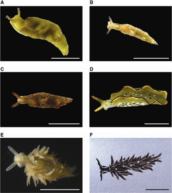
In July 1990, the second author collected a few specimens of the sacoglossan opisthobranch Aplysiopsis nigra (Baba, 1949) (Figure 1F) in Kominato, Pacific coast of Boso Peninsula on Honshu. When he observed these specimens alive under a stereomicroscope, a worm came out of a slug specimen. Later two other worms were obtained from the same slug specimen. More recently in February 2002, a similar parasitic worm was found from another sacoglossan species, Elysia trisinuata Baba, 1949 (Figure 1A) collected at Misaki, Sagami Bay, Pacific coast of Honshu. With careful searches for this parasitic worm in sacoglossan opisthobranchs more than 250 specimens have been found from several sacoglossan species since then.
By close observations of its morphology and life history, the parasitic worm was identified as a species of Turbellaria, Platyhelminthes with close similarity to the family Fecampiidae. Fecampiidae was established by Giard (Reference Giard1886), and now comprises three genera: Fecampia Giard, Reference Giard1886, Glanduloderma Jägersten, Reference Jägersten1940 and Kronborgia Christensen & Kanneworff, Reference Christensen and Kanneworff1964 (Williams, Reference Williams1988; Cannon, Reference Cannon and Rohde2005). All members of these genera are known to be obligate parasites, with Fecampia and Kronborgia associated with crustaceans while Glanduloderma associated with an annelid host.
While about 80% of the species are parasitic in Platyhelminthes due to a large, diverse, parasitic taxon, Neodermata, parasitic species account for only about 5% of the class Turbellaria (Bush et al., Reference Bush, Fernández, Esch and Seed2001). Fecampiidae is an unusual turbellarian group where all species are obligate parasites, and it is also interesting because it has several morphological similarities with Neodermata (Watson & Rohde, Reference Watson and Rohde1993; Rohde, Reference Rohde1994; Joffe & Kornakova, Reference Joffe, Kornakova, Littlewood and Bray2001).
While monophyly of Neodermata has been confirmed by DNA analyses (Baverstock et al., Reference Baverstock, Fielke, Johnson, Bray and Beveridge1991; Blair, Reference Blair1993; Rohde et al., Reference Rohde, Hefford, Ellis, Baverstock, Johnson, Watson and Dittmann1993), there is an intense debate on its origin. Fecampiidae was once regarded as a strong candidate for the direct sister group of Neodermata because of the morphological similarities (Watson & Rohde, Reference Watson and Rohde1993; Rohde, Reference Rohde1994). Although little support for this view has been obtained from DNA analyses (Littlewood et al., Reference Littlewood, Rohde and Clough1999a, Reference Littlewood, Rohde, Bray and Hernioub; Litvaitis & Rohde, Reference Litvaitis and Rohde1999; Lockyer et al., Reference Lockyer, Olson and Littlewood2003; Baguña & Riutort, Reference Baguña and Riutort2004), Fecampiidae is still among the important taxa to understand the origin of Neodermata and, hence, the evolution of parasitism in Platyhelminthes. The helminth parasite newly found from opisthobranchs may provide a new insight and significant advancement for phylogenetic studies of Platyhelminthes. Herein we describe the morphology, life history and host use of this new parasite and compare its features to described species of the family Fecampiidae.
MATERIALS AND METHODS
Sacoglossan opisthobranchs, potential hosts for the new parasitic worm, were collected at several localities of Japan including Misaki, Sagami Bay (35°2′N 139°6′E); Kominato, Boso Peninsula (35°1′N 140°2′E) and Tateyama, Boso Peninsula (35°0′N 139°8′E); Shimoda, Izu Peninsula (34°5′N 138°9′E); Mukaishima Island, Seto Inland Sea (34°5′N 133°2′E); Sobe, Okinawajima (26°4′N 127°7′E) by snorkelling or walking at low tides from February 2002 to April 2009. Collection details are shown in Table 1.
Number of the new parasite found from each host and field-collected cocoons.

Worms and cocoons were collected at Misaki unless marked with *. *1,*2,*3,*4 and *5 represents Shimoda, Mukaishima, Tateyama, Sobe and Kominato, respectively.
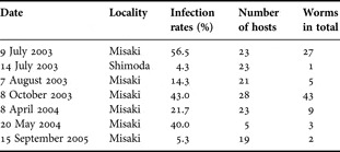
Collected sacoglossans were externally investigated for infection of the parasite. The parasites may be seen through translucent body surface of the host sacoglossans. When infection was detected, host specimens with parasites were anaesthetized in seawater mixed with 7.5% MgCl2 (1:1), and dissected to observe the parasites in situ under a stereomicroscope. Then the parasites were removed from the hosts. Some host specimens with obvious parasites were kept alive until the parasites came out of the hosts voluntarily. These host specimens were kept with their food algae in plastic containers filled with filtered seawater at 24°C in the laboratory. The seawater was changed every two or three days, and new algae were given when old ones were depleted. With sacoglossan species with high infection rates (e.g. Elysia trisinuata and E. ornata (Swainson, 1840)), potential host specimens with no obvious parasite were also kept in the same manner; when found, parasites from these hosts were also collected. Specimens of E. trisinuata collected at Misaki or Shimoda on seven occasions (the details are given in Table 2) were all investigated for infection by dissection to see what percentage of the slug specimens were actually inhabited by the parasite.
Infection rates of Elysia trisinuata.

Collected worms were transferred by a pipette to a new container and observed with a stereomicroscope. Several worms were mounted each on a slide for detailed observation of the external morphology with a compound microscope. All parasite specimens obtained by dissection were fixed for histological study right after these observations. A part of the parasites which voluntarily came out of hosts were also fixed for histological study while others were kept in the containers to observe ontogenetic changes.
Well-developed worms secreted a silky substance to make a cocoon around themselves on the bottom or the wall of the container within one or two days after emerging out of the hosts. These cocoons were kept in the same container for about a month to observe further ontogenetic changes of the worm. Cocoons with their contents were also fixed for histological study at several different times after cocoon formation.
Cocoons were also collected in the field at Misaki (Table 1). These cocoons were found on or near the green alga Codium fragile (Suringar) Hariot. Codium fragile is an important food of E. trisinuata, E. setoensis Hamatani, 1968 and Placida sp. (sensu Baba, Reference Baba1986) (Trowbridge et al., Reference Trowbridge, Hirano and Hirano2008). Field-collected cocoons were investigated for the contents. When egg capsules with eggs or embryos inside were found, they were transferred to a plastic container and kept alive to observe embryonic development to larval hatch.
For the histological study, worms (including those in cocoons) were anaesthetized by the same method for host sacoglossans, and fixed either with 5% formalin seawater, Bouin's fluid, or Holland's fluid. These three fixatives resulted in equally good fixations. After dehydration with ethanol series (70–100%) and xylene, specimens were embedded in paraffin. Serial sections were cut of these specimens with 5–7 µm thickness, stained with haematoxylin and eosin, mounted on slides with Entelan new (Merck Corp.), and observed under a compound microscope. Photographs of these sections were taken with the digital camera Olympus E330 attached to the microscope.
RESULTS
Associations
The newly discovered parasitic worm has been found from six sacoglossan species: Elysia trisinuata, E. setoensis, E. ornata, E. rufescens (Pease, 1871), Placida sp. and Aplysiopsis nigra (Figure 1). The most frequent host was E. trisinuata, followed by E. setoensis and E. ornata. The majority of collected worms were obtained from these three sacoglossan species (Table 1). Although Placida sp. was collected much more frequently than E. trisinuata, the worm was found only rarely from Placida sp. The two other host species, E. rufescens and A. nigra, were relatively uncommon and found only rarely during this study. The new parasitic worm was common at Misaki, where we found 269 living worms from four sacoglossan species and 51 cocoons in the field (Table 1). While the worm was found almost all year around, its occurrence at Misaki exhibited a major peak from May to August.
Host sacoglossans for the new parasite. (A) Elysia trisinuata; (B) Elysia setoensis; (C) Elysia rufescens, (D) Elysia ornata; (E) Placida sp.; (F) Aplysiopsis nigra. Scale bars = 10 mm for A, C, D and F. Scale bars = 5 mm for B and E.

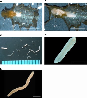
The parasitic worm inhabited the haemocoel of the hosts. In E. trisinuata, E. setoensis and E. ornata, the worms were most often found winding around digestive and reproductive organs which lie anteriorly to the pericardial region of the hosts (Figure 2A, B). Most often there was a single worm per host; however, multiple specimens were occasionally found (with the maximum of 19 worms of various lengths) in a single host (E. ornata). When many worms were found in a single host, some of them were parasitizing other parts of host body, including the parapodia (paired lateral outgrowths of the body) and the tail. These worms in unusual habitats were smaller than those found around digestive and reproductive organs. In Placida sp., the maximum number of worms found in a single host specimen was two, and none were found in the cerata on the slug dorsum. Large worms often caused a swelling in the head of sacoglossan hosts; however, these host slugs appeared to be normal in behaviour: they were able to feed on food algae and also spawned egg masses in the laboratory. Obvious atrophy of gonads was not found.
The new parasite. (A) Elysia trisinuata infected by many parasites; (B) same host specimen as Figure 2A, but after partial dissection; (C) worms of various sizes collected by dissection from a single host slug of Elysia ornata. The ruler at the bottom is graduated in millimetres; (D) young worm marked with ‘d’ in C; (E) adult worm marked with ‘e’ in C. Arrows in A and B show parasites in the host body. pa, parapodium; pe, pericardium; ph, pharynx; r, rhinophore. Scale bars = 10 mm for A and B. Scale bars = 1 mm for D and E.

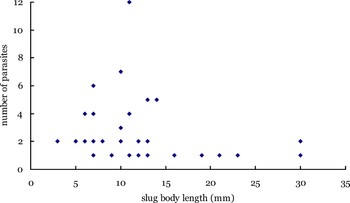
Infection rates of E. trisinuata on six collections at Misaki varied considerably ranging 5.3 to 56.5% while a single infection rate obtained for Shimoda was only 4.3% (Table 2). The number of parasites in a host slug was investigated for E. trisinuata in relation to slug body size. The maximum number for a single host was 12 for this slug species; no obvious correlation was found between the number of parasites per host and host body length (Figure 3). Most of the investigated hosts (more than 75%) were with one or two parasites.
Number of parasites found in Elysia trisinuata slugs and host slug sizes.

Morphology
The newly discovered parasitic worms obtained from different host species were all similar in morphology. They were cylindrical to sausage-shaped (Figure 2D, E), and 1 to 15 mm long. Worms which voluntarily emerged from hosts were longer than 5 mm. Worm sizes varied depending on host size. The small sacoglossans Elysia setoensis and Placida sp. contained smaller worms (<8 mm long) than larger slugs such as E. trisinuata and E. ornata did. The colour of worms differed with worm body size (Figure 2C). Worms ≤5 mm long were translucent white (Figure 2D). Those >5 mm were opaque, often with a slightly yellow tint. The yellow tint was darker in larger worms, and a narrow central cavity running along longitudinal axis was discernible in individuals longer than 8 mm (Figure 2E). The body surface of the parasitic worm was covered with cilia but had no mouth, eyes, or any noticeable organs or structures. Having no adhesive organs like suckers or hooks, the worms were easily removed from the host.
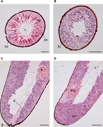
Internally there was central parenchymal tissue with a central cavity (Figure 4A). The central cavity ran along the longitudinal body axis and suddenly narrowed at both ends. On the surface of the worm, there was one-cell-layered epidermis deeply stained with eosin. Central to the epidermis was a thick subepidermal layer, where two types of subepidermal glands were distinguished (Figure 4A). In one type of gland the cytoplasm was uniformly stained with eosin. The other type was filled with rod-shaped structures with resemblance to glandular rhabdites of rhabdocoelans, which were not stained by eosin or haematoxylin. Both types of glands were elongate and gradually tapered towards the body surface to join with a pointed tip to the epidermis.
Only a few organs were discernible even internally. The most readily observed were a pair of ovaries: long cylindrical organs composed of many large cells (30–50 µm in diameter) each with a large nucleus which was deeply stained with haematoxylin (Figure 4A, D). The ovaries were situated in the subepidermal layer and ran between the anterior and posterior ends. Another distinctive organ was a fibrillar neuropile situated near the anterior end. It was H-shaped transversely, with a long neural fibre bridge about 300 µm long and 30–50 µm wide (Figure 4B). A distinctive accessory reproductive organ deeply stained with eosin was found in the subepidermal layer near the posterior end (Figure 4D). A gonopore with a duct was occasionally found at the posterior end of such specimens (Figure 4C), but we could not follow the duct to the anterior end. Testes or sperm were not found in any of the observed specimens (N = 120).
Internal morphology of the new parasite. (A) Transverse section of a mature specimen on its middle part; (B) transverse section of the anterior part of a specimen after retreating in a cocoon; (C, D) longitudinal sections at posterior end of a specimen after retreating in a cocoon. arg, accessory reproductive glands; c, central cavity; d, reproductive duct; fn, fibrillar neuropile; g, gonopore; ov, ovary; sg, subepidermal glands. Scale bars = 100 µm.

Histology of the subepidermal layer differed significantly, depending on the growth stage of the worm. In specimens found in cocoons, the characteristic subepidermal glands with rod-shaped structures were replaced by granules (Figure 4B, C, D). Subepidermal glands were not seen in worms ≤3 mm long. The subepidermal layer of these small specimens consisted of indistinct small cells which lined right inside the epidermis, and a large part of their body was empty.
Life history
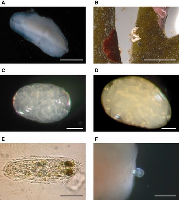
Within a day of emerging from a host, the parasitic worm secreted a white silky substance around its body to form a cocoon and confined itself in it. The cocoon was soft and thin in the beginning but gradually became tough and thick. It adhered to the container on the lower surface with its entire length and opened at one end on the upper surface (Figure 5A). Cocoons made in the laboratory were elongate oval and flattened a little having a diffuse outer layer. The central main part was more tubular and gradually tapering towards both ends. In a few days after retreating into the cocoon, worms were tinged with orange along their central axis and became thin. Almost all worms eventually died in the cocoon, showing no further ontogenetic change. One worm left two egg capsules in the cocoon. The egg capsules were oval and each measured about 2 mm long and 1 mm wide. About 20 eggs were counted in each capsule; however, these eggs did not undergo further embryonic development.
Cocoons, egg capsules and larvae. (A) A cocoon secreted on the surface of container in the laboratory; (B) three cocoons attached on the alga Codium fragile collected at Misaki; (C) egg capsule with early embryos; (D) egg capsule with larvae which are about to hatch; (E) larva just after hatching from an egg capsule; (F) larva intruding into a piece of host tissue (host: E. ornata). Scale bars = 5 mm for A and B. Scale bars = 200 µm for C and D. Scale bar = 100 µm for E. Scale bar = 300 µm for F.

Most of the life-history features after cocoon formation were obtained based on field-collected cocoons. Field-collected cocoons were more tubular than those obtained in the laboratory. They were cylindrical to sausage-shaped, and with an opening at one end (Figure 5B). They had no diffuse outer layer seen in cocoons formed in the laboratory. These cocoons contained a worm only, a worm and egg capsules with embryos, only egg capsules with embryos, or nothing. Worms found in the field-collected cocoons were with the same morphological features as those observed in cocoons formed in the laboratory. The number of egg capsules per cocoon varied from 1 to 42 averaging 16.9 ± 8.6 (SD) (N = 40). Each capsule contained 19 to 42 embryos with an average of 25.3 ± 2.6 (SD) (N = 22) per capsule.
Egg capsules in the same cocoon were similar in the developmental stage of the embryos they contained. Several developmental stages were discernible in different cocoons. At the earliest stage, capsules were closely packed with lipid-like granules and round eggs. The next stage was characterized by fewer granules and oval embryos. Embryos were almost transparent and about 150 µm long (Figure 5C). They became elongated and began to rotate in the capsule in about a week. After a few more days, two eye spots became visible. The eye spots were silvery white in colour and situated near the anterior end of the embryo (Figure 5D). These embryos with eyes moved actively in the capsule for a few days and then hatched as swimming larvae. The hatched larvae were elongate oval and 250–320 µm long. Their body surface was covered with long cilia (Figure 5E). Larvae actively swam, rotating spirally in the water, but often glided through water at the bottom of the container. They were negatively phototactic.
These larvae could not survive even for a day without a host. When given a piece of the parapodium of two host sacoglossans, Elysia ornata and E. setoensis, larvae gathered around the parapodial piece and crawled onto it. Then they stuck their head into the parapodial tissue (Figure 5F), gradually entered into it by repeated muscle contractions combined with frequent rotatory motions. They completed intrusion into the tissue within 2–8 minutes.
DISCUSSION
Biology
Few helminth parasites have been reported from opisthobranchs; reports suggest only a few species of hemiuroid digeneans including Brachyphallus crenatus (Rdpolphi, 1802), Hemiurus luehei Odhner, 1905 and Lecithocladium excisum (Rudolphi, 1819), the schistosome Gigantobilharzia huttoni (Leigh, 1953) and the monorchiid Asymphylodora demeli Markowski, 1935 use opisthobranchs as first intermediate hosts (Leigh, Reference Leigh1955; Køie, Reference Køie1990, Reference Køie1992; Cribb et al., Reference Cribb, Bray and Littlewood2001). However, the newly discovered parasitic worm seems rather common at our study sites. Over 250 specimens were found in total at six localities of temperate to subtropical waters in Japan (Table 1). The distributional range may be wider, expanding to colder and/or warmer waters where no surveys have been done. The concentration of searches on sacoglossan opisthobranchs may also have led underestimation of the occurrence of this parasitic worm. Similar worms were recently found from three nudibranch species of different families: Polyceridae, Dorididae and Chromodoridae (Sudo et al., unpublished data). Whether or not these worms from nudibranchs are conspecific with the parasitic worm from sacoglossans is yet to be elucidated, but they certainly indicate that helminth parasites are more common among opisthobranch hosts than are currently recognized.
Many more specimens of the new parasite were found at Misaki than other localities (Table 1). High densities of hosts and continuous host availability may be a reason for the richness of this parasite in this site. Three host species, Elysia trisinuata, E. setoensis and Placida sp. are common on Codium spp. at Misaki (Trowbridge et al., Reference Trowbridge, Hirano and Hirano2008). These host slugs are found almost always on these green macroalgae which are their food. The adherence to the food algae of these slugs often makes their density locally high. Infection rate of the parasitic worm's larvae can be high in such place.
Although some Codium slugs (e.g. E. trisinuata and E. setoensis) can overwinter at Misaki (Trowbridge et al., Reference Trowbridge, Hirano and Hirano2008), abundance of these slugs is significantly lower from late autumn to spring than in summer because important food Codium species for these host slugs, C. fragile and C. intricatum, are scarce in the cold seasons. By contrast, E. ornata, which preferentially feeds on Bryopsis spp. and related algae, but not on Codium (Jensen, Reference Jensen1992; Trowbridge et al., Reference Trowbridge, Hirano, Hirano, Sudo, Shimadu, Watanabe, Yorifuji, Maeda, Anetai and Kumagai2010), is frequently found from autumn to spring at Misaki. The new parasite has no dormant stage. Its larvae are competent for infection soon after hatching, and they cannot survive long without a host. Therefore continuous host occurrence must be important for successful infection of this parasite.
About 71% of these parasitic worms found during this study were obtained from E. trisinuata. Although E. setoensis and Placida sp. are often sympatric with and as common as E. trisinuata, they were much less parasitized by the worm than E. trisinuata. What may be responsible for the higher infection rate in E. trisinuata than in other slugs? Elysia trisinuata has a large body size (up to 30–40 mm long) compared with E. setoensis and Placida sp. (seldom reaches 20 mm with an average of about 10 mm). The larger body size of the host may be beneficial for the parasitic worms both spatially and nutritionally. However, less frequent occurrence of the worms in E. ornata specimens cannot be explained by the same token. Elysia ornata can reach 50 mm or more and is often larger than E. trisinuata. Most of food algae of E. ornata, namely Bryopsis spp. and related algae are small for this slug size and rapidly depleted of cytoplasm by these suctorial herbivores. Consequently, E. ornata is much less frequently found on its food algae than E. trisinuata, and often found solitary wandering on rock platforms. Different distributional patterns of these two sacoglossan species may lead to the differential infection rates between them.
Even though larvae of the worm parasite can actively swim, they have limited ability for dispersal because of their short survival time without a host as well as their small size. Larval success in infection must largely rely on host availability in and around their natal place. Aggregated distribution of E. trisinuata on its food algae may locally supply many hosts for the parasite larvae resulting in a high infection rate in this slug species, while wandering E. ornata may provide better opportunity for the larval infection than E. trisinuata in a larger spatial scale, and may be a target of mass infection by multiple larvae on occasions where other potential host slugs are scarce. In fact, the greatest number of parasites per host in this study was found in this slug species rather than in the most frequent host, E. trisinuata.
Elysia ornata as well as E. setoensis are also used as hosts by another endoparasite, a splanchnotrophid copepod (Sudo, Reference Sudo2004). This endoparasitic copepod has never been found from E. trisinuata. Both the new parasitic worm and the copepod parasite occupy basically the same body region of the hosts. No single slug of E. ornata or E. setoensis with both types of parasites has been found. There may be a competition for habitat between these two parasitic animals.
Study on parasites for opisthobranch gastropods has been rather neglected, while many parasites (especially of Trematoda) are known to utilize gastropods as hosts (Hanna & Halton, Reference Hanna, Halton, David, Jewzey and Ian2001). Having either no shell or a reduced shell, opisthobranchs are known to have special chemical defences against predation (Avila, Reference Avila1995; Wägele et al., Reference Wägele, Ballesteros and Avila2006). It is popularly thought that opisthobranchs have few predators but numerous studies have demonstrated that this paradigm is not well supported (Carefoot, Reference Carefoot1987; Trowbridge, Reference Trowbridge1994, Reference Trowbridge2002; Roger's et al., Reference Rogers, de Nys and And Steinberg2000; Arango & Brodie, Reference Arango and Brodie2003). Chemical defences may serve against parasite infection; however, this study suggests parasitic relationships with opisthobranch hosts may be more common than are currently expected.
Infection rates of the new worm parasite may be very high (56.5% of Elysia trisinuata slugs were inhabited by the new parasitic worm at Misaki on 9 July 2003) although infection rates vary considerably both spatially and temporally. High infection rates have also been reported for a few other opisthobranch hosts. Jensen (Reference Jensen1990) found an endoparasitic copepod, Arthurius elysiae (Jensen, Reference Jensen1990), in 7 out of approximately 25 slug specimens of Elysia australis (Quoy & Gaimard, 1832) collected in Eagle Bay, Western Australia (WA), although she could not find any parasite in hundreds of conspecific slugs in Princess Royal Harbour, WA. Schrödl (Reference Schrödl2002) reported very high infection rates (ranging from 70 to 100%) of another parasitic copepod, Ismaila aliena Haumayr & Schrödl, 2003, for Thecacera darwini Pruvot-Fol, 1950 in Bahía de Coliumo, Chile. He also found nearly 90% of Elysia patagonica Muniain & Ortea 1997 in Magellan Strait may be infected by I. magellanica Haumayr & Schrödl, 2003. Parasite–host interactions of opisthobranch hosts may be potentially important for opisthobranch biology and ecology, and merit further study.
Systematic position
This newly discovered parasite has no organ to attach to hosts' tissues on its body surface, neither mouth, pharynx nor intestine; in this sense it is a typical parasitic turbellarian. Thus, the worm must absorb soluble nutrients through their epidermis. Subepidermal gland cells which largely fill the internal space of the parasite are not seen in individuals collected from inside of the cocoon. This indicates the subepidermal glands may be in charge of secreting the material of the cocoon. Cocoons made in the laboratory differ slightly in shape from those found in the field in having the diffuse outer layer. Water movement in the field must prevent the cocoon from having the outer layer, continuously washing away the secretion on its outer surface. Neither testes nor sperm were found in any of the 120 worm specimens examined, which strongly indicates this worm parasite is not hermaphroditic but gonochoristic. We have not been successful in finding male specimens, and this could be the reason why only two egg capsules (with non-viable eggs) were obtained in the laboratory.
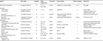
Possession of a central cavity, the H-shaped fibrillar neuropile and a pair of elongate ovaries, as well as the lack of eyes, mouth, pharynx and the intestine in the adult stage, suggest a close similarity of the new parasite to members of the family Fecampiidae, Turbellaria, Platyhelminthes. Cocoon formation for spawning is also consistent with the characteristic of this family, of which nine species classified into three genera have been described (Table 3).
Comparison between the new parasite and fecampiid species.

F, female; M, male; H, hermaphroditic; G, gonochoristic; *: no fully-developed larvae obtained but two oblong dark spots were seen anteriorly in young larvae by a histological observation
The new parasite is most similar to Fecampia species in body size and cocoon shape. However, known species of this genus are all hermaphroditic. The sexuality of the new parasite coincides only with that of Kronborgia species in the family. All Kronborgia species, where males are known, have dwarf males (about 1/5 of the length of females or shorter). If it is the case also in the new parasite, its male may be too small to find with the naked eye. Another possible explanation, though less plausible, may be differential host use between males and females.
The genus Kronborgia currently comprises five species of various body sizes and cocoon shapes (Table 3) and, thus, the new parasite may well be assigned to this genus. However, the different host use of the new parasite from all fecampiid species makes us cautious about identifying it as such. Hosts for almost all known fecampiid species are crustaceans (Table 3). The only non-crustacean host for this parasitic taxon is a myzostomatid (Annelida: Myzostomatida) for Glanduloderma myzostomatis Jägersten, Reference Jägersten1940. This parasite has been found only once and its systematic position is yet to be evaluated. In addition to the different host use, the newly discovered parasite has two characteristics different from Fecampiidae. One is the number of eggs in a capsule. In fecampiid species of which egg capsules have been observed, each capsule always contains two eggs, while the number of eggs per capsule is variable and much greater than two (19–42 eggs) in the new parasite (Table 3). The other difference is a larval behaviour for infection to hosts. Larvae of K. amphipodicola and K. pugettensis are known to make a cyst on the hosts' exoskeleton before intruding into their crustacean hosts. The crustacean exoskeleton may be a barrier for infection of these parasite species and it may be necessary for the parasites' larvae to make a cyst in order to firmly adhere to the host body surface to patiently break the barrier. Because the sacoglossan hosts of the new parasite have no hard covering, the parasite can enter into the hosts easily and quickly. Hence, they need not make a cyst.
The new parasite also shows some similarity to the family Urastomidae, although it substantially differs from urastomids in adult morphology (e.g. species of Urastomidae have a mouth, a pharynx and eye spots) (Zamparo et al., Reference Zamparo, Brooks, Hoberg and McLennan2000; Gonzàlez et al., Reference Gonzàlez, Alvarez, Dominguez, Bua, Iglesias, Fernandez and Estevez2005). The most significant similarity is host use. Known hosts for urastomids are all molluscs including the bivalves Mytilus and Crassostrea (Jennings, Reference Jennings1971; Gonzàlez et al. Reference Gonzàlez, Alvarez, Dominguez, Bua, Iglesias, Fernandez and Estevez2005). Cocoon formation for spawning is also known at least for one urastomid species, Urastoma cyprinae Graff, 1882 (Gonzàlez et al., Reference Gonzàlez, Alvarez, Dominguez, Bua, Iglesias, Fernandez and Estevez2005). Because high modification of morphology in parasitic life is the rule rather than exception, the new parasite may be included in the family Urastomidae. In fact, molecular analyses have suggested close affinity between Fecampiidae and Urastomidae (Littlewood et al., 1999a,b; Baguña et al., Reference Baguña, Carranza, Paps, Ruiz-Trillo, Riutort, Littlewood and Bray2001; Jaffe & Korkonova, 2001; Littlewood & Olson, Reference Littlewood, Olson, Littlewood and Bray2001). In order to determine the systematic position of the new parasite, a molecular analysis is essential.
Parasitism is of significant importance in the evolution of Platyhelminthes which contains Neodermata, a large monophyletic taxon diversified for obligate parasitism. Defining the sister group of Neodermata is a key to understand the origin of this large taxon; however, it seems a very difficult task. There has been much debate over it. Fecampiidae has attracted considerable attention as a candidate for the sister group of the Neodermata because of several morphological similarities (sperm ultrastructure, possession of reflector-type eyes, characteristic flame bulbs of the larval protonepheridium, etc.) (Rohde, Reference Rohde1994). However phylogenetic studies based on DNA analyses have suggested that Fecampiidae may not be the direct sister group of Neodermata but may be included in a large sister clade composed of several turbellarian families (Littlewood et al., Reference Littlewood, Rohde and Clough1999a, Reference Littlewood, Rohde, Bray and Hernioub, Reference Littlewood, Cribb, Olson and Bray2001; Baguñà et al., 2001; Lockyer et al., Reference Lockyer, Olson and Littlewood2003).
Another interest of parasitism in Platyhelminthes is the evolution of host use. Many species of Neodermata are associated with both vertebrates and invertebrates as definitive and intermediate hosts, respectively. It is a common scenario that an ancestral parasite which utilized an invertebrate as a single definitive host may have evolved into such parasite by being eaten together with the invertebrate host (Littlewood, Reference Littlewood, Maule and Marks2006). While molluscs are among important intermediate hosts for Neodermata, only a few molluscan hosts have been known for species included in the sister clade of Neodermata (Jennings, Reference Jennings1971; Littlewood et al., Reference Littlewood, Rohde, Bray and Herniou1999b). The new parasite from sacoglossan molluscs may contribute to the understanding of the evolution of host use in parasitic Platyhelminthes as well as the origin of Neodermata, the large, important parasitic clade of Platyhelminthes.
ACKNOWLEDGEMENTS
Special thanks are due to Dr K. Tajika and Dr K. Rohde for valuable advice on taxonomy and biology of Platyhelminthes, and to Dr C.D. Trowbridge for valuable advice on biology of host sacoglossan species, critically reading the manuscript and correcting English. We also thank Mr M. Sekimoto, Mr M. Sekifuji, Ms Sugii and staff of Misaki Marine Biological Station; Dr M. Aoki and staff of Shimoda Marine Biological Station; Dr K. Yasui, Dr M. Urata and staff of Mukaishima Marine Biological Laboratory; Mr K. Takiguchi and staff of Marine Biosystems Research Center, Chiba University for their kind assistance with collection of specimens. Thanks are also due to Mr Y. Ijima, Mr Y. Shimadu, Mr T. Maeda, Ms M. Yorifuji, Ms K. Kumagai, Mr K. Nishida, Mr T. Matsumine and Ms M. Sudo for their various help and support for this study.