A fundamental issue that has attracted attention in recent years is the role of morphology and interface-driven shape transitions in oxide-supported nanocrystals.[Reference Tersoff and Tromp1–Reference Marshall and Castell3] Understanding these morphological changes is important as they determine what crystallographic orientations bind the nanocrystals. Given the dominance of surface controlled properties in nanomaterials, it is these outer facets that take part in physio-chemical interactions. This has had a profound influence on the development of electrochemical and catalyst technologies.[Reference Armand and Tarascon4–Reference Gogotsi and Simon6] It follows that in order to fully understand the role of particular surfaces in such physio-chemical applications, we must first establish a detailed understanding of how a particular crystallographic surface may be formed.
Simply put, the specific equilibrium shape of a particular nanocrystal on a substrate is governed primarily by three factors; namely, the surface energy of a particular crystal facet, the interface energy between the crystal and its substrate, and the surface energy of the substrate itself.[Reference Silly and Castell2] Furthermore, it is shown that the particular crystallographic orientation can be tuned by controlling the substrate temperature and hence the thermodynamics of the crystal–substrate interface.[Reference Silly and Castell2, Reference Marshall and Castell3] The role of epitaxial strain and its effect on growth anisotropy has also been investigated.[Reference Silly and Castell7–Reference Mundschau, Bauer, Telieps and Świȩch9] In this regard, epitaxial nanocrystals, not only offer high surface-to-volume ratios, but also controlled crystallographic orientations (unlike nanocrystalline powders), and hence provide an ideal fundamental system for such studies.
Here we focus on the morphology and physical features of manganese oxide (Mn3O4) nanostructures deposited on single-crystalline strontium titanate (SrTiO3, STO) substrates. Mn3O4 has been identified by several reports as a potential replacement for (toxic) ruthenium oxide, a material commonly used as pseudocapacitors for rechargeable batteries.[Reference Broughton and Brett10–Reference Andreev, Sviridova, Chichkov, Volodin, Van Haesendonck and Mukovskii12] Capacitors made from porous manganese-oxide materials, particularly Mn3O4/MnO2 nanorods have achieved specific capacitance values as high as 256 F/g,[Reference Broughton and Brett10] where the porosity leads to an increased surface area thus improving capacitor performance.[Reference Davis13] For example, studies by Xia et al.[Reference Xia, Meng, Li, Yuan and Cui14] showed that by lithiating the dense Mn3O4 film precursors, specific capacitance as high as 314 F/g could be achieved.
Previously we have shown that nanocrystal shape, size, and orientation in Mn3O4 is controlled by the crystallographic orientation of the underlying oxide (STO) substrate.[Reference Bogle, Anbusathaiah, Arredondo, Lin, Chu, O'Neill, Gregg, Castell and Nagarajan15, Reference Bogle, Cheung, Chen, Liao, Lai, Chu, Gregg, Ogale and Valanoor16] The epitaxial relationship between the underlying oxide substrate and the growing Mn3O4 surfaces was exploited to achieve a range of morphologies with controlled crystallographic orientations. Specifically we demonstrated that (101)-oriented triangular Mn3O4 nanoplatelets were obtained when synthesized on the (111)-oriented STO and (112)-oriented Mn3O4 nanowires were obtained on the (110)-oriented STO substrates, respectively. More importantly it was shown that the specific capacitance of Mn3O4 is strongly dependent on the direction and polarity of each surface orientation.[Reference Liu, Ng, Okatan, Amal, Bogle and Nagarajan17] Although these morphological changes were touched upon, the details of the growth mechanism and interface structure have not been discussed before.
In this communication, we focus on the growth mechanisms that give rise to the (101)-orientated Mn3O4 nanoplatelets obtained on the (111) STO substrate and (112)-orientated Mn3O4 nanowires on the (110) STO. We use this system as an example to further understand substrate orientation control of nanocrystal morphology. Considering the (101) system first, spiral-terrace patterns are observed across the entire (101) Mn3O4 surface when viewed under high-magnification atomic force microscopy (AFM). These originate from linear defects at the sample–substrate interface and then expand via screw dislocation growth. Selected area electron diffraction (SAED) analysis reveals an in-plane relation of  $[2\overline {31} ]$Mn3O4//[100]STO, and an out-of-plane relation of
 $[\overline 1 01]$Mn3O4//
 $[\overline 1 11]$STO. Furthermore, 58° in-plane and 35° out-of-plane lattice misorientation have been found. The (112) nanowires, on the other hand, show a triangular cross-section under transmission electron microscope (TEM), hinting that similar to the (100) islands as seen by Bogle et al.,[Reference Bogle, Anbusathaiah, Arredondo, Lin, Chu, O'Neill, Gregg, Castell and Nagarajan15] the nanowires nucleate as pyramids. They have {022} and {202} as the binding surfaces with a much simpler epitaxial in-plane relationship of [110]Mn3O4//[100]STO, along with a 45° in-plane rotation. Analysis based on TEM diffraction also reveals that the longitudinal direction of the (112) nanowires is along the c-axis of Mn3O4, such that [001]Mn3O4//[110]STO.
The two different Mn3O4 nanostructure orientations were synthesized here following a two-step approach via pulsed laser deposition as detailed in Bogle et al.[Reference Bogle, Anbusathaiah, Arredondo, Lin, Chu, O'Neill, Gregg, Castell and Nagarajan15] Briefly, bismuth manganite (BiMnO3) was used as the starting target. A laser energy of 280 mJ was applied for 10,000 laser pulses at frequencies of 1, 3, 5, and 20 Hz for the growth of the (101)-oriented nanoplatelets and only 5 Hz frequency for the (112)-oriented nanowires. We used conditions of high temperature (875 °C) and oxygen partial pressures of 55 mTorr where in the perovskite BiMnO3 is unstable and hence spontaneously forms phase-separated bismuth oxide (Bi2O3) and Mn3O4 nanoislands. The growth process is thus based on the spontaneous phase separation of Bi2O3 and Mn3O4, where the Bi2O3 acts as a flux to separate the islands. Once the ablation is completed, the system was allowed to cool gradually at a rate of 5 °C/min under vacuum (typically 10−6 Torr). The Bi2O3 is highly volatile in such oxygen-deficient conditions therefore removed during the cooling stage, leaving behind a dispersed array of nanocrystals.
The Mn3O4 nanostructure topography and dimensions were analyzed using a JEOL JSPM 5400 environmental AFM. This was done in conjunction with scanning electron microscopy (SEM) using a FEI NOVA Nano SEM 230. θ−2θ X-ray diffraction measurements were carried out using an X-pert PANalytical MRD. In order to investigate the in-plane orientation of the Mn3O4 nanostructures grown on the STO substrate, SAED patterns were acquired from plan-view samples on a JEOL-2200F TEM at 200 kV. All samples were tilted to the STO [110] zone for TEM analysis. Specific orientation of each Mn3O4 plane has been indexed based on the SAED pattern.
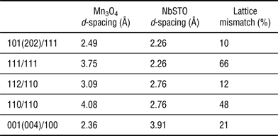
Table I tabulates the relevant misfits (at room temperature) for both the (101) and (112) Mn3O4 nanocrystals. It is clear that were the growing crystal to retain the orientation of the underlying STO, the excessively large lattice mismatches would dominate (neglecting the similar thermal coefficients of expansion) and thus render epitaxial growth completely unfavorable. This suggests that in-plane rotations must occur in order to achieve the resultant Mn3O4 nanostructures. Previously, Bogle et al.[Reference Bogle, Anbusathaiah, Arredondo, Lin, Chu, O'Neill, Gregg, Castell and Nagarajan15] showed for the (100)-orientated Mn3O4 the in-plane rotations played a critical role in maintaining the interface registry. High-resolution TEM (HRTEM) analysis coupled with SAED of (100)Mn3O4 grown on (100)STO confirmed that an in-plane epitaxy relation of [110]Mn3O4//[100]STO, with a 45° in-plane rotation. Such in-plane rotation dramatically reduces the lattice mismatch between the (100) Mn3O4 and (100) STO substrates from 21% to 4%. Applying the same logic here, in-plane rotation must account for the formation of both [101]Mn3O4//[111]STO and [112]Mn3O4//[110]STO systems.
We begin with the structural analysis of (101) Mn3O4 nanocrystals fabricated on (111) STO as shown in Fig. 1. θ−2θ scan of these structures have been previously published by Liu et al.,[Reference Liu, Ng, Okatan, Amal, Bogle and Nagarajan17] where we verified (101)-oriented growth on the (111) substrate with no secondary phase detected. AFM topography images presented in Fig. 1(a) show triangular nanoplatelets with clear stepped terrace layers. The mechanism of growth for these stepped-layers will be discussed later. Cross-section and plan-view TEM was carried out to investigate the interfacial relationship between the nanoplatelets and its underlying substrate. Figure 1(b) is a bright-field plan-view TEM image showing the (101) nanoplatelet to be equilateral triangles, with all three bounding surfaces confirmed to be along 〈111〉. Figure 1(c) is a low-magnification cross-section TEM image of (101) Mn3O4 nanoplatelets. The HRTEM image [shown as in Fig. 1(d)] suggests a rough interface between the Mn3O4 and the STO.[Reference Bogle, Anbusathaiah, Arredondo, Lin, Chu, O'Neill, Gregg, Castell and Nagarajan15] These incoherencies create “kinks” which would aid the formation of more dislocations[Reference Burton, Cabrera and Frank18, Reference Frank19] and could consequently further facilitate the growth of the terrace layers observed in AFM. From the inverse fast Fourier transform (IFFT) image [see Fig. 1(e)] of Fig. 1(d), this dislocation can now be clearly captured within the green square. The SAED pattern taken with electron beam aligned along the [110] direction of STO, is shown in Fig. 1(f). Clear lattice diffraction spots demonstrate the single-crystalline nature of each nanoplatelet. Indexing of diffraction patterns shows that the beam aligned along the STO (100) plane is also parallel to the  $[2\overline {31} ]$ zone axis of Mn3O4. This implies that the in-plane relationship of the triangular Mn3O4 must be
 $[2\overline {31} ]$Mn3O4//[100]STO, with a lattice misorientation of 58°. Furthermore, the diffraction pattern of the out-of-plane
 $(\overline 1 11)$ STO perpendicular to the (100) plane of STO, is simultaneously parallel to the
 $(\overline 1 01)$ plane of Mn3O4. This implies an out-of-plane relation of
 $[\overline 1 01]$Mn3O4//
 $[\overline 1 11]$STO with a 35° inclination. The registry of the atoms across the Mn3O4–STO interface for the nanoplatelets is illustrated in S1.
Structural analysis of (101) Mn3O4 nanoplatelets fabricated on (111) SrTiO3 STO substrate. (a) AFM topography shows triangular platelet structures, (b) plan-view TEM image shows the nanoplatelet that has facets and belongs to the {111} family, (c) cross-section TEM image shows the morphology of (101) Mn3O4 on (111) STO, (d) HRTEM image with its IFFT image (e) shows atomic registry of the Mn3O4–STO interface and the dislocation is indicated in the green square, (f) SAED pattern shows the epitaxial relationship of the nanoplatelet and the substrate. The SAED of STO is indexed by blue font and Mn3O4 by red font.

In order to further investigate the growth mechanism of the triangular islands (or nanoplatelets), higher magnification AFM was carried out. Figures 2(a) and 2(b) present the AFM images of the epitaxial Mn3O4 (101) nanostructures on the STO (111) substrate. The topographical view of each platelet reveals growth terraces spiralling outward from a center. The height between steps varies between 0.3 and 0.5 nm (see S2 for details), and remains constant within this range at different deposition frequencies (implications of which are discussed later). The schematic representation shown in Fig. 2(c) simulates the atomic structure of (101) Mn3O4, with purple and red atoms representing manganese and oxygen, respectively. From this atomic structure we observe that the structure of the (101)-cleaved Mn3O4 surface has a spiral character, which coincides with what we observe topographically.
AFM topography and height analysis of (101) Mn3O4 nanoplatelets. Panel (a) shows terrace step formation at each nanoplatelet surface, (b) height analysis of the AFM image shows total height of steps to be approximately 1.27 nm. Atomic structure presented in (c) shows the spiral growth mode thus explaining the morphology observed in the topography images. (d), (e) AFM images show the height of steps that are measured as 0.45 and 0.39 nm, respectively, at different magnifications.
A closer examination of the nanoplatelet topography in Figs. 2(d) and 2(e) reveals a dark spot at the center of the spirals. This hints that the growth spirals we observe originate from screw dislocations (both left-handed and right-handed) that are randomly distributed over the entire substrate.[Reference Andreev, Sviridova, Chichkov, Volodin, Van Haesendonck and Mukovskii12] We see this in Fig. 2(b), where the spirals appear to be growing on top of each other as well as expanding across the entire substrate. The screw dislocations are induced by linear defects introduced during the early stages of synthesis, which facilitate other dislocation sites to develop.[Reference Seiss, Ouisse and Chaussende20]
It has been shown that, when screw dislocations are present during growth, the crystal face always has exposed molecular terraces in order for growth to continue.[Reference Frank21] In our instance such dislocations are randomly distributed across the substrate surface, and terrace attached to these growth-promoting dislocations emerge in layers that are normal to the surface, thus forming the observed nanoplatelets. In addition, we observe that the height between each step corresponds to approximately half of the c-lattice parameter of Mn3O4 (0.944 nm), suggesting that the Burgers vector component of the screw dislocation perpendicular to the surface is approximately c/2[Reference Andreev, Sviridova, Chichkov, Volodin, Van Haesendonck and Mukovskii12] and that the advancement of each terrace layer is guided by the Burger's vector sweeping upward along the surface normal. This sweeping action would also give rise to a shear, and hence both in-plane and out-of-plane misorientations with respect to the substrate. Prior published spiral growth models for crystals suggest that the growing crystal develops curvature due to the presence of impurities. This leads to subsequent stress in the surface that demands a certain supersaturation at which point growth may continue if, and only if dislocations are formed.[Reference Andreev, Sviridova, Chichkov, Volodin, Van Haesendonck and Mukovskii12, Reference Burton, Cabrera and Frank18, Reference Frank21] Further growth is then guaranteed where a high concentration of kinks present during the movement of these dislocations.[Reference Cabrera and Burton22, Reference Burton and Cabrera23] The AFM images reveal that the distance between each (101) nanostructure is small, which implies a high concentration of kink sites. In such cases, the rate of advancement of each monolayer step is independent of its orientation, thus forming round spirals,[Reference Frank19, Reference Burton and Cabrera23] which can only advance with the addition of building units, i.e., adatoms that exist along the outer edges of each spiraling steps. In addition, we observe that in each case the turns of the spirals are more closely spaced near the center than further out, this is attributed to the inevitable supersaturation shortly before growth is ceased, caused by a fall in temperature during the cooling stage.[Reference Frank19] Thus for this (101) Mn3O4 on the STO (111) system, we may conclude that the mechanism of nanocrystal growth is dominated by screw dislocation dynamics.
To investigate the kinetics of this spiral-growth mode, depositions at 1, 3, 5, and 20 Hz were carried out under identical conditions as before. In each case, we ensured that the total number of pulses was kept constant, and thus the same amount of material is deposited. Average heights taken for each island decrease from 16.7 to 7.5 nm as a function of increasing frequency. Their progressive expansion in width with increasing frequencies is shown in Fig. 3. Figure 3(a) taken for the 1 Hz sample illustrates initial disjointed triangular platelets, which then proceeds to merge into elongated triangular islands at 3 Hz in Fig. 3(b). Figure 3(c) is the AFM image acquired for deposition carried out at 5 Hz. It reveals the triangles continue to conjoin and broaden into a single expansive surface. Importantly the average height of the triangles now reduces by ~ 40% compared with the 1 Hz and 10% compared with 3 Hz samples. It has been suggested that surface roughness directly relates to the diffusivity of the adatoms at the crystal surface.[Reference Ciesielski and Oleksy24, Reference Hata, Isu, Watanabe, Kajikawa and Katayama25] Lower frequency allows more time for adatom arrangement on the surface of the substrate. Individual atoms absorbed on the bottom layer of each Mn3O4 island can climb up to higher layers, creating smaller but taller island formation. Conversely, a higher frequency means that there is a higher abundance of adatom flux during deposition. We expect that lateral climb is favored in this case, as the large number of competing adatoms would render vertical ascent of individual adatoms more difficult, and thus explain the sharp decrease in the island height with increasing frequency. This trend is fully confirmed in Fig. 3(d), which is an AFM image for the deposition carried out at 20 Hz. As one proceeds to 20 Hz the nanostructures lose their individuality almost completely to become densely interconnected islands. Lateral expansion of the (101) nanoplatelets causes the path of these growth-promoting screw dislocations to widen, producing more terrace layers across the entire (101) surface with the addition of adatoms accessible along the edge of a spiral-shaped growth terrace.[Reference Andreev, Sviridova, Chichkov, Volodin, Van Haesendonck and Mukovskii12]
Height analysis of (101) Mn3O4 nanoplatelets grown at (a) 1 Hz, (b) 3 Hz, (c) 5 Hz, and (d) 20 Hz. The trend observed shows that the surface roughness directly relates to the diffusivity of the adatoms at the crystal surface. Slower frequency allows more time for adatom arrangement on the surface of the substrate. Bottom layer adatoms of each Mn3O4 island can climb up to higher layers creating smaller but taller island formation, and vice versa.
Having understood the growth mode of the (101) platelets in detail we turn our attention to the (112)-oriented nanowires obtained on (110) STO substrates. Previously we have reported that the (112) plane of Mn3O4 exhibits highest specific capacitance under standard cyclic voltammetry conditions.[Reference Liu, Ng, Okatan, Amal, Bogle and Nagarajan17] Here we aim to explain this phenomenon by understanding the complex faceting behavior of (112) Mn3O4. Figure 4(a) shows (112)-orientated Mn3O4 consisting of long nanowires approximately 32 nm in height growing at 54° with respect to the  $[1\overline {11} ]$ STO edges, obtained directly from AFM topography. Cross-sectional TEM shown in Figs. 4(b) and 4(c) reveal that these nanowires are in fact pyramidal in shape. Indexing of the diffraction pattern indicates that the electron beam aligned to the [100] direction of the STO substrate is also parallel to the [110] zone axis of Mn3O4. This implicates an in-plane crystallographic relationship of [110]Mn3O4//[100]STO (also confirmed by x-ray diffraction reciprocal space mapping shown in S3), which depicts the short axis that forms the base of the pyramidal nanowire. This also results in a calculated lattice misorientation of 45°. In addition, Figure 4(c) also shows that the Mn3O4 has two different facets, which have been identified via diffraction pattern [Fig. 4(d)] as the (022) and
 $(\overline 2 02)$ planes, each at 40° with respect to the base (110) plane of Mn3O4.
Structural analysis of (112) Mn3O4 grown on the (110) STO substrate. (a) AFM topography shows Mn3O4 nanowires on (110) STO. Panels (b), (c) show TEM of bulk and individual nanowires showing triangular cross-sections, suggesting that the nanowires are, in fact pyramidal in shape. (d) SAED pattern shows the epitaxial relationship of the nanowires (red font) and the substrate (blue font), which is also shown as schematic in (e).
Given the in-plane crystallographic relation of [110]Mn3O4//[100]STO, and out-of-plane relation of  $[\overline 1 12]$Mn3O4//
 $[0\overline 1 1]$STO from TEM, we can then deduce the directionality of the long-axis of the (112) Mn3O4 nanowires. To do this, we must first determine the interfacial registry between the various edges of Mn3O4 with its underlying STO substrate. The specific crystallographic relations have been schematically illustrated in Fig. 4(e). The y-axis of STO shown in Fig. 4(a) must be so that it is perpendicular to the out-of-plane
 $[0\overline 1 1]$ direction of STO. Having considered this, the index of the lowest order for this edge can only be [211]. Although [100] is also possible, this must be ruled out as the [100] direction of STO has already been defined via TEM diffraction, as shown in Figs. 4(d) and 4(e). Subsequently, the x-axis of the substrate in Figure 4(a), which lies perpendicular to both the [211] in-plane and
 $[0\overline 1 1]$ out-of-plane directions of STO, must be
 $[1\overline {11} ]$. From AFM topography we measured, the angle between the longitudinal axis of Mn3O4 nanowires makes an angle of approximately 54° with the now indexed
 $[1\overline {11} ]$ edge of STO. This means that the long axis of the (112) nanowire must be growing along the [110] direction of STO, which can only be of the [001] Mn3O4 registry. Hence, the in-plane relation for (112) nanowires along the longitudinal direction must be [001]Mn3O4//[110]STO, which corresponds to the longest axis, the c-axis of Mn3O4. This would also explain the anisotropic growth of the nanocrystal necessary to obtain the nanowires.
To summarize, we present detailed study on growth mechanism and crystallographic studies on various planes of Mn3O4. Based on the synthesis method previously outline by Bogle et al.,[Reference Bogle, Anbusathaiah, Arredondo, Lin, Chu, O'Neill, Gregg, Castell and Nagarajan15] we are able to achieve nanowires of (112) orientation and triangular platelets of (101) surface on (110) and (111) STO substrates, respectively. Subsequent SAED TEM studies provide in-depth studies on cross-sectional and in-plane relations. The (101) Mn3O4 plane displayed a complex growth mechanism, with in-plane relation of  $[2\overline {31} ]$Mn3O4//[100]STO, with misorientation of 58°, along with an out-of-plane relation of
 $[\overline 1 01]$Mn3O4//
 $[\overline 1 11]$STO with rotation of 35°. The (101)-orientated nanoplatelets also exhibit peculiar growth patterns, which consist of terraces growing attached to isolated dislocation sites and spirals normal to the substrate surface. The distance between each layer is controlled by the perpetual Burger's vector component which remains consistently below ½ of the c-axis.[Reference Andreev, Sviridova, Chichkov, Volodin, Van Haesendonck and Mukovskii12, Reference Burton, Cabrera and Frank18, Reference Seiss, Ouisse and Chaussende20, Reference Frank21] On the other hand, indexing of the diffraction pattern reveals that the in-plane relation for the (112) Mn3O4 system must be [110]Mn3O4//[100]STO, with 45° rotation. TEM diffraction pattern and subsequent calculations reveal that the long axis of (112) nanowires grow along its c-axis [001], which is also the longest axis of Mn3O4. We thus provide fundamental studies on oxide nanocrystal growth that shed light on how the epitaxial relationships govern the shape evolution in such transition metal oxide spinel materials systems.
Lattice spacing calculated for Mn3O4 growth on (111), (110), and (100) STO substrates, respectively. Mn3O4 (101) and (112) planes which we found to have formed on (111) and (110) STO exhibit lower d-spacing than the theoretical (111) and (110) orientations of Mn3O4. However, the overall large percentage of lattice mismatch of Mn3O4 and STO implies that the actual plane formation may be due to factors others than mechanical parameters.
Supplementary materials
For supplementary material for this article, please visit http://dx.doi.org/10.1557/mrc.2015.12
Acknowledgments
The TEM data were acquired at Australian Centre for Microscopy & Microanalysis (ACMM) in Sydney University, Australia. The authors specially thank Dr. Hongwei Liu for technical support and Mr. Henry Liu for the crystal structure graphics shown in S1. Research carried out in part at the Center for Functional Nanomaterials, Brookhaven National Laboratory, which is supported by the U.S. Department of Energy, Office of Basic Energy Sciences, under Contract No. DE-AC02-98CH10886.