Introduction
Microphytobenthos (MPB) of temperate intertidal and estuary mudflats is exposed to strong gradients in temperature and irradiance. Despite this, these areas are highly productive, and MPB can contribute up to 50% of the total ecosystem production (Underwood & Kromkamp, Reference Underwood and Kromkamp1999). MPB in estuaries, which are usually diatom-dominated (Ribeiro et al., Reference Ribeiro, Brotas, Rincé and Jesus2013), form dense photosynthetic biofilms on the sediment surface (Vieira et al., Reference Vieira, Cartaxan, Máguas and Da Silva2016). Environmental factors such as temperature and light contribute significantly to the species succession. Serôdio et al. (Reference Serôdio, Coelho, Vieira and Cruz2006), for example, reported that in an intertidal area of the Ria de Aveiro Canal, Portugal, more than 80% of the MPB communities during summer were diatom-dominated (Nitzschia perspicua, Parlibellus crucicula, Navicula gregaria and Nitzschia draveillensis), however in winter, this percentage dropped to 41.5%. Laviale et al. (Reference Laviale, Barnett, Ezequiel, Lepetit, Frankenbach, Méléder, Serôdio and Lavaud2015) likewise suggested species composition changed in response to differing light and temperature conditions, and different taxa responded differently to these changes. Apart from the influences of temperature and light, MPB is also affected by the presence of grazers, whereby locations with higher presence of grazers had better stratification of biofilms (Morelle et al., Reference Morelle, Claquin and Orvain2020).
Microphytobenthos in temperate intertidal areas exists in highly dynamic environments and experiences an extensive seasonal temperature range. In Tasmania, the average midday water temperature in Browns River (a small coastal lagoon) varies from a minimum of 9.4°C (winter) to 21.7°C (summer) (Jordan et al., Reference Underwood and Kromkamp2008) and at nearby Ralphs Bay, it ranges from 7.7 to 21.8°C from winter to summer (Cook et al., Reference Cook, Roberts, Hallegraeff and McMinn2007). Importantly, these temperature differences are thought to be responsible for a drop in photosynthesis of more than 50% in winter (Jordan et al., Reference Jordan, McMinn and Wotherspoon2008). In Browns River, higher temperatures in summer caused an increase in the maximum photosynthetic rate (rETRmax) from 27.0 ± 2.8 in winter to 65.4 ± 3.8 in summer (Jordan et al., Reference Jordan, McMinn and Wotherspoon2008). These seasonal temperature changes also affect the MPB biomass and species composition (An et al., Reference An, Choi and Noh2020). Generally, MPB production is higher in spring/summer than in winter due to sufficient light and moderate temperatures (Scholz & Liebezeit, Reference Scholz and Liebezeit2012). In winter, the production is generally significantly lower. For example, in a muddy-sand macrotidal estuary on the northern coast of France, primary production in winter can be as low as 6.7 mg C m−2 h−1 while in summer, it can increase to 97.7 mg C m−2 h−1 (Migné et al., Reference Migné, Spilmont and Davoult2004). However, in some locations, higher biomass has been recorded in winter, which is thought to be due to lower grazing pressure (Pierre et al., Reference Pierre, Zhao, Orvain, Dupuy, Klein, Graber and Maugard2014).
The light environment of intertidal habitats is highly variable, primarily due to tidal events, which expose the cells to a wide range of irradiances with significant spatial and temporal variability (Perkins et al., Reference Perkins, Mouget, Lefebvre and Lavaud2006, Du et al., Reference Du, Yan, Liu and Mao2018). MPB needs to cope with these conditions to minimize energy imbalances within the photosynthetic apparatus of the cells and maintain an optimal photosynthetic rate (Consalvey et al., Reference Consalvey, Jesus, Perkins, Brotas, Underwood and Paterson2004a, Reference Consalvey, Paterson and Underwood2004b; Laviale et al., Reference Laviale, Frankenbach and Serôdio2016). However, these communities have evolved photoprotection mechanisms, which optimize photosynthetic efficiency. At low light levels, these mechanisms ensure that maximum photosynthetic rates are maintained, whilst at high light, they prevent photodamage or photoinhibition. Non-photochemical quenching (NPQ) is often activated at high light levels to protect cells from excess energy (Blommaert et al., Reference Blommaert, Huysman, Vyverman, Lavaud and Sabbe2017). During this process, the xanthophyll cycle is activated, and the pigment diadinoxanthin (DD) is rapidly and reversibly converted to the energy dissipating pigment diatoxanthin (DT) (Lavaud & Goss, Reference Lavaud, Goss, Demmig-Adams, Garab, Adams III and Govindjee2014; Laviale et al., Reference Laviale, Barnett, Ezequiel, Lepetit, Frankenbach, Méléder, Serôdio and Lavaud2015). When the capacity of the xanthophyll cycle is exceeded due to light or temperature stress, the excess energy can damage photosystem II (PSII) and induce photoinhibition. NPQ of chlorophyll fluorescence is indicative of the level of excess energy dissipation in the light-harvesting antenna of photosystem II (PSII) (Maxwell & Johnson, Reference Maxwell and Johnson2000; Goss & Lepetit, Reference Goss and Lepetit2015). Alternatively, rather than invoking a physiological photoprotective mechanism, such as activation of the xanthophyll cycle, some diatoms invoke a behavioural response and migrate into the sediment (Consalvey et al., Reference Consalvey, Paterson and Underwood2004b, Reference Consalvey, Perkins, Paterson and Underwood2005; Laviale et al., Reference Laviale, Frankenbach and Serôdio2016) to avoid photodamage (Du et al., Reference Du, Yan, Liu and Mao2018), particularly during low tide when the surface of the sediment is exposed to high light.
Temperature also affects photosynthesis and growth rates in microalgal species from the intertidal zone (Vieira et al., Reference Vieira, Ribeiro, da Silva and Cartaxana2013). Studies have shown that thermal stress is responsible for decreasing photosynthetic rates at high and low temperatures and a reduction in the efficiency of the photosynthetic electron transport carrier (Anning et al., Reference Anning, Harris and Geider2001; Falkowski & Raven, Reference Falkowski and Raven2007; Walter et al., Reference Walter, Peters, Van Beusekom and St. John2015). High temperature stress reduces variable fluorescence (F v), maximum quantum yield (F v/F m) and the effective quantum yield (ΔF/F m), thus suggesting that structural and functional disorders of the photosynthetic apparatus occur and PSII (Photosystem II) is damaged (Salleh & McMinn, Reference Salleh and McMinn2011; Laviale et al., Reference Laviale, Barnett, Ezequiel, Lepetit, Frankenbach, Méléder, Serôdio and Lavaud2015). A decrease in variable fluorescence (F v) indicates a reduction in the number of open PSII reaction centres. Reductions in F v/F m and ΔF/F m under elevated temperature stress indicate that a significant proportion of the PSII reaction centres have been damaged (El-Sabaawi & Harrison, Reference El-Sabaawi and Harrison2006; Wu et al., Reference Wu, Zhang, Li, Xu and Beardall2020). Temperature has a major effect on photosynthetic parameters, particularly electron transport rate (ETR) because they are controlled by enzyme reactions (Underwood & Kromkamp, Reference Underwood and Kromkamp1999). As most photosynthetic reactions originate in PSII, this is the most heat-sensitive part of the photosynthetic apparatus (Davison, Reference Davison1991).
Intertidal sediment temperature variations can occur on long (days) and short (hourly) time scales, depending on factors such as local meteorological conditions and tidal inundation. Most studies on MPB have examined the responses of monocultures of diatoms. Results may thus not represent the photosynthetic characteristics of organisms in an intact MPB biofilm. Hence the work herein is intended to show the combined effects of high light and high temperature on the photophysiology of intertidal diatom-dominated MPB communities from Browns River, Tasmania, specifically how they respond to elevated temperatures in summer and low temperatures in winter. In addition, the recovery of MPB from light and temperature stressed communities was examined and an assessment made of whether these communities could acclimate to the given conditions.
Materials and methods
Field sampling
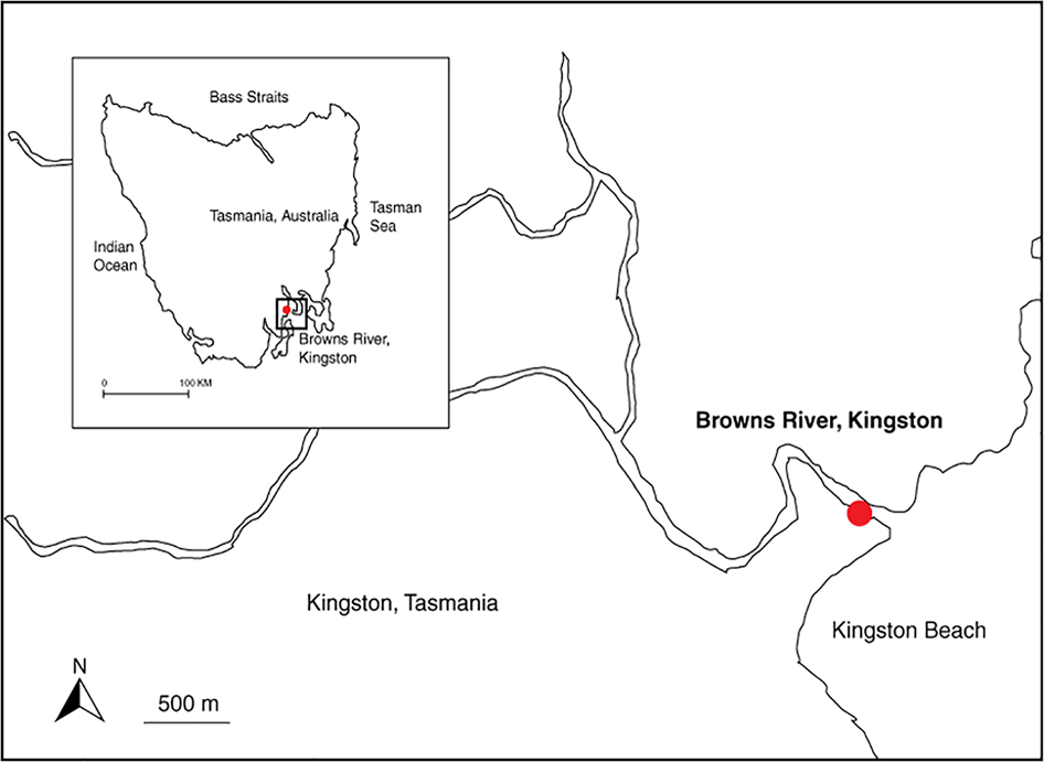
Browns River (42°96′S 147°51′E) is situated in Kingston, south-eastern Tasmania (Figure 1). The river runs from Mount Wellington to Kingston Beach and flows into the estuary of the Derwent River. This ecosystem, which has a small tidal range of ~2 m, is exposed during low tide and submerged during high tide. The benthic chlorophyll a, in-situ photosynthetic parameters (see below) and environmental parameters were sampled in both winter (August 2007), and summer (January 2008); the temperature experiments were also conducted in both summer and winter (0–15°C in winter and 20–40°C in summer). Sampling was performed at noon during low tide when the surface sediment was exposed. The surface irradiance (PAR) was measured as μmol photons m−2 s−1 using a Biospherical QP radiometer with 2π sensor, whereby the probe was placed on the sediment surface. Sediment temperature was measured by inserting a temperature probe (HI 766C) (K-type thermocouple thermometer, HI935005, Hanna Instruments, Woonsocket, Rhode Island, USA) vertically into the sediment. Triplicate measurements were taken of all parameters.
Sampling location in Browns River, Kingston, Tasmania (marked in red).

Species composition
As diatoms are usually the main contributors to the primary productivity in these benthic ecosystems, relative diatom abundance was determined at each sampling to provide a general idea of the benthic diatom diversity. One 150 mm long sediment core (diameter 45 mm) was collected at each sampling period, and the top 10 mm was removed and preserved in Lugol's iodine solution (10 ml). Samples were later cleaned in hydrogen peroxide (H2O2) and mounted onto slides using the Naphrax mounting medium. Diatom identity was assessed using a Zeiss Light Microscope (Zeiss, Germany). A minimum of 400 individuals was counted per sample using a 100× oil immersion lens. Abundances are expressed as relative abundance (% total diatoms) of frustules counted. Diatom identification was based on selected references (Round et al., Reference Round, Crawford and Mann1990; Cooper, Reference Cooper1995; Castrejon, Reference Castrejon1999; Witkowski et al., Reference Witkowski, Lange-Bertalot and Metzeltin2000).
Chlorophyll a and photosynthetic parameters of the microphytobenthos
The sediment samples for both chlorophyll a and in-situ photosynthetic parameter analysis were collected in both winter and summer. Six sediment cores (45 mm diameter) were taken; three for chlorophyll analysis and three for fluorescence analysis. The cores were taken at water depths of ~0.3 m. Samples were placed in an ice-filled light-proof container and immediately transferred to the laboratory for chlorophyll a analysis. The top 10 mm of sediment was re-suspended in 10 mL methanol and measured on a Turner Designs 10 AU fluorometer using the acidification method (Holm-Hansen et al., Reference Holm-Hansen, Lorenzen, Holmes and Strickland1965). The fluorometer was calibrated against a chlorophyll a standard (Sigma Chemical Co., St. Louis, MO, USA).
The photosynthetic parameters were measured on-site using a Water PAM (Walz, Effeltrich, Germany). The PAM fluorometer comprises a computer-operated PAM-Control Unit and a WATER-ED-Universal emitter-detector unit (Walz, Effeltrich, Germany). The top 10 mm of each sediment core was collected and shaded (kept in the dark plastic bag) to avoid direct sunlight exposure during collection and handling. Filtered seawater was added to re-suspend each 10 mm sediment sample and transferred rapidly to the Water PAM. Rapid light curves (RLC) were taken under software control (WinControl, Walz GmbH, Effeltrich, Germany) to obtain values for maximum quantum yield (F v/F m), relative ETRmax (rETRmax), photosynthetic efficiency (α) and photoacclimation index (E k).
Temperature and irradiance experiments
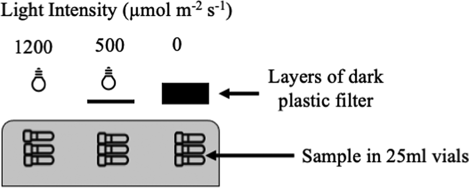
Sediment samples were collected from Browns River during both summer and winter for the temperature and irradiance incubation experiments. Samples were always taken from the same area, which was always submerged. The top 0.5 cm of the sediment was collected during low tide by hand using a clear polycarbonate core and wrapped in a dark plastic bag prior to analysis. The cores were kept upright and immediately returned to the lab for experimentation. The top 0.5 cm of the sediment cores was placed in a beaker and excess seawater decanted. The concentrated sediment samples were then transferred to transparent 20 ml glass vials. Filtered seawater was added to each vial and placed in three different light intensity chambers in a temperature-controlled water bath (Lauda 20 L, Brinkmann Lauda Inc. Konigshoven, Germany). Each chamber had a constant light intensity (Phillips Horticultural Lamp, SON-T 400 AGRO, 400W, Belgium) controlled by layers of dark plastic filters. The irradiance in each chamber was measured with a LI-COR Quantum-Light Meter (Model L1-189). The cells were exposed to three different irradiance treatments, A: 1200 μmol photons m−2 s−1, B: 500 μmol photons m−2 s−1 and C: 0 μmol photons m−2 s−1 with three replicates for each treatment. Samples were placed in the water bath for 1 h for each temperature treatment. The start of each experiment was carefully planned to ensure samples were not exposed for more than 1 h. The temperatures selected were 0, 5, 10, 15°C for winter and 20, 25, 30, 35 and 40°C for the summer experiments (Figure 2). Lower temperatures (0–15°C) were chosen for winter samples because the community had naturally been growing at these lower temperatures in winter and at higher temperatures (20–40°C) in summer. Freshly collected sediment samples were used for each temperature change. After each temperature treatment, the chlorophyll fluorescence and photosynthetic recovery were determined (Ralph & Gademann, Reference Ralph and Gademann2005; McMinn & Hattori, Reference McMinn and Hattori2006) using a PAM fluorometer (Water PAM, Walz).
Schematic representation of experimental set-up. Freshly collected samples were placed in the water bath for 1 h for each temperature treatment. The temperature of the water bath was adjusted to 0–15°C (winter) and 20–40°C (summer) experimental set-up.

Chlorophyll fluorescence measurement
Photosynthetic parameters of each treatment were measured using a Water PAM, following Ralph & Gademann (Reference Ralph and Gademann2005), Jordan et al. (Reference Jordan, McMinn and Thompson2010), Salleh & McMinn (Reference Salleh and McMinn2011). To calculate the maximum quantum yield (F v/F m), samples were dark-adapted in the field for 15 min by wrapping the containers in aluminium foil and placing them in a dark container. Rapid light curves (RLC) were taken under software control (Wincontrol, Walz) to obtain values for F v/F m, NPQ, α and E k (Ralph & Gademann, Reference Ralph and Gademann2005). Red light-emitting diodes (LED) provided the actinic light used in the RLC at levels of 0, 85, 125, 194, 289, 413, 517, 1046 and 1554 μmol photons m−2 s−1. The saturation pulse was set at 3000 μmol photons m−2 s−1 for 0.8 s. Samples were exposed to each light level for 10 s. Photomultiplier gain (PM-gain) was adjusted to between 6 and 12 before each measurement to keep the measurements consistent between samples. The relative electron transport rate (rETR) was calculated by multiplying the irradiance (PAR) by the quantum yield measured at the end of that interval (Genty et al., Reference Genty, Briantais and Baker N1989). PAR vs rETR curves was described using the model of Jassby & Platt (Reference Jassby and Platt1976) using multiple non-linear regression curve fitting protocols on SPSS software (IBM SPSS V18.0). The initial slope of the function is termed α, which is a measure of the plant cells' ability to utilize light. The maximum photosynthetic rate (rETRmax) occurs when the function reaches a plateau. E k is the light saturation parameter and calculated from the intercept between rETRmax and α (Falkowski & Raven, Reference Falkowski and Raven1997).
Non-photosynthetic quenching (NPQ) measures photoprotection activity, processes that protect against over-reduction of the photosynthetic electron transport chain by dissipation of excess absorbed light energy in the PSII antenna system as heat (Ruban et al., Reference Ruban, Lavaud, Rousseau, Guglielmi, Horton and Etienne2004). The short light exposure time in this experiment (10 s duration per light step in the RLCs), would have been insufficient for maximum NPQ development and so are only a relative measure of the ability of the treatments to develop NPQ (Ralph & Gademann, Reference Ralph and Gademann2005). NPQ was determined as NPQ = (F m−F m’)/F m’ (Schreiber, Reference Schreiber, Papageorgiou and Govindjee2004).
Recovery from photoinhibition
The method of determining and analysing the rate of recovery was adapted from McMinn & Hattori (Reference McMinn and Hattori2006) and Salleh & McMinn (Reference Salleh and McMinn2011). The measurements of recovery from photoinhibition were made using the pre-installed routine RLC + Recovery of the Water PAM. After the last actinic light period of a RLC, the sample was kept in the dark, while F v/F m was determined after 30, 60, 180, 300 and 600 s. The rate of recovery from photoinhibition was calculated following Oliver et al. (Reference Oliver, Whittington, Lorenz and Webster2003):

Where Φt is the given value of F v/F m at time t (s), Φ I is the initial value of F v/F m before recovery measurements have commenced, ΦM is the fully recovered value and r is an exponential rate constant for the recovery of F v/F m from photoinhibition (s−1). This formula assumes that the recovery rate of F v/F m is dependent on the degree of damage (McMinn & Hattori, Reference McMinn and Hattori2006).
Statistical analysis
The mean values and standard deviations of all parameters were calculated from three (N = 3) independent replicates. Significant differences between the photosynthetic parameters measured in winter and summer were determined using a Student's t-test. To evaluate the effects of temperature and irradiance (fixed factor) on photosynthetic parameters (F v/F m, α, rETRmax and E k), NPQ and recovery, a two-way analysis of variance (ANOVA) was used. A significance level of P < 0.01 was considered to be strongly significant, while P < 0.05 was treated as significant. Before analyses, the data were checked for normality and homogeneity of variances using Shapiro–Wilk's and Levene's tests, respectively. Data were transformed whenever necessary to comply with ANOVA assumptions. Differences were accepted as significant at P < 0.05 unless otherwise stated. All statistical analyses were performed using SPSS V18.0.
Results
Environmental conditions
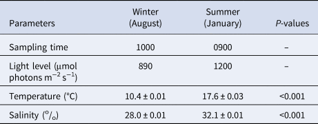
The surface irradiance recorded during the winter sampling was 890 μmol photons m−2 s−1 at 10:00 h and 1200 μmol photons m−2 s−1 at 09:00 h in the summer sampling. The seawater temperature ranged from 10.4–17.6°C between winter and summer. Salinity in summer was significantly higher (32.1 ± 0.01) by comparison to that in winter (28.0 ± 0.01) (Table 1).
Environmental parameters of benthic diatoms collected during winter and summer in Browns River, Kingston

Values are mean ± SD (N = 3).
Species composition
The benthic diatom community in Browns River was mostly dominated (abundance (%) given in parentheses) by species from the genera Achnanthes, Amphora, Cocconeis, Gyrosigma, Navicula, Nitzschia and Pleurosigma; species from these collectively compromised >70% of the community. In winter Achnanthes brevipes (25.9%), Cocconeis peltoides (17.8%), Navicula punctulata (15.8%) and Amphora ovalis (10.8%) were the most abundant. While in summer, the community diversity was evenly distributed between Amphora coffeaeformis (23.4%), Navicula zostereti (16.7%), Nitzschia dubia (15.4%) and Navicula gregaria (14.7%) being the most common.
Chlorophyll a and in-situ photosynthetic parameters of the microphytobenthos
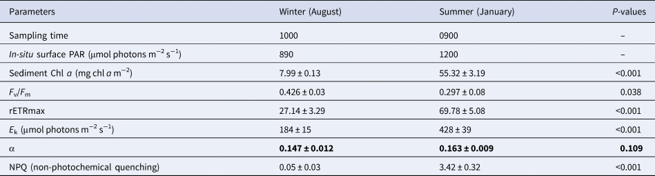
The sediment surface chlorophyll a concentration was measured during both summer and winter. In winter, average biomass was 7.99 ± 0.13 mg Chl a m−2, while in summer, the concentration increased to 55.32 ± 3.19 mg Chl a m−2. Significant increases in rETRmax, E k and α values were seen between summer and winter. rETRmax in winter was 13.76 ± 0.99, but this increased significantly to 36.76 ± 2.28 in summer. E k value in winter was 224.9 ± 15.16, and it increased to 534.3 ± 49.7 in summer. Significant declines, however, were observed in both the effective and maximum quantum yields. As surface PAR and temperature increased in summer, a significant decline in F v/F m was observed. NPQ was activated as a photoprotective mechanism during summer (3.42 ± 0.32) but was almost inactive in winter (0.05 ± 0.03). The photosynthetic parameters (F v/F m, rETRmax, E k and NPQ) were significantly different between the two seasons (Table 2).
Photosynthetic parameters of microphytobenthos from Browns River in the surface 10 mm (depth ~ 0.3 m)

Values are mean ± SD. rETRmax (relative maximum photosynthetic rate); E k (light saturation parameter); α (photosynthetic efficiency); F v/F m, maximum quantum yield. (N = 3). Bold indicates a non-significant difference (P > 0.05).
Temperature and irradiance experiments
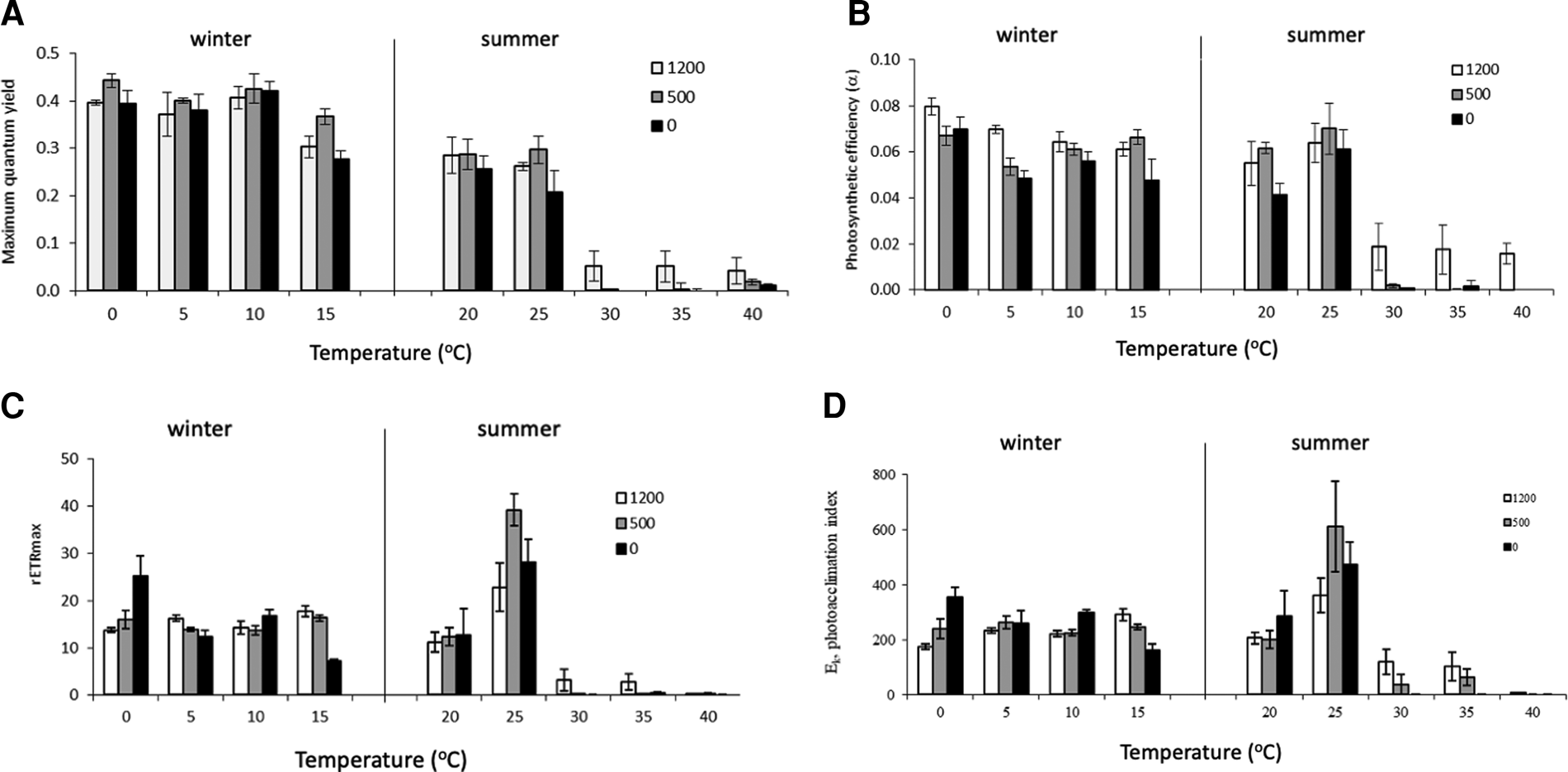
In general, increased experimental temperatures had a more significant impact on the photosynthetic parameters in summer than in winter (Table 3; Figure 3). RLCs taken in summer showed more evidence of photoinhibition in those exposed to temperatures above 25°C than those exposed to lower temperatures (0–15°C) in winter (Supplementary Figures 1a–d and 3a–e). A sharp decline was observed in all photosynthetic parameters at each light level (1200 μmol photon m−2 s−1, 500 μmol photon m−2 s−1 and 0 μmol photon m−2 s−1 in summer and winter) when incubation temperatures were increased beyond 25°C (Figure 3). The cells were apparently only able to photosynthesize at temperatures up to 25°C. At the low temperatures during the winter sampling (0–10°C) F v/F m increased by 5–10% at all light levels. Above 10°C, F v/F m decreased and at 40°C, the F v/F m values were reduced to only 0.05–0.06 (Figure 3A).
(A) Maximum quantum yield (F v/F m), (B) Photosynthetic efficiency (α), (C) Relative electron transport rate (rETRmax) and (D) Photoacclimation index (E k). Photosynthetic parameters for microphytobenthos incubated for 1 h at different temperatures (0–15°C in winter and 20–40°C in summer) and three different irradiance levels (1200, 500 and 0 μmol photons m−2 s−1). Values are means ± SD (N = 3).

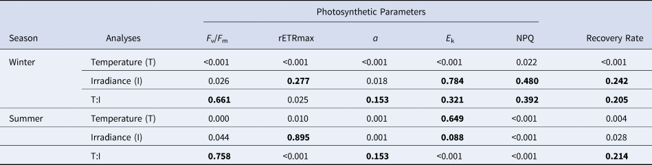
Two-way ANOVA of effects of temperature (0–15°C in winter and 20–40°C in summer) and irradiance level (1200, 500 and 0 μmol photons m−2 s−1) on the photosynthetic parameters (F v/F m, maximum quantum yield; α, photosynthetic efficiency; rETRmax, relative electron transport rate; Ek, photoacclimation index), NPQ (non-photochemical quenching) and recovery rate of the microphytobenthos after exposure to temperatures treatment for 1 h

Bold indicates a non-significant difference (P > 0.05).
There was a significant decrease in α at temperatures between 0–15 °C at different light intensities for the winter MPB, while a significant decline was observed (by 80%) as the temperature increased from 25–40 °C during the summer sampling. As anticipated, high light (1200 μmol photon m−2 s−1) produced a higher photosynthetic efficiency at temperatures of 0–10°C (Figure 3B). The relative maximum photosynthetic rate (rETRmax) was highest at 25°C regardless of the irradiance level. The rETRmax at 25°C, ranged from 39.18 ± 3.42 (500 μmol photons m−2 s−1) to 22.83 ± 1.05 (1200 μmol photons m−2 s−1) (Figure 3C), which was lower than values recorded during summer (Table 2). rETRmax values decreased to almost zero when incubated at 40°C. The E k (photoacclimation index) was highest at 25°C (500 μmol photons m−2 s−1), although it is likely that these values are problematic. If these outlying values are eliminated, the highest values were at 25°C, and the lowest was at 40°C (Figure 3D).
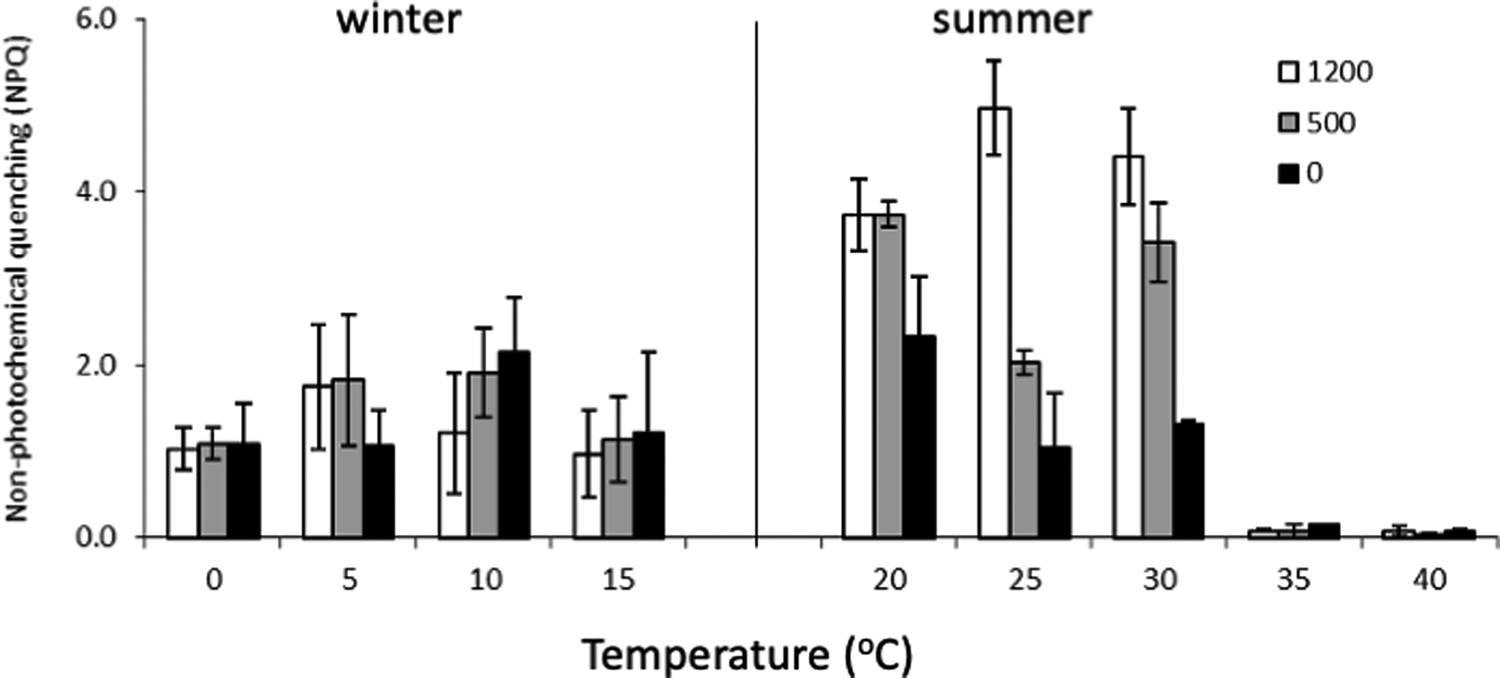
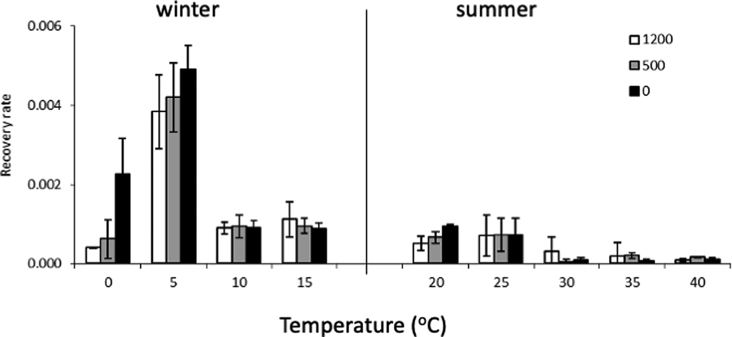
High temperature and irradiance levels caused more significant NPQ activity in the summer MPB compared with the winter incubations. At an irradiance level of 1200 μmol photon m−2 s−1, the highest NPQ values were at 25°C, at 500 μmol photon m−2 s−1 and 0 μmol photon m−2 s−1, samples incubated at 20°C had the highest NPQ values (Figure 4). Significant differences were observed between the temperature treatments (Table 3). NPQ was not activated at any light level in samples incubated at 35 and 40°C. The ability of cells to recover was not affected by the light intensity, but temperature had significant effects (Table 3) on the recovery rate. The recovery rates were highest at 5°C and decreased significantly when samples were exposed to 30–40°C (Figure 5). Incubations at temperatures between 10–25°C (all irradiances) showed a similar recovery rate.
The NPQ of microphytobenthos incubated for 1 h at different temperatures (0–15°C in winter and 20–40°C in summer) and three irradiance levels (1200, 500 and 0 μmol photons m−2 s−1). The NPQ values were obtained at the end of RLC with actinic irradiance of 1520 μmol photons m−2 s−1. Values are means ± SD (N = 3).

The recovery rate s−1 of F v/F m after the RLC. Microphytobenthos incubated for 1 h at different temperatures (0–15°C in winter and 20–40°C in summer) and three different irradiance levels (1200, 500 and 0 μmol photons m−2 s−1). Values are means ± SD (N = 3).

Discussion
In this study, the combined effects of higher temperature and irradiance significantly increased susceptibility to photoinhibition. The fluorometric analyses of the samples incubated at irradiances of 0, 500 and 1200 μmol photon m−1 s−2 showed that significant photosynthetic activity was only present at temperatures up to 25°C. Above 25°C, all photosynthetic parameters declined significantly, suggesting damage to the photosynthetic apparatus, while temperatures below the natural ambient minimum (~9.0°C) did not affect the photosynthetic activities. Since the MPB in Browns River naturally experiences temperatures below 0°C (Lee & McMinn, Reference Lee and McMinn2013) in winter, the MPB incubated at temperatures down to 0°C did not show any sign of photoinhibition, despite an increase in light levels. Similarly, Lee & McMinn (Reference Lee and McMinn2013) found that the photosynthetic parameters (Water PAM) of MPB from Browns River were not adversely affected by freezing conditions. Elsewhere, however, it has been shown that temperatures below the optimal can disrupt the enzymatic reactions and completely inhibit photosynthesis (Morris & Kromkamp, Reference Morris and Kromkamp2003; Falkowski & Raven, Reference Falkowski and Raven2007). A possible explanation for the observations at Browns River is that the natural populations of MPB grew in a highly fluctuating temperature regime (Jordan et al., Reference Jordan, McMinn and Wotherspoon2008), unlike those grown at a stable growth temperature (20°C) (Morris & Kromkamp, Reference Morris and Kromkamp2003).
In summer, when experimental temperatures rose above ambient (>25°C), the 1-h incubations used herein were sufficient to cause damage to PSII regardless of light intensity. Temperature optima of 25–30°C have been reported in some other studies of temperate benthic diatom cultures (e.g. Claquin et al., Reference Claquin, Probert, Lefebvre and Veron2008; Salleh & McMinn, Reference Salleh and McMinn2011; Gleich et al., Reference Gleich, Plough and Glibert2020), which are similar to those used in the summer MPB incubations presented here. However, others (Hancke & Glud, Reference Hancke and Glud2004) have found a lower temperature optimum of 12–15°C for intact subtidal MPB communities exposed to short-term changes in temperature during winter months. Vieira et al. (Reference Vieira, Ribeiro, da Silva and Cartaxana2013) observed a higher temperature optimum (up to 35°C) for intact MPB communities, similar to those observed naturally in summer at midday low tides in the Tagus estuary, Portugal. This indicates that experimental optimal temperatures for intact MPB correlate largely with in-situ (natural habitat) temperature ranges and may provide valuable information regarding the potential photosynthetic responses of these organisms while part of an MPB biofilm.
The maximum quantum yield (F v/F m), obtained when all reaction centres are open, is proportional to the fraction of reaction centres capable of converting absorbed light to photochemical energy. Thus, a low F v/F m can be used to indicate stress. A decrease in F v/F m at constant irradiance generally implies damage to PSII or approaching photoinhibition when exposed to adverse temperatures (Béchet et al., Reference Béchet, Laviale, Arsapin, Bonnefond and Bernard2017). It suggests that high temperatures affect the PSII electron transport and NADPH and ATP generation, leading to inhibition of carbon fixation and assimilation by the primary photochemical reactions. This will modulate cellular concentrations of RUBISCO and the Calvin Cycle enzymes that directly affect F v/F m (Davison, Reference Davison1991). F v/F m values were highest at 0–10°C and then declined as the temperature increased. The values obtained at incubation temperatures of 0–10°C were most similar to the in-situ winter values (0.421 ± 0.034) when the ambient temperature was also 10°C. Above 10°C, F v/F m decreased gradually.
In summer, when samples were exposed to higher temperatures, a large decrease in F v/F m was observed when they were incubated at 30°C. F v/F m decreased by almost 85% at all light levels when the temperature was increased from 25 to 30°C. A further increase in temperature caused F v/F m to decline to almost zero. In natural environments, when the summer sea temperature was 17.6°C, lower in-situ F v/F m (0.297 ± 0.028) values were obtained compared with those in winter (temperature: 10.4°C/F v/F m: 0.427 ± 0.031). This was mainly due to the lower irradiance and cooler temperatures during winter not being at a level to impair photosynthesis. Thus, if ambient water temperatures were to increase, F v/F m values of this community would be significantly affected. During the summer months, the highest in-situ temperatures recorded were less than 25°C (Jordan et al., Reference Jordan, McMinn and Wotherspoon2008), which suggests that temperatures greater than 25°C would also adversely affect the photosynthetic capacity of these communities.
Although MPB in estuarine environments are frequently exposed to very high irradiances and temperatures, photoinhibition is rarely recorded. Photoinhibition is prevented by both behavioural (Blommaert et al., Reference Blommaert, Lavaud, Vyverman and Sabbe2018) and photophysiological responses, such as the activation of NPQ (non-photochemical quenching). NPQ is considered to reflect a mechanism of photoprotection that prevents the over-reduction of the photosynthetic electron transport chain by dissipating excess absorbed light energy in the PSII antenna system as heat (Pniewski et al., Reference Pniewski, Richard, Latała and Blanchard2017). It has been suggested that exposure to direct sunlight at low temperatures can be particularly damaging to the photosynthetic apparatus, and this is expected to slow down the photoprotective response under high light (Serôdio et al., Reference Serôdio, Cruz, Vieira and Brotas2005). At Browns River, it was found that NPQ was constant at temperatures between 0–15°C ranging from 1.0 to 2.2 at all three light levels. This implies that the low temperatures did not affect the capability of the MPB to use light for photosynthesis. Furthermore, the high maximum quantum yields did not indicate any sign of photoinhibition. However, the NPQ values increased significantly at temperatures between 20 and 30°C for samples exposed to 500 and 1200 μmol photons m−2 s−1, implying that the photoprotective mechanism had been induced. Activation of high NPQ also occurred at higher light intensities (500 and 1200 μmol photons m−2 s−1). At 35 and 40°C, NPQ was not activated and damage was probably occurring as indicated by the significant decrease in F v/F m and other photosynthetic parameters. Thus, indicating photoinhibition resulting from an increase in temperature and high light exposure. High temperature (>35°C) is known to reduce temperate MPB motility and to deactivate their photoprotective capacities to avoid photoinhibition under high light (Laviale et al., Reference Laviale, Barnett, Ezequiel, Lepetit, Frankenbach, Méléder, Serôdio and Lavaud2015). This, however, is inconsistent with the results of Anning et al. (Reference Anning, Harris and Geider2001) and El-Sabaawi & Harrison (Reference El-Sabaawi and Harrison2006), who demonstrated that there was a decrease in photoprotective pigments associated with the xanthophyll cycle in temperate diatoms exposed to supra-optimal temperatures. Maxwell & Johnson (Reference Maxwell and Johnson2000) suggested that an increase in NPQ could occur either as a protective mechanism or as the damage itself. Jordan et al. (Reference Jordan, McMinn and Thompson2010) confirmed the presence of xanthophyll activity in Browns River MPB.
During chronic photoinhibition, when most of the photosynthetic apparatus is inactive, the excess excitation energy is no longer dissipated harmlessly and this affects the recovery process (Cartaxana et al., Reference Cartaxana, Domingues, Cruz, Jesus, Laviale, Serôdio and da Silva2013). At Browns River, the highest recovery rates were observed at 5°C and 0 μmol photon m−2 s−1, suggesting that at these temperature and light levels, damage was minimal (Lee & McMinn, Reference Lee and McMinn2013). The ability to recover rapidly implied the presence of a protective action that prevented PSII damage (Xu et al., Reference Xu, Wang, Wu and Li2014). This conclusion is consistent with the high maximum quantum yield values of these samples. At 10°C and above, the recovery rate decreased as the temperature increased. The lack of recovery of MPB exposed to temperatures above 30°C suggests that photosynthetic enzymes may have been either de-activated or damaged. The reduction in the photosynthetic parameters and low rate of recovery at higher temperatures are both characteristics of photoinhibition. Degradation of D1 protein may have occurred in the higher temperature treatments and a 10-min recovery period may not have been sufficient for repair. At Browns River, the low maximum quantum yields and the deactivation of NPQ at higher temperatures suggests that the damage was irreversible. Slow recovery rates may result in reduced carbon-fixation under natural light regimes and subsequently lead to photoinhibition when exposed to prolonged heat stress (Lambrev et al., Reference Lambrev, Miloslavina, Jahns and Holzwarth2012; Blommaert et al., Reference Blommaert, Huysman, Vyverman, Lavaud and Sabbe2017). Recovery rates are dependent on the repair of damaged reaction centres and both the rate and extent of recovery are light and temperature dependent.
The photosynthetic efficiency (α) measures the cells' ability to utilize light. At temperatures between 0 and 25°C, α remained constant at all light levels suggesting that these temperatures did not affect the ability of the cells to harvest light for photosynthesis. Furthermore, the short-term (1-h) incubations may not have been sufficiently long to affect the light-harvesting efficiency at ambient temperatures (0 and 25°C) (Morris & Kromkamp, Reference Morris and Kromkamp2003). It is known that the temperature-based acclimation of light-harvesting properties in the form of pigment complexes involves an adjustment in both the number and ratio of several photosynthetic pigments (Davison, Reference Davison1991). If incubated for a longer period, the α value may have been affected by temperature, thereby giving the cells enough time to acclimate to a change in temperature, as observed in other studies (Mock & Hoch, Reference Mock and Hoch2005; Claquin et al., Reference Claquin, Probert, Lefebvre and Veron2008). For example, in response to low temperatures, algae can decrease their pigment concentrations and their ratio of accessory pigments to chlorophyll a, thereby reducing the light-harvesting capability (Casper-Lindley & Björkman, Reference Casper-Lindley and Björkman1998; Anning et al., Reference Anning, Harris and Geider2001; Claquin et al., Reference Claquin, Probert, Lefebvre and Veron2008).
However, in this study, at temperatures above 30°C, damage occurred, and light-harvesting activity stopped. This suggests that at temperatures beyond optima, α is linked to photochemical reactions that are largely independent of temperature (Davison, Reference Davison1991; Kirk, Reference Kirk1994). Furthermore, α values obtained at temperatures between 0 and 25°C were similar to in-situ values obtained during summer and winter in this study and in those of Jordan et al. (Reference Jordan, McMinn and Wotherspoon2008). A similar trend was also seen in the diatom Cylindrotheca closterium, where a decrease in α only occurred at the most extreme temperatures of 5 and 35°C (Morris & Kromkamp, Reference Morris and Kromkamp2003). It is widely accepted that relationships between α and temperatures are species-specific. For example, using a Water PAM, Claquin et al. (Reference Claquin, Probert, Lefebvre and Veron2008) have shown that there was no relationship between α and temperature from 5–35°C in Skeletonema marinoi, although seven other phytoplankton species did have a relationship with temperature. At Browns River, whether the effects of temperature on α were species-specific or not could not be determined since multiple species were present in all samples.
The maximum electron transport rate (rETRmax) is a measure of the relative maximum photosynthetic capacity. rETRmax, and subsequently the rate of photosynthesis, increases with both increasing temperature and light (up to light saturation irradiance) (Kirk, Reference Kirk1994). At Browns River, irradiances up to 1200 μmol photon m−2 s−1 and temperatures between 0 and 15°C did not affect the photosynthetic performance of the intertidal MPB in winter. The rETRmax values obtained in this study suggest that Browns River MPB were productive at temperatures up to 25°C. Similarly, in-situ rETRmax values obtained in summer were within the range obtained for samples incubated at 25°C regardless of light intensity. In a monthly monitoring experiment at Browns River, Jordan et al. (Reference Jordan, McMinn and Wotherspoon2008) obtained their highest rETRmax values (65.4 ± 3.8) at in-situ temperatures between 20 and 25°C, whereby measurements were also done using a Water PAM. This is consistent with our results that showed a maximum photosynthetic rate at 25°C. Temperatures beyond this affected the capability of MPB to photosynthesize. While most results from temperate regions have photosynthetic optima between 15 and 25°C (Coles & Jones, Reference Coles and Jones2000), Blanchard et al. (Reference Blanchard, Guarini, Gros and Richard1997) found that diatoms from intertidal mudflats of the temperate Bay of Marennes-Oleron in France had the highest photosynthetic rate at a temperature of 30°C. rETRmax values will usually increase as the temperature increases. However, beyond an optimal temperature, the values will decrease significantly (Ibelings, Reference Ibelings1996). Yun et al. (Reference Yun, Lee and Chung2010) noted that the large diatom species Pleurosigma elongatum and Amphora coffeaeformis had higher rETRmax values (Diving PAM) under increased temperature (up to 35°C). Although both species were present in the Browns River (and among the dominant species), such temperature tolerance in the summer experiments was not observed. The cumulative responses of the photosynthetic characteristics (Vieira et al., Reference Vieira, Ribeiro, da Silva and Cartaxana2013) of the overall MPB communities in Browns River are likely responsible for the observed result, hence providing differences in stress responses. Optimal temperature ranges depend on the composition of the communities and locations investigated, as different species or groups differ in their optimal temperature ranges.
Studies have shown that low temperatures are likely to significantly impact cell growth with cells grown at colder temperatures absorbing less light than those grown at a warmer temperature (Mock & Hoch, Reference Mock and Hoch2005; Gleich et al., Reference Gleich, Plough and Glibert2020). However, using very short exposure steps of different irradiances (such as in RLCs) may lead to an overestimation of rETRmax (Serôdio et al., Reference Serôdio, Cruz, Vieira and Brotas2005; González-Guerrero et al., Reference González-Guerrero, Vásquez-Elizondo, López-Londoño, Hernán, Iglesias-Prieto and Enríquez2022), unlike using O2 or CO2 gas exchange methods. In particular, rETRmax measurements generated from RLCs do not achieve steady-state condition during each light step, and down-regulation of photosynthesis can occur. Hence rETRmax should be interpreted with care as temperature tolerance may have more pronounced effects on actual ETR than is reported in our results (Torres et al., Reference Torres, Ritchie, Lilley, Grillet and Larkum2014). Nevertheless, many studies have provided a useful estimation of photoadaptation of MPB living in the variable intertidal sediments (e.g. Laviale et al., Reference Laviale, Barnett, Ezequiel, Lepetit, Frankenbach, Méléder, Serôdio and Lavaud2015; Blommaert et al., Reference Blommaert, Lavaud, Vyverman and Sabbe2018).
Ek is commonly used as a parameter to characterize the photoacclimation status (Kirk, Reference Kirk1994; Consalvey et al., Reference Consalvey, Perkins, Paterson and Underwood2005). Since the light-saturated rate of photosynthesis is strongly affected by temperature, Ek was expected to decrease as the temperature decreased. At a temperature optimal of 25°C, when rETRmax was the highest, the MPB communities were photoacclimated to higher irradiances (higher Ek). Ek showed no clear response to temperatures between 0 and 20°C but was affected at temperatures of 30–40°C. At low temperatures, the photosynthetic dark reactions work slowly, thus less light is required. However, at Browns River, this process was not observed, as the E k values remained similar below 25°C, suggesting that the MPB were able to adjust their metabolism to the given light environment. Furthermore, samples exposed to low irradiance at ambient temperatures (below 25°C) were acclimated to the conditions and able to perform photosynthesis. This is indicated by the low E k values and suggests that the MPB could acclimate to the limited light availability. Richardson et al. (Reference Richardson, Beardall and Raven1983) suggested that the differences between E k values and ambient irradiance were primarily due to an underestimation of photosynthetically active radiation (PAR) within benthic sediments, which was caused by strong backscattering. They concluded that PAR within the benthic habitat might be 50% lower than the down-welling PAR measured at the benthic surface. This is likely to be the case in this current study as the MPB were partly buried or shaded by the sediment.
This study aimed to assess the combined effects of light and temperature stress on the photosynthesis of diatom-dominated MPB communities. A 1-h incubation period is environmentally relevant as it replicates the rapid changes that occur during tidal cycles in natural environments. Furthermore, photosynthetic responses also happen on a scale of minutes to hours (Falkowski & Raven, Reference Falkowski and Raven1997; Zhang et al., Reference Zhang, Yuan, Guan, Wang, Wang, Jiang, Cao and Zhang2019). As temperature is anticipated to rise due to global change (IPCC, 2013), longer-term experiments are also needed to understand the importance of temperature in regulating MPB photosynthetic activity in its natural habitat. Our results identify that the communities were more significantly impacted by short-term elevated temperatures than by light. Unlike many other studies (Vieira et al., Reference Vieira, Ribeiro, da Silva and Cartaxana2013; Laviale et al., Reference Laviale, Barnett, Ezequiel, Lepetit, Frankenbach, Méléder, Serôdio and Lavaud2015) suggesting that MPB photosynthetic activities may not be affected by increases in temperature (up to 35°C), MPB photosynthesis in Browns River was impacted at temperatures above 25°C. The MPB were able to tolerate short-term exposure to temperatures between 0 and 25°C and light conditions up to 1200 μmol photons m−2 s−1. However, temperatures above 25°C led to irreversible photoinhibition even at low light intensities. If ambient water temperatures exceed 25°C, the photosynthetic capabilities of the MPB will likely be reduced, which will potentially affect the total primary production of temperate shallow intertidal ecosystems such as the Browns River. MPB in Browns River could suffer from the short-term change in temperatures, especially at higher light during the summer months. Using natural MPB communities in this experiment provided valuable insight into how MPB will respond to future environmental change and differs from experiments using monocultures. This study only examined physiological responses to changing temperature and light. To fully appreciate the effect on natural benthic ecosystems, concurrent behavioural (i.e. migration) studies will also need to be undertaken, emphasizing the comparison of individual species and community responses.
Supplementary material
The supplementary material for this article can be found at https://doi.org/10.1017/S0025315422000686
Acknowledgements
The authors would like to thank the University of Tasmania and Universiti Sains Malaysia for the logistical and financial support.
Author contributions
Salleh S. designed the experiments, analysed and interpreted the data. McMinn A. supervised this study and provided research materials. Salleh S and McMinn A. wrote the paper.
Financial support
This work was supported by funding from the Universiti Sains Malaysia (1001/CDASAR/8011046 & 304/CDASAR/6311108) and University of Tasmania.
Conflict of interest
The authors declare no conflicts of interest.
Ethical standards
No animal testing was performed during this study.
Data availability
The data that support the findings of this study are available from the corresponding author upon reasonable request.