Introduction
Intensity-modulated fields have the potential to deliver dose distributions that result in greater dose uniformity in the target and greater sparing of normal tissue as compared with three-dimensional conformal radiotherapy. When using intensity-modulated radiation therapy (IMRT), the dose distribution calculated by the treatment planning system (TPS) has to be verified before treatment during patient-specific quality assurance (QA). Many systems permit transferring of a plan to a quality control phantomReference Alber, Broggi and De Wagter 1 , Reference Ezzell, Galvin and Low 2 in order to facilitate a comparison with results from that phantom. There are also several methods to verify the dose distribution calculated using two-dimensional arrays of ionisation chamber systems, electronic portal imaging dosimetry, thermoluminescent dosimeters and film dosimetry.Reference Saminathan, Manickam, Chandraraj and Supe 3 – Reference D’Agostino, Bogaerts, Defraene, de Freitas Nascimento, Van den Heuvel, Verellen, Duchateau, Schoonjans and Vanhavere 7
Film dosimetry is a well-established method to verify dose distributions in a phantom. It has high spatial resolution but requires a lengthy calibration procedure. For this reason, it is usually used for relative dosimetry.Reference Sancar, Ayyangar and Nehru 8 Radiochromic films are widely used for patient-specific dose verifications. They contain a sensitive continuous medium and offer a very high spatial resolution. The films do not present any volumetric averaging effect, as opposed to ionisation chamber (IC) arrays. In contrast with diode and chamber arrays, radiochromic films provide an isotropic response to radiation and do not exhibit an over-response to low-energy photons owing to their near water equivalency. The films can be irradiated in any position in any (water) phantom and minimally perturb the beam fluence. Moreover, they are insensitive to visible light and do not require well-controlled wet chemical processing in a dedicated darkroom facility. Ashland Inc., Covington, KY, USA (formerly International Specialty Products) is the only supplier of radiochromic films (Gafchromic films) dedicated to therapeutic beam dose measurements. In the early days of 2009, the well-investigated EBTReference van Battum, Hoffmans, Piersma and Heukelom 9 – Reference Bouchard, Lacroix, Beaudoin, Carrier and Kawrakow 13 was replaced by the model EBT2.Reference Hartmann, Martisiková and Jäkel 14 – Reference Carrasco, Perucha, Luis, Baeza and Herrador 17 By the end of 2011, a new EBT3 film was released. The EBT3 radiochromic film was introduced as the next generation of EBT films, maintaining the composition of the active layer. Absorption spectral measurements were collected for the EBT3 radiochromic film by exposing to the 6 MV photon beams in a dose range from 0 to 50 Gy. Spectral analysis showed that in addition to the two main absorption bands, the absorption spectrum in the spectral range from 400 to 700 nm contains nine additional absorption bands than the EBT. Because of the symmetrical structure of the EBT3 radiochromic film, we assume that the additional absorption bands in the spectral range 400–540 nm are due to an interference pattern which is the characteristic of a Fabry-Perot cavity.Reference León-Marroquín, Camacho-López, García-Garduño, Alfredo Herrera-González, Eduardo Villarreal-Barajas, Gutiérrez-Fuentes and Contreras-Bulnes 18 , Reference Sorriaux, Kacperek and Rossomme 19
Since IMRT is a complex dose delivery process involving a number of beam segments, dose fluence obtained from a TPS is well suited to be measured with EBT3 film and evaluated using computer software using γ analysis, which relies on percentage dose difference (DD) and distance-to-agreement (DTA) criteria. These methods have long been developed and implemented commercially.Reference Low and Dempsey 20 – Reference Ezzell, Burmeister and Dogan 22 The verification of IMRT using film dosimetry is usually a qualitative process, and it is relatively hard to find an acceptable comparison. In this context, γ analysis acts as a quantitative and reliable method used to discourse this problem. This is done by using dose comparison software and importing a planned dose matrix into the software. The comparison between two dose distributions is made by using standard tools such as difference maps, profile overlay and isodose overlay.Reference Van Dyk 23 – Reference Wendling, Zijp and McDermott 25
The γ comparison is widely used to evaluate the agreement between measurements and TPS calculations in patient-specific IMRT QA. The γ index has been extended to create gamma histograms (GHs) which represent the agreement between two data points graphically, using volume and area plots. These histograms show the relationship between γ value versus area or volume of the irradiated volume. Using dose volume histograms (DVHs) and discrete two-dimensional γ plots for each slice of irradiated volume, GAHs and gamma volume histograms are generated. GHs show the number of pixels passing rate or percentage of irradiated area for a definite γ value on a plot rather than single passing rate percentage. Graphically, GAH signifies the treated area that satisfies the given γ criteria in percentage. These histograms could be used to locate any offensive difference at any point, between calculated and measured dose distributions. Nowadays, GHs are widely used for the IMRT QA.Reference Spezi and Lewis 26 – Reference Stasi, Bresciani, Miranti, Maggio, Sapino and Gabriele 30
In this work, the dose distributions calculated by the Eclipse TPS (Varian Medical Systems, Palo Alto, CA, USA) were compared with dose distributions measured by EBT3. The verification of dose distribution for all treatment plans was done using 3%/2 mm, 3%/3 mm and 5%/3 mm γ evaluation criteria. Moreover, the effect of beam energy on the quality of IMRT plans was systematically studied to optimise the delivery either using the 6 or 15 MV photon beams, to ensure the sensitivity of γ criteria on low- or high-energy photon beam fluence maps for the brain IMRT plans. The aim of this work is to suggest a fast, reliable and useful dosimetric method for IMRT QA using γ criteria to evaluate IMRT treatment plans and thus establish a protocol for the brain IMRT.
Materials and Methods
Treatment planning
Treatment plans were created using the Eclipse TPS, and the anisotropic analytical algorithm was used for dose calculations. Treatments were delivered using a Varian DHX linear accelerator (Varian Medical System) equipped with a 120-leaf millennium multi-leaf collimator using the 6 and 15 MV photon beams.Reference Almond, Biggs and Coursey 31
A total of 16 brain patients were treated using the step-and-shoot IMRT which was found to be requiring only fewer monitor units than the sliding window technique to deliver similar dose distribution.Reference Iqbal, Isa, Buzdar, Gifford and Afzal 32 The treatment plan used photon beams at angles of 60°, 105°, 180°, 250° and 300°, and dose–volume constraints were applied to satisfy the minimum dose objectives to the critical organs without compromising a planning target volume coverage of at least 95% of dose to 95% of volume.
Treatment verification
An ESTRO project known as “QUASIMODO (quality assurance of intensity-modulated radiation oncology)” discovered that it is more trustworthy to verify a composite plan than verifying a long series of single beams.Reference Gillis, De Wagter, Bohsung, Perrin, Williamms and Mijnheer 33 – Reference Bohsung, Gillis and Arrans 34 Film dosimetry as a reliable tool allows to detect dosimetric deviations between calculated and measured dose distribution along with appropriate data analysis methods. Under this consideration, Gafchromic EBT3 films with sheet dimensions of 20.3 × 25.4 cm2 from LOT#: A10171102 were used and fingered according to the procedure in the AAPM TG-55 report for verification of IMRT plans. The films were irradiated with the prescribed plan and energy separately with a dual-energy DHX linear accelerator. The irradiated films were scanned with an Epson Expressions 10000 XL (Seiko Epson Corporation, Nagano, Japan) flatbed colour scanner, and were analysed with the Film QA pro software using triple channel dosimetry. A pixel-to-pixel comparison of dose grid of DICOM images and Gafchromic EBT3 film was made using pixels passing rate and GAHs.
QA tool
Dose-to-distance agreement criteria
The γ index is based on the DD between the measured dose using the EBT3 film and calculated dose from the TPS. The DTA γ criteria suggested by Low et al. was calculated as per Equation (1):Reference Low, Harms, Mutic and Purdy 35
 ${\gamma }_{\rm{norm}}={({D}_{\rm{norm}}^{2}({L}_{\rm{dose}})+{L}_{\rm{norm}}^{2})}^{\raisebox{1ex}{$1$}\!\left/ \!\raisebox{-1ex}{$2$}\right.}$
For comparison of measured dose using the EBT3 film and calculated dose from the TPS, two tests were executed. The first test comprised the comparison of percentage of pixel passing rate, whereas the second test compared the absorbed DD between the measured and calculated dose.
Absorbed DD
For each measured point r m and calculation point r c , the evaluation between the measured dose distribution D m and calculated dose distribution D c was determined by the following equation:
 ${{\Gamma }} = \sqrt {{{{\boldsymbol{r}} ^{2} \left( {\mathop{\to}\limits_{{{\boldsymbol{r}} _{{\boldsymbol{m}} } {{ }} }} {{ }} ,{{ }} \mathop{\to}\limits_{{{\boldsymbol{r}} _{{\boldsymbol{c}} } }} } \right)} \over {\Delta {\boldsymbol{d}} _{{\boldsymbol{M}} }^{2} }}+{{{{\delta }} ^{{2{{ }} }} \left( {\mathop{\to}\limits_{{{\boldsymbol{r}} _{{\boldsymbol{m}} } }} {{ }} ,{{ }} \mathop{\to}\limits_{{{\boldsymbol{r}} _{{\boldsymbol{c}} } }} } \right)} \over {\Delta {\boldsymbol{D}} _{{\boldsymbol{M}} }^{2} }} } $
where
 $\delta( {\mathop{\to}\limits_{{{{r}} _{{{m}} } {{ }} }} {{ }} ,{{ }} \mathop{\to}\limits_{{{{r}} _{{{c}} } }} } )=D_c( {\mathop{\to}\limits_{{{{r}} _{{{c}} } {{ }} }} {{ }}} )-D_m( {\mathop{\to}\limits_{{{{r}} _{{{m}} } {{ }} }} {{ }}} )$
=
 $| {\mathop{\to}\limits_{\vskip -3pt{r_{c} }} \_\mathop{\to}\limits_{\vskip -3pt{r_{m} }} } \right|$
is the square of difference between the distance of the measured and calculated points.
 $\delta ( {\mathop{\to}\limits_{{r_{m} }} ,\mathop{\to}\limits_{{r_{c} }} } \right)= D_{c} ( {\mathop{\to}\limits_{{r_{c} }} \right){\minus}D_{m} ( {\mathop{\to}\limits_{{r_{m} }} } \right)$
is the variance between the measured and calculated dose.
A passing criterion (3 mm distance between two dose points) termed as DTA was used. In addition, a dose <x% was not considered, where x% was the small amount of dose in order to eliminate the influence from the low-dose regions.Reference Low, Harms, Mutic and Purdy 35 – Reference Elawady, Attalla, Elshemey, Shouman and Alsayed 38
For all brain IMRT plans delivered by the 6 and 15 MV photon beams, the DD calculated between the TPS and Gafchromic EBT3 film was measured at the isocentre and the 30, 60 and 90% area extended after isocentre, in the selected regions of interest (ROI). We made a condition as accepting criteria for the DD as <1, <3, <5 and <10% at the isocentre, and 30, 60 and 90% area extended from the isocentre, respectively, for all IMRT plans. The choice of area was made to locate the dose distribution at the edges of the area, more than half of the area, one half of the area and at the isocentre of the selected ROI.
The DTA criteria of 3%/2 mm, 3%/3 mm and 5%/3 mm were compared (pixels passing rate and GAHs) to analyse which DTA criteria were most suitable for comparing DD among TPS and Gafchromic EBT3 film according to the choice of photon beam energy to check sensitivity of γ criteria.
We selected these combinations of DD% and DTA for the brain IMRT plans as suggested by several authorsReference Smilowitz, Das, Feygeiman, Fraas, Kry and Marshall 39 , Reference Spezi and Lewis 40 and generated the GAHs to measure the DD in the selected ROI as suggested by Spezi et al.Reference Spezi and Lewis 40 GAH is an exceptional tool for the quantitative assessment of dose distribution.
Results and Discussion
In IMRT technique, most of the treatments are isocentrical setup and it is necessary to verify these treatment plans because of the inherent complexity of the TPS. For IMRT QA using the Gafchromic EBT3 film dosimetry, it requires an efficient data analysis tool.Reference Chaikh, Giraud, Perrin, Bresciani and Balosso 41 , Reference Chaikh and Balosso 42
GAH is an efficient tool to calculate DD for IMRT QA because it can indicate the region where calculated dose differs from the measurement. The DDs calculated by GAHs have to ensure that the dimensions are properly registered with planned dose within the analysing software so that we can approximate the limits of dose beam modelling and calculation algorithms.
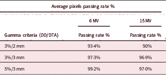
GAHs generated with tighter DTA criteria (brain: 3%/3 mm) showed fewer DD for the 6 MV photon beams, whereas for the 15 MV a relaxed DTA criterion (brain: 5%/3 mm) was found. The average passing rate for these γ criteria is given in Table 1.
Average pixels passing rate % for all brain IMRT plans treating at 6 and 15 MV photon beams for different γ criteria.

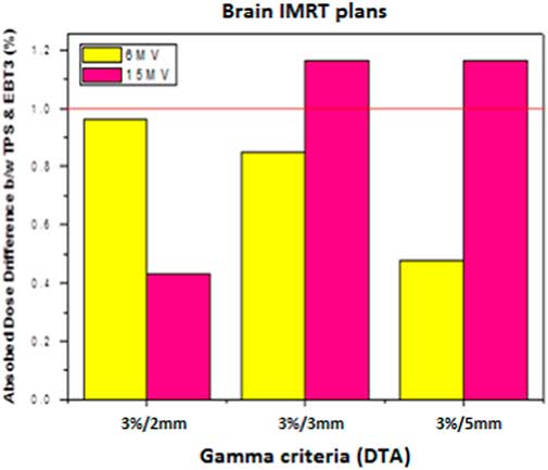
We calculated an average of these DDs to optimise the total results. For IMRT plans using the 6 MV photon beams, DDs at the isocentre for all selected DTA criteria were found to be within the acceptable range. For the 15 MV photon beams, only a criterion of 3%/2 mm shows <1% DD, as shown in Figure 1.
Dose difference at the isocentre analysed with different γ criteria for all brain IMRT plans and treated using the 6 and 15 MV photon beams.

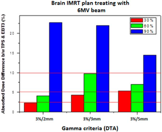
For verification of computed (TPS) and measured dose (EBT3 film), a pixel-to-pixel comparison was carried out based on the DDs calculated at the isocentre, and the 30, 60 and 90% area of the ROI as determined radially away from the isocentre for each gamma criteria. The acceptability criteria for the isocentre, 30, 60 and 90% area DDs are <1, <3, <5 and <10%, respectively, for all brain IMRT plans, as shown in Figure 2.
Dose difference for area away from the isocentre analysed with different DTA criteria for all brain IMRT plans using the 6 MV photon beams.

At the isocentre, only a small DD was observed relatively <1%, which was also found by Fenoglietto et al.Reference Fenoglietto, Laiberte, Ailleres, Riou, Dubois and Azira 43 When moving away from the isocentre, it is found that the DDs increased, a feature also demonstrated by Mohammedi et al.Reference Mohammadi, Miri-Hakimabad and Rafat-Motavalli 44 The DDs between the calculated and measured doses are proportional to the distance from the isocentre.
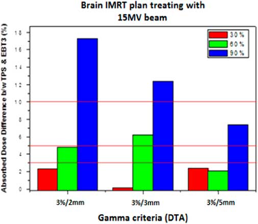
For the 30% area away from the isocentre using the 6 MV photon beams, DTA criteria of 3%/2 mm showed a DD <3%. However, for the 15 MV beams, all the chosen DTA criteria were reliable to the acceptability criteria. For the 60% area, only 3%/2 mm for the 6 MV, and both 3%/2 mm and 5%/3 mm for the 15 MV were reliable to the acceptability criteria. For the 90% area, not a single criterion passed the acceptability criteria for the 6 MV beams. When the 15 MV photon beams were used, the 5%/3 mm showed <10% DD, as shown in Figure 3.
Dose difference for area away from the isocentre analysed with different DTA criteria for all brain IMRT plans using the 15 MV photon beams.

Overall, we found that the 6 MV photon beams showed accurate results for tumour coverage over the 15 MV, which was also determined by Wang et al.Reference Wang, Yorke, Desobry and Chui 45 They demonstrated this for the three-dimensional conformal radiation therapy, but our results of IMRT plans also agreed to their results. With the increase of photon beam energy, there is a widening of the beam penumbra. This results in an increase in lateral dose heterogeneity for the 15 MV photon beams. Spiteful to this fact, for a relaxed criterion, the 15 MV photon beam showed quite fair agreement between the calculated and measured dose. This might be due to the fact that for high-energy (>10 MV) photon beams may have neutron contamination because their high energy photons exceed the threshold energy of the photon–neutron reaction. There is also pair and triplet production that play a role in how the photons interact with matter, something that is not well modelled in most TPS.Reference Allen and Chaudhri 46 – Reference Hideghéty, Szabo and Polanek 49
Conclusions
When considering passing rate % and DDs, the DTA criterion of 3%/2 mm may be used for the IMRT QA with low-energy (6 MV) photon beams. For high-energy (15 MV) photon beams, DTA criterion of 5%/3 mm is reliable for the QA purposes. Moreover, low-energy (6 MV) photon beams showed agreement when considering pixels passing rate % and less DD with tighter criteria than the 15 MV. High-energy photon beams should use relaxed DTA criteria to maintain a comparison of planned dose in an acceptable level.
Acknowledgements
The authors would like to thank Dr Daniel Markel from Princess Margaret Cancer Centre/University of Toronto, in discussing and editing this work. The authors would also like to thank Shaukat Khanum Cancer Hospital and Research Center, Lahore, Pakistan, for accommodating students for research work.