Introduction
Poor nutritional status can result in a decreased quality of life and influence a variety of effects on the endocrine system.Reference Cónsole, Jurado and Oyhenart1,Reference Ramos, Teixeira and Passos2 In adult rats, malnutrition can reduce the testes, epididymis, and prostate weights and decrease the serum concentrations of Luteinizing hormone (LH), Follicle-stimulating hormone (FSH), and testosterone.Reference De Souza Santos, Ferraz, Teixeira, Sampaio and da Fonte Ramos3–Reference Chik, Ho and Brown6
The consumption of sugars has increased considerably during the past years, especially in the developed countries. A commonly consumed added sugar is fructose. Fructose is a monosaccharide and a natural component of fruits and vegetables. It is sweeter than glucose or sucrose; therefore, it is commonly used as a sweetener in corn syrup, soft drinks, fruits, and other derivatives. Its consumption in the form of soft drinks is considered a public health problem.Reference Parker, Salas and Nwosu7
The consumption of fructose that is naturally present in food has beneficial effects and provides energy, while high intake as a sweetener in industrialized products is unhealthy. The increase in consumption of foods with high levels of fructose is considered as one of the main factors responsible for the development of metabolic syndrome, insulin resistance, obesity, and diabetes in children and adolescents.Reference Malik, Popkin, Bray, Després and Hu8–Reference Elliott, Keim, Stern, Teff and Havel10 Unlike glucose, the effect of fructose to stimulate insulin secretion from pancreatic β-cells is low, causing a decrease in leptin levels, thereby leading to a high caloric intake favoring weight gain.Reference Shapiro, Mu and Roncal11–Reference Bantle, Laine and Castle13
The concept of developmental origins of health and disease (DOHaD) defines the environment in utero or during early development that can affect the health and increase the susceptibility to diseases in adulthood.Reference Gage, Munafò and Davey Smith14 Previous studies by our research group reported that diet caloric restrictionReference De Oliveira, Lisboa and de Moura15 or excess intakeReference Ibáñez, Erthal and Ogo16 during adolescence leads to cardiometabolic dysfunction in adult rats. The testis and epididymis mature due to postnatal development of the reproductive system during pubertyReference Picut, Remick and de Rijk17; therefore, an unhealthy lifestyle can result in alterations in adulthood leading to impaired fertility.Reference Sharpe, Woodruff, Janssen, Guillette and Giudice18
Some studies have shown negative correlations between fructose intake and fertility. In female rats, fructose consumption induces changes in the length of the estrous cycle, ovarian, and uterine histology.Reference Ko, Kim, Kim, Kim and Lee19 In adult male rats, a high-fructose diet causes testicular degeneration with increased expression of inflammatory factors,Reference Yildirim, Sumlu and Aslan20 destruction of the germinal epithelium, lipid accumulation in Sertoli cells,Reference Meydanli, Gumusel and Ozkan21 and necrosis and atrophy in the seminiferous tubules.Reference Dokumacioglu, Iskender and Yenice22 However, the role of a high-fructose diet and its effects on puberty and the reproductive system have not been explored.
Thus, the aim of the present study was to evaluate the influence of fructose intake (20%) on the reproductive system in pubertal rats and investigate the changes that can occur in adulthood even after ceasing consumption.
Methods
Ethics approval
The experimental procedures described in this study were performed in accordance with the Ethical Principles in Animal Research adopted by the Brazilian College of Animal Experimentation and were approved by the Ethics Committee on Animal Use of the State University of Maringá (CEUA/UEM protocol no. 5669210917).
Animals and experimental design
Male Wistar rats (n = 60; 25 d old) were obtained from the Central Animal House of the State University of Maringá. The rats were allowed to acclimatize to their new environment for 5 d. The animals were randomly divided into four experimental groups consisting of 15 animals each: two groups received fructose supplementation (20%) diluted in water (corresponding to 0,2 g/ml) for 30 d and were euthanized at postnatal day (PND) 60 (Fr30 group) or PND 120 (recuperation for 60 d without fructose treatment, Re-Fr30 group), while the other two control groups received only the vehicle (water) and were euthanized at PND 60 (C30 group) or PND 120 (Re-C30). PND 30 to PND 60 period corresponding to the end of juvenile period (around PND 35) to the peripuberal period that ends at about PND 55 to 60 d, according to the authors Ojeda and SkinnerReference Ojeda, Skinner, Knobil and Neill23 and Clegg.Reference Clegg24 Fructose was obtained commercially from Lighsweet Industries (Lowçucar, Paraná, Brazil), and the fructose solution (20%) was prepared as per Jearapong et al.Reference Jearapong, Chatuphonprasert and Jarukamjorn25 and Yildirim et al.Reference Yildirim, Sumlu and Aslan20 The rats were weighed and then euthanized by guillotine decapitation, and blood was collected from the ruptured cervical vessels. The reproductive organs were removed and weighed. The testes and epididymis were used for histological analysis and sperm count.
Daily sperm production per testis and transit time in the epididymis
Decapsulated right testis and epididymis were weighed and homogenized as described previously by Robb et al.,Reference Robb, Amann and Killian26 with the adaptations described previously.Reference Siervo, Vieira and Ogo27 After dilution of homogenates, a small aliquot of sample was transferred to a Neubauer chamber (four fields per animal) for counting homogenization-resistant spermatids (that corresponds to the stage 19 of the spermatogenesis) in the testis and spermatozoa in epididymis. To calculate the daily sperm production (DSP), the concentration of homogenization-resistant spermatids per testis was divided by 6.1, which is the number of days during which mature spermatids remain in the seminiferous epithelium. To calculate sperm transit time through the epididymis, the number of sperm in each epididymal portion was divided by DSP.
Sperm motility
Sperm motility was evaluated using the methods of Siervo et al.Reference Siervo, Vieira and Ogo27 Briefly, the left vas deferens was rinsed with 1.0 ml modified human tubal fluid (HTF) medium with gentamicin (Irvine Scientific) at 34°–37°C to obtain spermatozoa. At the same temperature, a Makler counting chamber (Sefi-Medical) was loaded with a 10 µL aliquot of the sperm solution prepared previously. Sperm motility was assessed by visual estimation (100 spermatozoa per animal) under a light microscope (Motic) at 100× magnification and was performed by the same person throughout the study. Spermatozoa were classified as motile or immotile.
Sperm morphology
The contents of the vas deferens were removed via internal rinsing with 1.0 ml of 10% formol-saline. Histological slides were prepared from this solution and observed using an Opton photomicroscope (400× magnification). One hundred spermatozoa were analyzed per animal. Morphological analysis was classified into three general categories: normal morphology, head abnormalities (without characteristic curvature or isolated form, i.e., no tail attached), and tail abnormalities (broken, rolled into a spiral and isolated, i.e., no head attached). This analysis was performed as described by Fernandes et al.Reference Fernandes, Arena and Fernandez28
Histopathological analyses
Testis and epididymis were fixed in methacarn solution (10% acetic acid, 60% methanol, and 30% chloroform) and then put in alcohol 70%. Next, they were embedded in Paraplast® wax and cut into 5 µm sections and stained with haematoxylin and eosin (H&E). The tissue was evaluated using a Motic microscope (Motic®, Richmond, Canada) (100× and 400× magnification). For the testes, random seminiferous tubular sections (100 per animal) in three nonconsecutive testis cross sections were analyzed to detect the presence of abnormalities in these tubules: immature germ cells in the lumen, acidophil cells, vacuoles, and tubular degeneration. In the epididymis, histopathological inspection was performed qualitatively in the caput–corpus and cauda of each animal as described by Favareto et al.Reference Favareto, de Toledo and Kempinas29
Number of Sertoli cells
The number of Sertoli cell nuclei was determined in 20 cross sections of the seminiferous tubules (stages VII–VIII of spermatogenesis) per testis in each rat, under a light microscope at 400× magnification.Reference Nassr, Arena and Toledo30
Kinetics of spermatogenesis
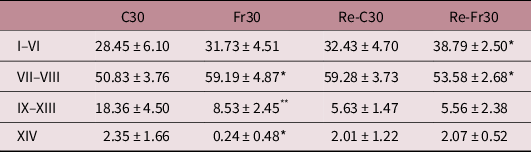
Random seminiferous tubular sections (100 per animal) in three nonconsecutive testis cross sections were classified into four stages: I–VI, VII–VIII, IX–XIII, and XIV of the seminiferous epithelial cycle, according to Leblond and ClermontReference Leblond and Clermont31 using a photomicroscope at 400× magnification.Reference Fernandes, Fernandez and Campos32 This analysis allows the assessment of the proportion of staging of the seminiferous tubules.
Seminiferous tubule diameters and seminiferous epithelium height
For evaluation of the seminiferous tubule diameter and epithelium height, 40 random testicular cross sections per animal in stage IX of the seminiferous epithelium cycle were examined. Seminiferous tubule diameters were measured using an Opton photomicroscope (400× magnification) and BELview software (version 6.2.3.0 for Windows). Likewise, the seminiferous epithelium height was measured using the same tubules and methodology as mentioned above. In each seminiferous tubule, the mean of four measures for the diameters and heights was calculated and used in the statistical analysis.Reference Favareto, de Toledo and Kempinas29
Stereology analyses of the epididymis
For stereological analysis of the epididymis, 10 random epididymal cross sections per animal were captured and analyzed. This analysis was performed by means of Weibel’s multipurpose graticulate, with 168 points were superimposed on the images. By counting the overlapping dots in the seminiferous cord or interstitial compartment, it was possible to establish the respective proportions of each of these components in the epididymis for each experimental group as described by Favareto et al.Reference Favareto, de Toledo and Kempinas29
Testosterone assay
Blood samples were collected in heparinized tubes and centrifuged at 2400 × g for 20 min (4°C). The plasma was separated and stored at −20°C until the assay. Commercial testosterone ELISA kit (Cayman Chemical, Ann arbor, MI, USA – #582701) was used for testosterone measurement, according to the manufacturer’s protocol. All samples were included in the same assay to avoid inter-assay errors. The intra-assay coefficient of variation is 4.4%.
Statistical analysis
All parameters were submitted to the Shapiro–Wilk test for normality and thus classified into parametric and nonparametric data. Data were compared using unpaired t-test or the nonparametric Mann–Whitney test. Differences were considered significant when P < 0.05. Statistical analyses were performed using GraphPad Prism (version 5.0).
Results
Body weight and weight of the reproductive organs
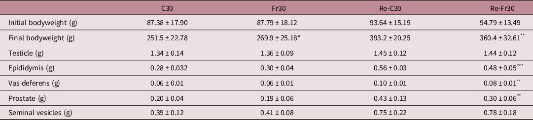
Body and reproductive organ (testis, epididymis, seminal vesicle, prostate, and vas deferens) weights are shown in Table 1. The Fr30 group had a significant increase in body weight compared to the C30 group (P = 0.0446). However, after discontinuing the treatment for 60 days (Re-Fr30), there was a reduction in body (P = 0.0036), epididymis (P = 0.0002), vas deferens (P = 0.0013), and prostate gland (P = 0.0022) weights compared with their respective controls.
Body and reproductive organs weight

Values are expressed as mean ± SD, Student t-test (n = 15 per group).
C30 – control 30 days; Fr30 – fructose intake for 30 d; Re-C30 – control recuperation for 60 d; Re-Fr30 – fructose recuperation for 60 d.
* Significantly different to the respective control (P < 0.05).
** P < 0.01.
*** P < 0.001.
Sperm morphology and motility
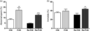
The percentage of abnormal sperms was significantly greater in the Fr30 and Re-Fr30 groups than in the control C30 (P = 0.0031) and Re-C30 (P < 0.0001) groups, respectively (Fig. 1a). The animals that received fructose and underwent a recovery time of 60 d had a significant increase in the percentage of immobile sperms compared to those in the control (P = 0.0032) (Fig. 1b).
Sperm morphology and motility. (a) Percentage of abnormal sperm and (b) percentage of immobile sperm. Values are expressed as mean ± SD, Student t-test (n = 15 per group). C30 – control 30 d; Fr30 – fructose intake for 30 d; Re-C30 – control recuperation for 60 d; Re-Fr30 – fructose recuperation for 60 d. *Significantly different to the respective control (P < 0.05); **P < 0.01; ***P < 0.001.

Histopathological analysis, kinetics of spermatogenesis, and Sertoli cell count
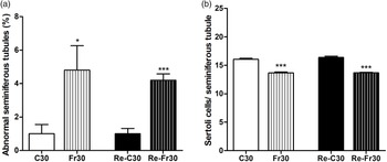
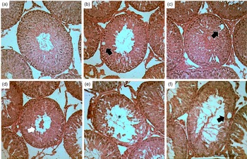
Histopathological analysis of the testes from rats treated with fructose revealed a significant increase in the number of abnormal tubules in the Fr30 and Re-Fr30 groups compared with the control C30 (P = 0.0410) and Re-C30 (P = 0.0002) groups, respectively (Fig. 2a). Certain structural changes, such as immature germ cells in the lumen, tubular degeneration, and presence of vacuoles, were observed in the seminiferous tubules (Fig. 3). Moreover, fructose consumption led to a significant reduction in the Sertoli cell number per seminiferous tubule in the animals from the Fr30 and Re-Fr30 groups when compared to the animals from control groups C30 (P < 0.0001) and Re-C30 (P < 0.0001), respectively (Fig. 2b). Analysis of the kinetics of spermatogenesis (Table 2) revealed a significant increase in the number of seminiferous tubules in stages VII–VIII in the Fr30 group compared to the control group (P = 0.0162). Conversely, in the same groups, there was a significant reduction in stages IX–XIII (P = 0.0027) and XIV (P = 0.0453). In the Re-Fr30 group, there was an increase in the number of seminiferous tubules in stages I–VI (P = 0.0286) and a reduction in stages VII–VIII (P = 0.0244) compared with the control.
Testicular histopathological analysis and Sertoli cells counts. (a) Percentage of abnormal seminiferous tubules and (b) number of Sertoli cells per seminiferous tubules. Values are expressed as mean ± SD, Student t-test (n = 5 per group). C30 – control 30 d; Fr30 – fructose intake for 30 d; Re-C30 – control recuperation for 60 d; Re-Fr30 – fructose recuperation for 60 d. *Significantly different to the respective control (P < 0.05); ***P < 0.001.

Testicular histopathological analysis. (a) C30 group, (b–d) Fr30 group, (e–f) Re-Fr60 group. Vacuolization (black arrows), immature germ cells in the lumen (white arrow), and epithelial degeneration (asterisks). Haematoxylin and eosin staining (original magnification ×100).

Spermatogenesis kinetics

Values are expressed as mean ± SD, Student t-test (n = 5 per group).
C30 – control 30 d; Fr30 – fructose intake for 30 d; Re-C30 – control recuperation for 60 d; Re-Fr30 – fructose recuperation for 60 d.
* Significantly different to the respective control (P < 0.05).
** P < 0.01.
Seminiferous tubule diameter and epithelium height in the testis
Morphometric analysis revealed a significant increase in the diameter of seminiferous tubule in the Fr30 group (P = 0.0130) and the seminiferous epithelium height in the Re-Fr30 group (P = 0.0269) compared with their respective control groups (Table 3).
Seminiferous tubule diameter and seminiferous epithelium height

Values are expressed as mean ± SD, Student t-test (n = 15 per group).
C30 – control 30 d; Fr30 – fructose intake for 30 d; Re-C30 – control recuperation for 60 d; Re-Fr30 – fructose recuperation for 60 d.
* Significantly different to the respective control (P < 0.05).
Stereological analysis
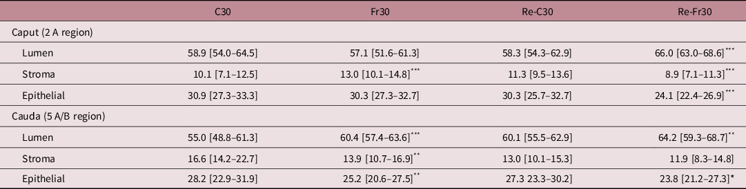
The results of the stereological analysis are presented in Table 4. There was a significant increase in the stromal compartment from the 2 A region of the caput epididymis in the Fr30 group compared to the control group (P = 0.0008). Fructose treatment (Fr30) increased the luminal compartment from the 5 A/B region of the cauda epididymis (P = 0.0005), followed by a decrease in the stromal (P = 0.0075) and epithelial (P = 0.0092) compartments, compared with the control group. Furthermore, after 60 d, a significant increase was observed in the luminal compartment (P < 0.0001), followed by reduction in the stromal (P = 0.0007) and epithelial (P < 0.0001) compartments from the 2 A region of the caput epididymis in the Re-Fr30 group compared to the control group. Moreover, in the 5 A/B cauda region, fructose treatment caused a significant increase in the luminal compartment in the Re-Fr30 group (P = 0.0087) and a significant decrease in the epithelial compartment when compared to the control (P = 0.0158).
Epididymal stereological analysis

Values are expressed as median [Q1–Q3], Mann–Whitney test (n = 5 per group).
C30 – control 30 d; Fr30 – fructose intake for 30 d; Re-C30 – control recuperation for 60 d; Re-Fr30 – fructose recuperation for 60 d.
* Significantly different to the respective control (P < 0.05).
** P < 0.01.
*** P < 0.001.
Daily sperm production per testis and transit time in the epididymis
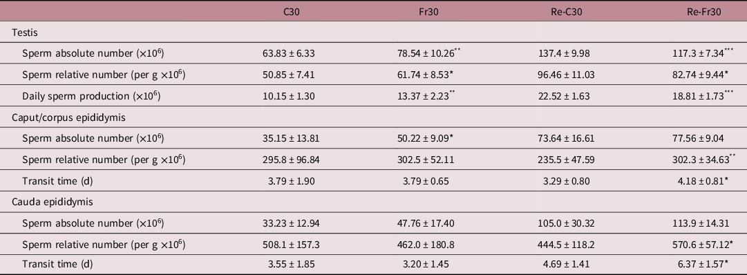
The sperm count parameters are shown in Table 5. Analysis of the sperm parameters showed that fructose treatment increased the DSP (P = 0.0024), number of sperm in the testis (absolute and relative) (P = 0.0051 and P = 0.0117, respectively), and caput and corpus of the epididymis (absolute) (P = 0.0114) in the Fr30 group. The sperm transit time through the epididymal caput and corpus remained unchanged. However, the Re-Fr30 group showed a decrease in the number of sperms (P = 0.0002) and DSP (P = 0.0003) in the testis, but the relative number (P = 0.0033) and sperm transit time in the epididymal caput, corpus (P = 0.0345), and cauda (P = 0.0252) were significantly higher than those in the control group.
Sperm parameters

Values are expressed as mean ± SD, Student t-test (n = 10 per group).
C30 – control 30 d; Fr30 – fructose intake for 30 d; Re-C30 – control recuperation for 60 d; Re-Fr30 – fructose recuperation for 60 d.
* Significantly different to the respective control (P < 0.05).
** P < 0.01.
*** P < 0.001.
Plasma testosterone concentrations
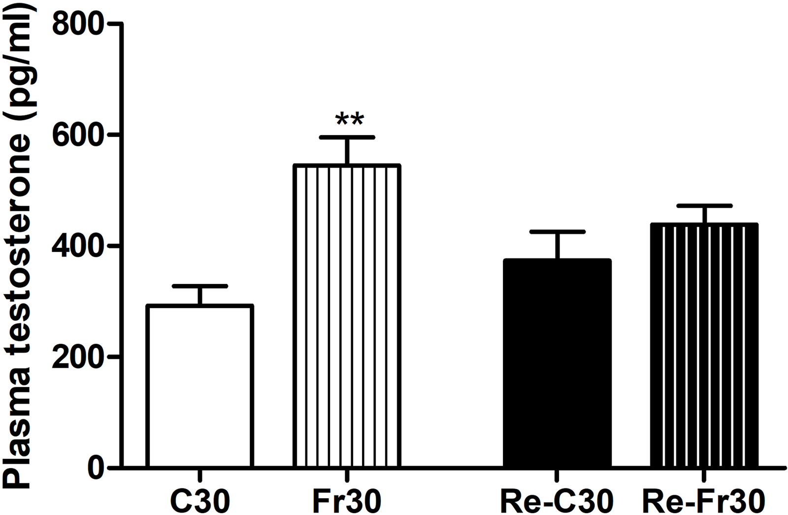
The plasma testosterone concentrations increased in the Fr30 group compared to the C30 group (P = 0.0017) but remained unchanged in the Re-C30 and Re-Fr30 groups (Fig. 4).
Serum testosterone concentrations. Values are expressed as mean ± SD, Student t-test (n = 9 per group). C30 – control 30 d; Fr30 – fructose intake for 30 d; Re-C30 – control recuperation for 60 d; Re-Fr30 – fructose recuperation for 60 d. **Significantly different to the respective control (P < 0.01).

Discussion
The results from the present study indicate that high-fructose consumption during puberty impairs testicular and epididymal development and the quality of male gametes in pubertal and adult life. Therefore, the consumption of sugar as fructose should be avoided during the postnatal developmental phase of the male reproductive system. Poor nutritional status can result in a decreased quality of life and influence a variety of effects on the endocrine system.Reference Cónsole, Jurado and Oyhenart1,Reference Ramos, Teixeira and Passos2 In adult rats, malnutrition can reduce the testes, epididymis, and prostate weights and decrease the serum concentrations of LH, FSH, and testosterone.Reference De Souza Santos, Ferraz, Teixeira, Sampaio and da Fonte Ramos3–Reference Chik, Ho and Brown6
Fructose is more lipogenic than glucose and can decrease leptin levels and favor weight gain.Reference Elliott, Keim, Stern, Teff and Havel10,Reference Shapiro, Mu and Roncal11 However, ceasing consumption can alter this phenomenon, as shown by the increase in body weight after 30 d of fructose intake, followed by a reduction in the body weight and weight of few reproductive organs after 60 d without sugar consumption. Interestingly, this effect was not observed in the testes.
In humans, Chen et al.Reference Leblond and Clermont31 explained how the consumption of calories affects the body weight of adults. The authors associated the initial weight gain with a large amount of fructose in beverages that favor fat storage and an increase in lipogenesis. Although fructose is related to weight gain,Reference Elliott, Keim, Stern, Teff and Havel10,Reference Shapiro, Mu and Roncal11,Reference Chen, Appel and Loria33,Reference Bocarsly, Powell, Avena and Hoebel34,Reference Huynh, Luiken, Coumans and Bell35 this association was not observed in few studies.Reference Yildirim, Sumlu and Aslan20,Reference Meydanli, Gumusel and Ozkan21
In the present study, high-fructose intake led to an increase in the percentage of abnormal seminiferous tubules, which corroborates the findings of Yildirim et al.,Reference Yildirim, Sumlu and Aslan20 Meydanli et al.,Reference Meydanli, Gumusel and Ozkan21 and Dokumacioglu et al.Reference Dokumacioglu, Iskender and Yenice22 Yildirim et al.Reference Yildirim, Sumlu and Aslan20 observed the presence of vacuolar and intratubular degeneration in the testes of Wistar rats (PND 30) treated with fructose (20%) for 15 weeks. The Wistar rats (6–8 weeks old) treated with fructose (30%) for 8 weeks showed degeneration, necrosis, and atrophy of seminiferous tubules, vacuolization of Sertoli cells, and spermatogonia, as reported by Dokumacioglu et al.Reference Dokumacioglu, Iskender and Yenice22 In addition, under the same experimental conditions, Meydanli et al.Reference Meydanli, Gumusel and Ozkan21 observed disorganization and destruction of germinal epithelium with immature germ cells in the lumen, accumulation of lipids within the cytoplasm of Sertoli cells, and germ cell apoptosis.
The presence of epithelial vacuoles indicates the absence of Sertoli cells or loss of germ cells. Sertoli cells provide nutritional and structural support to the seminiferous tubules.Reference Boekelheide, Neely and Sioussat36,Reference Johnson, Thompson and Varner37 The absence of these cells impairs the structural organization of the epithelium, thereby releasing immature cells in the lumen. In the present study, this impairment was observed even after discontinuing the treatment in the Re-Fr30 group. The decrease in Sertoli cells may signal germ cell apoptosis,Reference Boekelheide, Fleming, Johnson, Patel and Schoenfeld38 as well as changes in the stages of spermatogenesis.Reference Johnson, Thompson and Varner37
In the current study, the increase in the number of seminiferous tubules at stages VII–VIII of spermatogenesis in the Fr30 group is associated with an increase in sperm number and DSP in the testes. This increase in sperm production indicates a decrease in gamete quality since the sperm morphology is impaired after fructose consumption. After 60 d without fructose exposure, the number of seminiferous tubules at stages VII–VIII of spermatogenesis, as well as DSP, reduced significantly, which indicated the loss of gamete quality and persistence of this damage. In this context, it is noted that sperm motility is affected only after the recuperation period, indicating that some imminent factors associated with sperm motility were affected during fructose exposure; however, this damage was not observed upon immediate evaluation. Despite normalization of the testosterone concentrations after 60 d without treatment, the initial damage influenced by fructose in spermatogenesis persisted, thereby decreasing the DSP and sperm number.
The spermatogenic cycle in rats is approximately 58 dReference Xiao, Mruk, Wong and Yan Cheng39 and more than 8–10 d for the sperm transit time.Reference França, Avelar and Almeida40 Therefore, the spermatozoa were stimulated since the beginning of the spermatogenic cycle, and the influence of fructose consumption on quality of gamete could have been different if the recuperation period was longer.
In the present work, fructose consumption for 30 d led to an increase in the absolute sperm number in the epididymis (caput/corpus), while in the transit time the relative sperm number remained unchanged. This result is due to the high amount of sperm produced in the testes that travel to the epididymis with no significant increase in the cauda region. Nevertheless, after 60 d, the reduction in DSP and absolute sperm number in testes and epididymis and the increase in relative number in epididymis (caput and cauda) indicate the negative effects of fructose intake. Therefore, this suggests that the increase in relative sperm number in the caput and cauda of epididymis, in fact, was due to a reduction in the epididymis weight.
In the current study, high-fructose intake led to tissue remodeling in the epididymis at puberty and adulthood. Thus, after discontinuing fructose consumption for 60 d, the epididymal alteration remains associated with decreased sperm motility since epididymal epithelial cells produce components for sperm maturation.Reference Kohane, Cameo, Pineiro, Garberi and Blaquier41
Based on the results of testosterone concentration in the present study, we inferred that the increase in this hormone in animals from the Fr30 group is related to the damage observed in the sperm, histopathological, and morphometric parameters of the testes and epididymis. Furthermore, despite the normalization of testosterone concentrations after a period of 60 d without treatment (Re-Fr30 group), it is still possible to observe alterations in these parameters when compared to those observed in the Fr30 group. These results show the influence of testosterone on the testicular and epididymal postnatal development since the increase in plasma testosterone levels in the pubertal phase was crucial to cause immediate damage and reprogrammed the characteristics of these tissues in adulthood. Dokumacioglu et al.Reference Dokumacioglu, Iskender and Yenice22 showed an increase in serum testosterone concentrations caused by high-fructose intake (30%) for 8 weeks in adult male rats. In conclusion, the present study shows the negative effects of high-fructose intake in peripubertal rats, leading to alterations in the spermatozoa, testis, and epididymis in the pubertal and adult periods by increasing testosterone concentrations in the pubertal phase.
Acknowledgments
The authors are grateful to State to CAPES (Coordinating Body for the Improvement of Postgraduate Studies in Higher Education) for providing a PhD scholarship to D.S. Alvarenga-Medaglia. This paper represents part of the PhD thesis by D.S. Alvarenga-Medaglia (Graduate Program in Experimental Pathology, State University of Londrina – Brazil) under the supervision of GSA Fernandes.
Financial Support
This research was supported by CAPES – PROEX (Coordinating Body for the Improvement of Postgraduate Studies in Higher Education, Brazil).
Conflicts of interest
The authors declare no conflicts of interest.
Ethical standards
The authors assert that all procedures contributing to this work comply with the ethical standards of the relevant national guides on the care and use of laboratory animals, Law 11.794 of October 8, 2008, Decree 6899 of July 15, 2009, as well as with the rules issued by the National Council for Control of Animal Experimentation (CONCEA) and has been approved by the institutional Ethic Committee on Animal Use of the State University of Maringá (CEUA/UEM) in the meeting of 12/13/2017.