Congenital ventricular diverticulum is a rare cardiac malformation that is postulated to be a consequence of impaired development of the endocardial tube in the 4th week of embryologic development. It constitutes 0.02–0.4% of all cardiac malformations and occurs with other cardiac, vascular, or midline thoracoabdominal abnormalities in approximately 70% of cases.
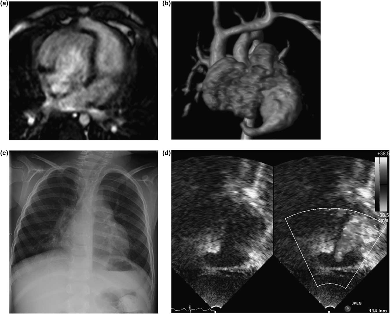
The images depict the heart of a 3-year-old boy who was referred to the outpatient clinic with the diagnosis of multiple restrictive muscular ventricular septal defects, mesocardia, and a cutaneous midline supraumbilical defect. He was asymptomatic. A thoracoabdominal resonance was performed and a finger-shaped chamber was found in the left ventricular apex (Figs 1a and b); no intra-abdominal abnormalities were found. Chest X-ray showed mesocardia (Fig 1c) and oriented transthoracic echocardiography showed the same finger-shaped chamber in the left ventricular apex (Fig 1d, Online Video 1). He was started on aspirin and he remains asymptomatic and with no major changes in echocardiography 2 years later.
(a) Four chamber view of cardiac MRI imaging demonstrates the left ventricle diverticulum. (b) Coronal anteroposterior view of a 3D volume rendering reconstruction depicts a finger-shaped chamber in the left ventricular apex corresponding to the left ventricle diverticulum. (c) Chest X-ray shows mesocardia and a remarkable prominent bulging of the left side of the cardiac shape. (d) Modified apical four chamber view of a two-dimensional transthoracic echocardiography reveals a left ventricular diverticulum located in the anteroapical region, color Doppler illustrates blood flow entering the diverticulum from the left ventricle.

Ventricular diverticulum, mesocardia, and cutaneous midline defect have all a common origin in the endocardial tube. Owing to the fact that this is an uncommon malformation, with a scarce number of reported cases, information is limited, and this interferes with its detection with imaging diagnostic methods. Treatment of congenital ventricular diverticulum is controversial, especially if the patient remains asymptomatic. Surgical resection to avoid the risk of rupture and antiplatelet therapy or chronic anticoagulation to prevent systemic thromboembolism have been proposed.