Total anomalous pulmonary venous connection is a rare congenital anomaly and accounts to 1.5–2% of all CHD.Reference Ferencz, Rubin and McCarter 1 This anomaly is classified into various types depending upon the site of anomalous drainage of the pulmonary veins. The clinical presentation, haemodynamic consequences, and the prognosis depend on whether the pulmonary venous flow is obstructed or unobstructed. Although obstruction may occur with any anatomic type of total anomalous pulmonary venous connection, the highest incidence is encountered with the infracardiac type.Reference Seale, Uemura and Webber 2 Obstructed total anomalous pulmonary venous connection is a surgical emergency and is associated with high morbidity and mortality. This report describes a rare finding of flow reversal in the upper descending aorta, which is probably an indicator of extremely low left ventricular output.
Case report
In this study, we report the case of a 26-day-old neonate. Born at term by normal vaginal delivery to a second gravida mother. The baby was asymptomatic for the first 10 days of life. Thereafter, he developed tachypnoea followed by marked respiratory distress by next day or which he was hospitalised. He was suspected to have CHD in view of persistent desaturation, and thus he was referred to a tertiary-care centre. Subsequently, he was diagnosed as a case of obstructed total anomalous pulmonary venous connection with severe pulmonary hypertension. He was also diagnosed to have sepsis (total leucocyte count=36,700 cumm, neutrophils=75%, procalcitonin=21 ng/ml) and renal dysfunction (blood urea nitrogen=56 mg/dl, creatinine=3.2 mg/dl).
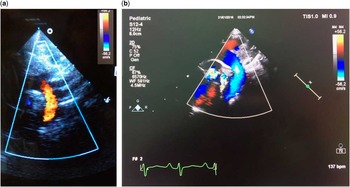
On examination, he had tachycardia (heart rate=170/minute), tachypnoea (respiratory rate =52/minute), and cyanosis. Saturation on oxygen was 62%, and there were mild subcostal retractions. Echocardiographic evaluation revealed obstructed total anomalous pulmonary venous connection with all four pulmonary veins forming a common chamber and draining into the hepatic veins with obstruction at the site of entry into the hepatic vein (Fig 1). A large patent ductus arteriosus shunted right to left along with right ventricular systolic dysfunction. There was presence of pandiastolic flow reversal in the upper descending thoracic aorta (Fig 2a). There was no evidence of any cause for aortic flow reversal, such as aortic regurgitation or hepatic or intracranial arteriovenous malformations.
Obstruction to pulmonary venous flow at the entry into the hepatic veins.

( a ) Pandiastolic flow reversal in the upper descending aorta and arch preoperatively; ( b ) no evidence of flow reversal in the arch after surgery.

The patient was operated within 4 hours of admission. Surgical findings confirmed the echocardiography diagnosis, and the pulmonary veins were re-routed to the left atrium, the descending channel was ligated, and the interatrial communication was closed. The cardiopulmonary bypass time was 58 minutes, and cross-clamp time was 32 minutes. The postoperative course was stormy and prolonged. He had significantly high lactate levels (6.2) and deranged mixed venous oxygen saturation (58%), for which dobutamine and milrinone were administered, and he required prolonged ventilatory and inotropic support for haemodynamic instability. Pre-discharge echocardiography revealed laminar pulmonary venous flow to the left atrium, no interatrial shunt, and normal biventricular function. There was no flow reversal in the arch after the surgery (Fig 2b). He was finally discharged on the 19th postoperative day in stable condition.
Discussion
In 1798, Wilson described total anomalous pulmonary venous connection as a “monstrous formation of the heart”.Reference Anderson and Macartney 3 The incidence of total anomalous pulmonary venous connection is 0.008% of live births and it accounts to 1.5–2% of CHD.Reference Ferencz, Rubin and McCarter 1 Survival depends on the presence of a right-to-left intracardiac shunt, which maintains the cardiac output and almost always occurs through a patent foramen ovale. Darling and colleagues have classified total anomalous pulmonary venous connection into four subgroups depending on the site of anomalous drainage of the pulmonary veins. type I is supracardiac (45%), type II is cardiac (26%), type III is infracardiac (24%), and type IV is mixed drainage(5%).Reference Delisle, Ando and Calder 4 Pathphysiologically, total anomalous pulmonary venous connections are classified as obstructed or unobstructed. Obstructed total anomalous pulmonary venous connections have higher morbidity and mortality and the infracardiac variety is often obstructed.
This case was unusual because of the presence of pandiastolic flow reversal in the upper descending aorta. The patient had associated severe pulmonary hypertension with right ventricular dysfunction and a large patent ductus arteriosus shunting right to left. Such aortic flow reversal has not been observed in our previous total anomalous pulmonary venous connection patients. A review of the literature did not reveal any such documentation either. There is no documented cause for this phenomenon, but we hypothesise that the presence of flow reversal in the descending aorta in obstructed total anomalous pulmonary venous connections probably suggests an extremely low left ventricular output. Consequent to the markedly compromised left ventricular output, flow reversal occurs in the descending aorta to provide adequate blood flow to the brain. The patient had a prolonged and stormy postoperative course with marked haemodynamic instability in the early postoperative period. Thus, a descending aortic flow reversal may portend a worse prognosis.
Acknowledgement
None.
Financial Support
This research received no specific grant from any funding agency, commercial or not-for-profit sectors.