Introduction
Generalized anxiety disorder (GAD) is a condition characterized by excessive and persistent worry and anxiety accompanied by physical symptoms such as restlessness, fatigue, irritability, difficulty in concentrating, muscle tension, and sleep disturbance (American Psychiatric Association, 2013). Intolerance to uncertainty, a generalization of fear and alterations in emotional processing and regulation seem to characterize this disorder (Mochcovitch et al. Reference Mochcovitch, Da Rocha Freire, Garcia and Nardi2014). This condition leads to significant distress and impairment in everyday life, as well as to increased risk for other mental and physical problems, including alcohol and drug abuse, being also frequently comorbid with major depression. Lifetime prevalence of GAD is estimated around 4.7% (Kessler et al. Reference Kessler, Petukhova, Sampson, Zaslavsky and Wittchen2012), resulting more common among women, almost twice as high as men (McLean et al. Reference Mclean, Asnaani, Litz and Hofmann2011). Female sex, low socioeconomic status, and exposure to childhood adversity are considered as risk factors for the disease (Fonzo et al. Reference Fonzo, Ramsawh, Flagan, Simmons, Sullivan and Allard2016). The heritability of GAD has also been studied, suggesting a moderate contribution of additive genetics (estimated to be around 32%) in its pathogenesis (Hettema et al. Reference Hettema, Neale and Kendler2001a).
Previous neuroimaging studies have suggested functional and structural brain abnormalities in patients suffering from GAD (see (Mochcovitch et al. Reference Mochcovitch, Da Rocha Freire, Garcia and Nardi2014) and (Hilbert et al. Reference Hilbert, Lueken and Beesdo-Baum2014) for a review), involving both subcortical nuclei – especially the amygdala - and different cortical regions. In synthesis, GAD seems to be associated with altered functional connectivity in the amygdala (Etkin et al. Reference Etkin, Prater, Schatzberg, Menon and Greicius2009; Makovac et al. Reference Makovac, Meeten, Watson, Herman, Garfinkel and DC2016b) and prefrontal cortex (Andreescu et al. Reference Andreescu, Sheu, Tudorascu, Walker and Aizenstein2014). These areas, together with portions of temporal and parietal cortices, were reported as abnormally activated during emotional tasks (Palm et al. Reference Palm, Elliott, Mckie, Deakin and Anderson2011; Greenberg et al. Reference Greenberg, Carlson, Cha, Hajcak and Mujica-Parodi2013; Holzel et al. Reference Holzel, Hoge, Greve, Gard, Creswell and Brown2013; Fonzo et al. Reference Fonzo, Ramsawh, Flagan, Sullivan, Letamendi and Simmons2015; Moon & Jeong, Reference Moon and Jeong2015; Park et al. Reference Park, Kim, Jeong, Chung and Yang2016). Structural studies reported neuroanatomical alterations in prefrontal (Abdallah et al. Reference Abdallah, Coplan, Jackowski, Sato, Mao and Shungu2012; Strawn et al. Reference Strawn, Wehry, Chu, Adler, Eliassen and Cerullo2013; Cha et al. Reference Cha, Greenberg, Carlson, Dedora, Hajcak and Mujica-Parodi2014; Strawn et al. Reference Strawn, John Wegman, Dominick, Swartz, Wehry and Patino2014; Canu et al. Reference Canu, Kostic, Agosta, Munjiza, Ferraro and Pesic2015), temporal (De Bellis et al. Reference De Bellis, Keshavan, Shifflett, Iyengar, Dahl and Axelson2002; Abdallah et al. Reference Abdallah, Coplan, Jackowski, Sato, Mao and Shungu2012; Strawn et al. Reference Strawn, John Wegman, Dominick, Swartz, Wehry and Patino2014), parietal (Abdallah et al. Reference Abdallah, Coplan, Jackowski, Sato, Mao and Shungu2012; Makovac et al. Reference Makovac, Meeten, Watson, Garfinkel, Critchley and Ottaviani2016a) and cingulate cortices (Strawn et al. Reference Strawn, John Wegman, Dominick, Swartz, Wehry and Patino2014; Makovac et al. Reference Makovac, Meeten, Watson, Garfinkel, Critchley and Ottaviani2016a).
Considering the most recent magnetic resonance imaging (MRI) studies on adult GAD patients, prefrontal cortex (PFC) appears to be widely involved in the illness. Structural MRI (sMRI) studies found in GAD patients v. healthy controls (HC) increased gray matter volume (GMV) in dorsomedial PFC (Schienle et al. Reference Schienle, Ebner and Schafer2011), as well as reduced white matter volume (Hilbert et al. Reference Hilbert, Pine, Muehlhan, Lueken, Steudte-Schmiedgen and Beesdo-Baum2015; Moon & Jeong, Reference Moon and Jeong2016) and cortical thickness (CT) (Abdallah et al. Reference Abdallah, Coplan, Jackowski, Sato, Mao and Shungu2012; Cha et al. Reference Cha, Greenberg, Carlson, Dedora, Hajcak and Mujica-Parodi2014; Canu et al. Reference Canu, Kostic, Agosta, Munjiza, Ferraro and Pesic2015) in dorsolateral PFC. Functional connectivity studies have shed light on how, in GAD patients more than in HC, lateral PFC is richly connected to the amygdala (Etkin et al. Reference Etkin, Prater, Schatzberg, Menon and Greicius2009; Andreescu et al. Reference Andreescu, Sheu, Tudorascu, Walker and Aizenstein2014). Overall, the functional MRI (fMRI) studies on GAD showed an abnormal prefrontal activation in response to different tasks (Blair et al. Reference Blair, Shaywitz, Smith, Rhodes, Geraci and Jones2008; Palm et al. Reference Palm, Elliott, Mckie, Deakin and Anderson2011; Schlund et al. Reference Schlund, Verduzco, Cataldo and Hoehn-Saric2012; Greenberg et al. Reference Greenberg, Carlson, Cha, Hajcak and Mujica-Parodi2013; Holzel et al. Reference Holzel, Hoge, Greve, Gard, Creswell and Brown2013; Cha et al. Reference Cha, Greenberg, Carlson, Dedora, Hajcak and Mujica-Parodi2014; Moon & Jeong, Reference Moon and Jeong2015; Park et al. Reference Park, Kim, Jeong, Chung and Yang2016).
Moreover, temporal and parietal cortices were shown to be more active in adult GAD patients than in HC during working memory tasks involving anxiety-inducing stimuli (Park et al. Reference Park, Kim, Jeong, Chung and Yang2016) and during face emotion recognition tasks (Blair et al. Reference Blair, Shaywitz, Smith, Rhodes, Geraci and Jones2008; Fonzo et al. Reference Fonzo, Ramsawh, Flagan, Sullivan, Letamendi and Simmons2015). The findings from the sMRI studies are less consistent: studies investigating CT found deficits in GAD compared with HC in right postcentral and left inferior parietal gyri (Abdallah et al. Reference Abdallah, Coplan, Jackowski, Sato, Mao and Shungu2012) and left temporal pole and right fusiform gyrus (Canu et al. Reference Canu, Kostic, Agosta, Munjiza, Ferraro and Pesic2015), whereas GMV studies reported increased volume in right superior temporal pole (Hilbert et al. Reference Hilbert, Pine, Muehlhan, Lueken, Steudte-Schmiedgen and Beesdo-Baum2015) and reduced volume in portions of the parietal cortex, specifically supramarginal, precentral, and postcentral gyri (Makovac et al. Reference Makovac, Meeten, Watson, Garfinkel, Critchley and Ottaviani2016a).
The above heterogeneous findings could be explained in light of a recent theory, suggesting that symptoms and features of GAD reflect complex network alterations, in turn affecting the functionality of several cortical structures and deep brain nuclei (Etkin et al. Reference Etkin, Prater, Schatzberg, Menon and Greicius2009; Sylvester et al. Reference Sylvester, Corbetta, Raichle, Rodebaugh, Schlaggar and Sheline2012). The main circuits that are thought to be implicated in anxiety disorders are the frontoparietal, the default mode, the cingulo-opercular and the ventral attention networks (Sylvester et al. Reference Sylvester, Corbetta, Raichle, Rodebaugh, Schlaggar and Sheline2012). Even if the reasons for these alterations are yet to be understood, the involvement of different perturbing factors during brain development is conceivable.
Nowadays, the sMRI studies on GAD mainly focused on gray and white matter volumes (De Bellis et al. Reference De Bellis, Keshavan, Shifflett, Iyengar, Dahl and Axelson2002; Mohlman et al. Reference Mohlman, Price, Eldreth, Chazin, Glover and Kates2009; Schienle et al. Reference Schienle, Ebner and Schafer2011; Strawn et al. Reference Strawn, Wehry, Chu, Adler, Eliassen and Cerullo2013; Hilbert et al. Reference Hilbert, Pine, Muehlhan, Lueken, Steudte-Schmiedgen and Beesdo-Baum2015; Makovac et al. Reference Makovac, Meeten, Watson, Garfinkel, Critchley and Ottaviani2016a; Moon & Jeong, Reference Moon and Jeong2016), whereas only a few of them (Abdallah et al. Reference Abdallah, Coplan, Jackowski, Sato, Mao and Shungu2012; Cha et al. Reference Cha, Greenberg, Carlson, Dedora, Hajcak and Mujica-Parodi2014; Strawn et al. Reference Strawn, John Wegman, Dominick, Swartz, Wehry and Patino2014; Canu et al. Reference Canu, Kostic, Agosta, Munjiza, Ferraro and Pesic2015) considered other cortical features, specifically thickness, reflecting the cellular organization of the brain and, indirectly, the brain development. To our knowledge, no data about the cortical surface area (CSA) and gyrification patterns in GAD are available.
The main objective of the present MRI study is to quantitatively compare cortical morphology between GAD patients and HC in terms of thickness, volume, curvature or gyrification, and surface area. The identification of specific morphological differences between GAD patients and HC can shed new light on the neurobiological determinants of GAD, with relevant implications for the management and treatment of anxiety disorders.
Methods and materials
Subjects
Thirty-one adult individuals (11 males, aged 21–70 years, 43.8 ± 14.9 years) who met the DSM-IV criteria for GAD underwent a sMRI session. The patients were recruited in the outpatient Psychiatric Clinic of the University Hospital of Udine, Italy. Patients with a history of traumatic head injury with loss of consciousness and neurological or medical illness were excluded from the study. The diagnosis of GAD was assessed using the Structured Clinical Interview for Axis I DSM-IV disorders (SCID-I) and confirmed by the clinical consensus of two expert psychiatrists, who discussed about any major symptom and psychopathological discrepancies emerged from interviews and SCID-I scores, finally agreeing on the presence of the illness.
Eighteen patients were taking one or more psychotropic drugs at the time of the MRI scanning. Fifteen were receiving regular antidepressants: four were taking escitalopram, three paroxetine, two sertraline, two mirtazapine, two citalopram, one venlafaxine and one duloxetine. One patient was taking an antipsychotic drug (amilsulpride). In addition, eight patients were taking benzodiazepines when needed. Given the variety of therapies administered to the patients, their possible contribution to brain morphology was not considered in the study design. Three patients met the criteria for a current comorbid anxiety or mood disorder, one major depressive disorder (MDD), one Specific Phobia (SP), and one SP and past MDD, whereas 16 patients had no history of other psychopathologic conditions, as determined by the SCID-I. Of the remaining twelve patients, four were diagnosed as having only past MDD, three only past panic disorder (PD), one only past eating disorder (ED), two past PD and MDD, one past MDD and ED and one past alcohol abuse and depressive episode induced by substance abuse. Since just a few patients presented a comorbidity at the time of the MRI acquisition and almost half of the sample had no history of other mental illnesses, in this work we chose not to consider the effect of an actual or past comorbid psychopathologic condition.
Thirty-one healthy subjects (14 males, aged 22–65 years, 39.7 ± 13.5 years) with no significant differences in terms of age, gender, handedness, socioeconomic status and ethnicity (p < 0.05) were also included in the study for reference. They were recruited from the local community and had no history of alcohol or substance abuse, head trauma or neurological or major medical illnesses, or psychiatric axis I disorders determined using a brief modified version of the SCID-I (non-patient version). They also had no history of psychiatric disorders among first-degree relatives. All the participants signed a written informed consent to the study protocol, which was approved by the competent ethical committee and was in accordance with the Declaration of Helsinki.
Psychopathological and cognitive assessment
The Brief Psychiatric Rating Scale (BPRS, 24-item version) (Ventura et al. Reference Ventura, Nuechterlein, Subotnik, Gutkind and Gilbert2000), the Hamilton Rating Scale for Anxiety (HARS) and Depression (HDRS) (Hamilton, Reference Hamilton1959; Hamilton, Reference Hamilton1960) and the Global Assessment Functioning (GAF) scales (Endicott et al. Reference Endicott, Spitzer, Fleiss and Cohen1976) were administered to all participants. Taken together, these gold standard instruments allowed to comprehensively investigate the variety of symptoms experienced by GAD patients. The socioeconomic status and level of education were also assessed. Handedness was determined by the Edinburgh Handedness Inventory (Oldfield, Reference Oldfield1971). The subjects’ cognitive performances were evaluated by administering the Raven Matrices Test (RMT) (Raven, Reference Raven2000).
MRI data acquisition
MRI data were acquired using a Philips Achieva 3.0 T scanner (Best, The Netherlands). An 8-channel head coil was used for radiofrequency transmission and reception. The images were obtained with a magnetization prepared rapid acquisition gradient echo T1-weighted three-dimensional (3D) turbo field echo sequence using the following parameters: echo time = 3.7 ms, repetition time = 8.1 ms, field of view = 240 × 240 mm2, matrix size = 240 × 240, 190 axial slices with no gap, isotropic voxel size = 1 mm3. Scan parameters were consistent across all the imaging sessions of this study.
MRI data processing
All the MRI scans were processed on the same workstation using the Freesurfer (FS) image analysis suite, v5.3.0 (http://surfer.nmr.mgh.harvard.edu/). Technical details regarding the use of FS are described elsewhere (Fischl, Reference Fischl2012). Briefly, T1-weighted images were used to segment brain tissues and to estimate the gray matter–white matter interface, which was used as the starting point for the 3D reconstruction of the cortical surfaces. The segmentation output and reconstructed surfaces were visually inspected and manually corrected when necessary. Each subject's cortical model was parceled into regions of interest (ROIs) according to the Desikan–Killiany atlas (Desikan et al. Reference Desikan, Segonne, Fischl, Quinn, Dickerson and Blacker2006), and surface-based measures, specifically CT, CSA, GMV and local gyrification index (LGI), were estimated at the ROI level for all participants. More details on their computation are in the Supplementary section. Based on the clinical knowledge of anxiety disorders, 19 cortical ROIs of the Desikan–Killiany atlas (i.e. caudal and rostral anterior cingulate, isthmus cingulate, posterior cingulate, caudal and rostral middle frontal, lateral and medial orbitofrontal, superior frontal, entorhinal, inferior parietal, superior parietal, supramarginal, lateral occipital, superior, middle and inferior temporal, fusiform, and insula) were selected for morphological comparison between GAD patients and HC.
Statistical analyses
The demographic, socioeconomic and clinical data were analyzed using the Statistical Package for the Social Sciences (SPSS) (IBM Corp. Released 2012. IBM SPSS Statistics for Windows, Version 21.0. Armonk, NY, USA), whereas the FS cortical measures were compared among patients and HC using Matlab R2015b in-house scripts (The Mathworks, Inc. Natick, MA, USA).
Group differences in sociodemographic and clinical variables were examined using the chi-squared test or the Mann–Whitney U test for independent samples, as appropriate. The level of significance was placed at 0.05. The choice to use the Mann–Whitney U test was motivated by the fact that most variables were not normally distributed in either one or both groups, based on the Shapiro–Wilk normality test.
The neuroimaging data were analyzed using General Linear Model (GLM) designs, which compared one by one the regional morphological measures between GAD patients and HC while accounting for possible confounding factors. A total of 152 GLM analyses (one for each cortical parameter of each ROI) were performed. Age and gender were included as covariates in all the GLM analyses. No additional regressors were added in the analyses of gyrification and thickness, whereas the total intracranial volume and total hemispheric surface area were added as covariates in the analyses focusing on GMV and CSA, respectively. To limit the false positive rates, a correction for multiple comparisons was applied throughout the regional analyses by using the Bonferroni method (considering the 19 different regions within one hemisphere).
Within the GAD group, the relationship between neuroanatomical and clinical variables was also investigated. To this end, we conducted Pearson linear partial correlation analyses between each regional parameter (GMV, CSA, CT, and LGI) and the score of the clinical tests (GAF, BPRS, HDRS, and HARS) while accounting for the contribution of age and gender.
Results
Sample characteristics
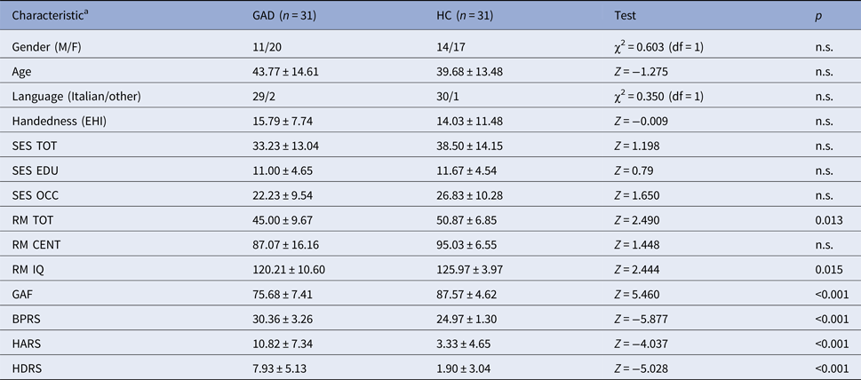
The sample characteristics are reported in Table 1. There were no significant differences in age, gender, handedness, socioeconomic status and ethnicity between the two groups. Compared to HC, GAD patients showed lower intelligence quotient (IQ) as calculated with the RMT (p < 0.05), as well as higher scores in all the clinical tests that measured anxiety or depressive symptoms (p < 0.001).
Characteristics of the sample

M, male; F, female; GAD, General Anxiety Disorder; HC, healthy controls; χ2, chi-squared test value; Z, z-statistic value from the Mann–Whitney U test; P, p value of the test; df, degrees of freedom; EHI, Edinburgh handedness inventory; SES EDU, socioeconomic status, level of education; SES OCC, socioeconomic status, occupation level; SES TOT, socioeconomic status, total; RM TOT, Raven Matrices, total score; RM CENT, Raven Matrices, percent score; RM IQ, Raven Matrices, Intelligence Quotient; BPRS, Brief Psychiatric Rating Scale; GAF, Global assessment of functioning; BPRS, Brief Psychiatric Rating Scale; HDRS, Hamilton Depression Rating Scale; HARS, Hamilton Anxiety Rating Scale.
a For continuous variables, mean ± s.d. is given.
Surface area
Although no significant CSA differences emerged between GAD patients and HC after the Bonferroni correction, some tendencies were observed, as GAD patients presented reduced CSA in the left inferior parietal, fusiform and lateral orbitofrontal regions (p < 0.05, uncorrected). The statistical details are reported in Table 2.
Results of the surface area comparison

GAD, General Anxiety Disorder; HC, Healthy Controls; ROI, region of interest.
a in mm2; mean ± s.d. is given.
b General Linear Model: ROI area ~ 1 + Diagnosis + Gender + Age + Total Hemispheric Area.
c Level of significance after Bonferroni correction: p < 0.0026.
Cortical thickness
Compared to HC, GAD patients showed significantly reduced CT in the right caudal middle frontal gyrus (p < 0.05, Bonferroni corrected). An additional tendency was observed in the superior parietal region of the right hemisphere, whose CT was lower in GAD compared with HC (p < 0.05, uncorrected). The statistical details are reported in Table 3.
Results of the cortical thickness comparison

GAD, General Anxiety Disorder; HC, Healthy Controls; ROI, region of interest.
a in mm; mean ± s.d. is given.
b General Linear Model: ROI thickness ~ 1 + Diagnosis + Gender + Age.
c Level of significance after Bonferroni correction: p < 0.0026.
Gray matter volume
No significant GMV differences between patients and HC were found.
Gyrification
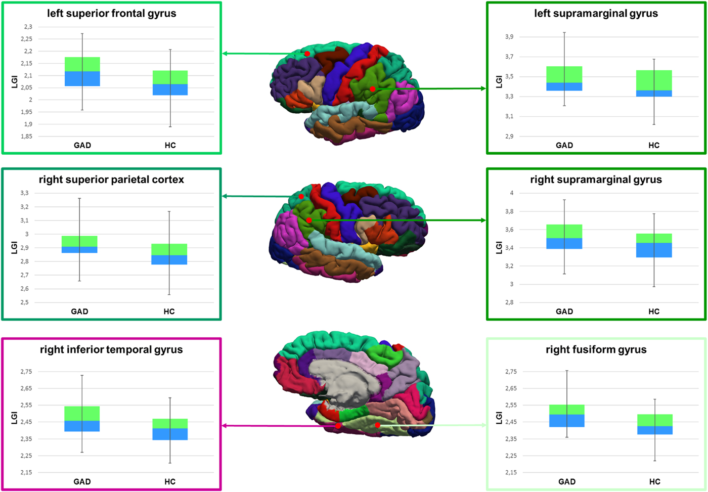
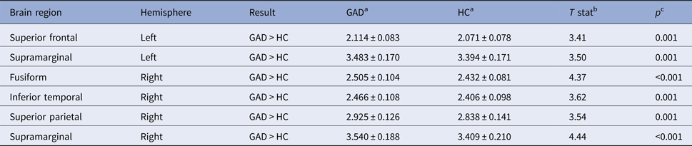
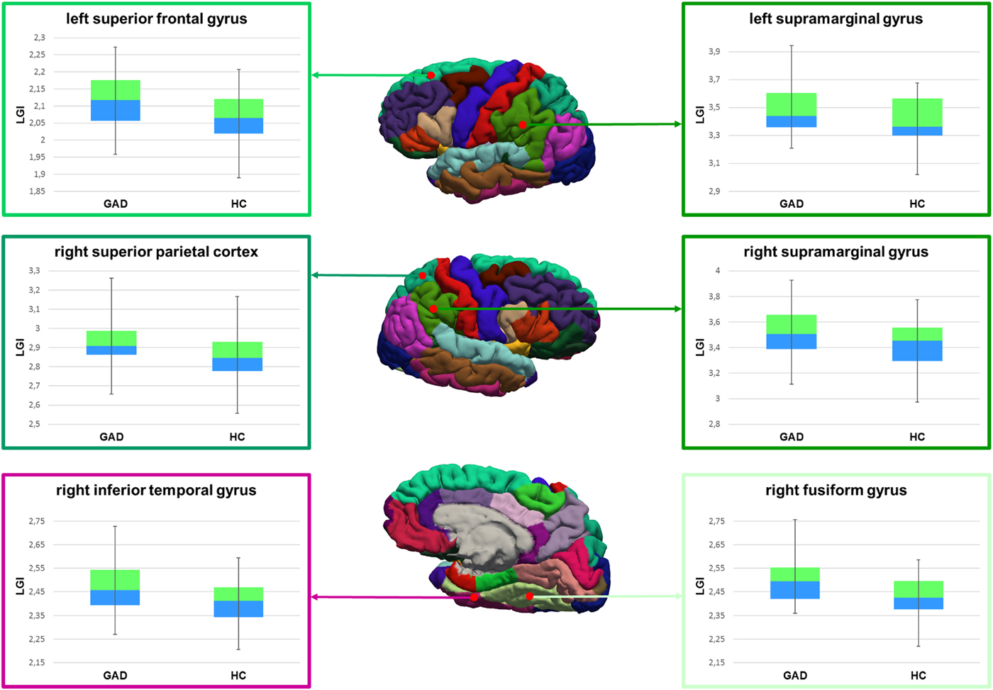
The significant results of the LGI comparison are shown in Fig. 1. Compared with HC, GAD patients were characterized by significantly higher LGI in the fusiform, inferior temporal and supramarginal gyri and in the superior parietal cortex of the right hemisphere, as well as in the supramarginal and superior frontal gyri of the left hemisphere (p < 0.05, Bonferroni corrected). The analysis details are reported in Table 4.
Results of local gyrification index (LGI) comparison. The regions with significant differences between generalized anxiety disorder (GAD) patients and healthy controls (HC) are highlighted with red dots on the cortical surface parceled according to the Desikan–Killiany atlas. The diagrams show the LGI minimum, maximum, median, and 25th and 75th percentiles in the two groups.
Results of the local gyrification index comparison

GAD, general anxiety disorder; HC, healthy controls; ROI, region of interest.
a mean ± s.d. is given.
b General Linear Model: ROI LGI ~ 1 + Diagnosis + Gender + Age.
c Level of significance after Bonferroni correction: p < 0.0026.
Correlation analyses
Within the GAD group, the neuroanatomical parameters were not significantly correlated to the clinical scores (p < 0.05, Bonferroni corrected). However, a few tendencies were observed (p < 0.01). Specifically, we found positive correlations between (1) BPRS score and CSA in the right anterior cingulate cortex (rostral section) (rho = 0.554, p = 0.004), (2) HDRS score and CT in the left isthmus cingulate cortex (rho = 0.564, p = 0.003), (3) HARS score and CT in the left middle temporal cortex (rho = 0.563, p = 0.003).
Discussion
The aim of the present work was to comprehensively investigate the cortical anatomy of patients affected by GAD, providing a robust basis for understanding the pathophysiology of this disabling illness. To this end, regional cerebral morphological parameters (CT, CSA, GMV, and LGI) were estimated and compared between GAD and HC. Overall, our results suggest the presence of neuroanatomic underpinnings of GAD, affecting specific morphological parameters. Indeed, we observed significant CT deficits and hyper-gyrification mainly in the parietal, frontal and temporal cortices of the right hemisphere, but no differences in surface area and volume of the cortex.
Among the available literature, the present work investigated the neuroanatomy of GAD in one of the largest samples of patients. Besides confirming the most consistent literature knowledge on brain structure in GAD, our work identified its neuronal bases with improved morphological specificity compared to previous ones, as it considered different cortical determinants in several regions. The sample characteristics, methods, and findings of previous anatomical and functional MRI studies on GAD and other anxiety or depressive disorders are summarized in online Supplementary Table S1. Among the fMRI studies, only the ones whose results are related to our structural findings are reported. It should be noticed that only in a couple of studies the patients’ sample size was similar to ours (Greenberg et al. Reference Greenberg, Carlson, Cha, Hajcak and Mujica-Parodi2013; Cha et al. Reference Cha, Greenberg, Carlson, Dedora, Hajcak and Mujica-Parodi2014), while the majority of previous works was conducted on smaller samples. Two out of four studies investigating CT were based on less than 15 patients (Abdallah et al. Reference Abdallah, Coplan, Jackowski, Sato, Mao and Shungu2012; Strawn et al. Reference Strawn, John Wegman, Dominick, Swartz, Wehry and Patino2014).
Cortical thickness
The present study found CT deficits in the right caudal middle frontal gyrus of GAD patients compared with HC. Since CT is thought to reflect phenomena taking place in cortical maturation during intrauterine life (asymmetric division of founder cells) (Rakic, Reference Rakic2009) and during childhood and adolescence (synaptic pruning, myelination and apoptosis) (Bourgeois & Rakic, Reference Bourgeois and Rakic1993; Morrison & Hof, Reference Morrison and Hof1997; Sowell et al. Reference Sowell, Thompson, Leonard, Welcome, Kan and Toga2004; Shaw et al. Reference Shaw, Kabani, Lerch, Eckstrand, Lenroot and Gogtay2008; Vanderhaeghen & Cheng, Reference Vanderhaeghen and Cheng2010), the CT alterations in GAD may result from neurodevelopmental factors.
As summarized in online Supplementary Table S1, just a few studies quantitatively investigated CT in anxiety, and fewer of them focused specifically on GAD (Abdallah et al. Reference Abdallah, Coplan, Jackowski, Sato, Mao and Shungu2012; Cha et al. Reference Cha, Greenberg, Carlson, Dedora, Hajcak and Mujica-Parodi2014; Strawn et al. Reference Strawn, John Wegman, Dominick, Swartz, Wehry and Patino2014). Among them, (Abdallah et al. Reference Abdallah, Coplan, Jackowski, Sato, Mao and Shungu2012) and (Canu et al. Reference Canu, Kostic, Agosta, Munjiza, Ferraro and Pesic2015) observed in GAD patients CT deficits in the rostral middle frontal cortex, adjacent to the area emerged from our study. Frontal neuroanatomical changes emerged also from studies on social anxiety (Kuhn et al. Reference Kuhn, Schubert and Gallinat2011; Syal et al. Reference Syal, Hattingh, Fouche, Spottiswoode, Carey and Lochner2012; Frick et al. Reference Frick, Howner, Fischer, Eskildsen, Kristiansson and Furmark2013).
The just mentioned neuroanatomical deficits may underlie intolerance to uncertainty and emotional dysregulation characterizing GAD patients (Ladouceur et al. Reference Ladouceur, Gosselin and Dugas2000). Indeed, fMRI studies reported altered activity of the middle frontal gyrus in GAD patients during face emotion processing tasks. Taken together, these results strongly support the role of the dorsolateral PFC for the neurobiological underpinnings of GAD.
It should be noticed that a study from Strawn and colleagues on a pediatric population (Strawn et al. Reference Strawn, John Wegman, Dominick, Swartz, Wehry and Patino2014) also identified regions with higher CT in GAD than in HC, mainly in frontal, temporal, and occipital cortices. However, CT seems to be widely affected by brain maturation: for example, the relationship between prefrontal CT and anxiety/depressive symptoms has been shown to change throughout the course of adolescence (Ducharme et al. Reference Ducharme, Albaugh, Hudziak, Botteron, Nguyen and Truong2014). Therefore, the discrepancy between our findings and the ones of Strawn et al. may be due to the younger sample of patients characterizing the latter study.
Future longitudinal studies are needed to shed light on the CT developmental changes in GAD patients in comparison with normally developing subjects.
Gyrification
One of the main novelties of our study concerns the investigation of cortical gyrification in GAD. We found significantly altered gyrification in GAD patients compared to HC in regions belonging to frontal, temporal and parietal lobes of both hemispheres.
Gyrification is believed to reflect the cortical connectivity during neurodevelopment, as the patterns of cortical folding are thought to result from different growth mechanisms of cell layers during brain maturation (Van Essen, Reference Van Essen1997). Recent works put forward the hypothesis that abnormalities in cortical folding could be linked to altered connectivity (Schaer et al. Reference Schaer, Ottet, Scariati, Dukes, Franchini and Eliez2013; Nixon et al. Reference Nixon, Liddle, Nixon, Worwood, Liotti and Palaniyappan2014).
The abnormalities emerged from our study may be interpreted in terms of neuronal development. Indeed, although the development of the cerebral cortex takes mainly place during life in utero and early childhood, structural modifications, especially in gyrification patterns, have been observed also during adolescence and young adulthood, suggesting that cortical structure encounters different levels of change during lifetime (White et al. Reference White, Su, Schmidt, Kao and Sapiro2010; Raznahan et al. Reference Raznahan, Shaw, Lalonde, Stockman, Wallace and Greenstein2011; Hogstrom et al. Reference Hogstrom, Westlye, Walhovd and Fjell2013; Mutlu et al. Reference Mutlu, Schneider, Debbane, Badoud, Eliez and Schaer2013; Su et al. Reference Su, White, Schmidt, Kao and Sapiro2013). Since anxiety disorders often emerge during adolescence and early adulthood, the presence of cortical folding abnormalities in GAD suggests a link between late brain maturation and neurobiological bases of anxiety.
Specifically, our results suggest that GAD is characterized by hyper-gyrification in bilateral supramarginal, left superior frontal and right fusiform, inferior temporal and superior parietal gyri. The absence of gyrification studies on GAD prevents us from making any direct comparison between our and previous findings. Nonetheless, structural and functional alterations in these regions have previously been observed in GAD. Literature evidence suggests abnormal activity of superior frontal, superior parietal, supramarginal, fusiform and inferior temporal gyri during emotional processing in GAD patients (Blair et al. Reference Blair, Shaywitz, Smith, Rhodes, Geraci and Jones2008; Strawn et al. Reference Strawn, Bitter, Weber, Chu, Whitsel and Adler2012; Fonzo et al. Reference Fonzo, Ramsawh, Flagan, Sullivan, Letamendi and Simmons2015), which may explain the poorer performances in working memory tasks with emotional distracters (Moon & Jeong, Reference Moon and Jeong2015; Park et al. Reference Park, Kim, Jeong, Chung and Yang2016).
The involvement of bilateral supramarginal gyri in GAD is further supported by recent evidence of (1) GMV deficits in this region (Makovac et al. Reference Makovac, Meeten, Watson, Garfinkel, Critchley and Ottaviani2016a) and (2) decreased functional connectivity between right supramarginal gyrus and right amygdala (Makovac et al. Reference Makovac, Meeten, Watson, Herman, Garfinkel and DC2016b).
Interestingly, the hyper-gyrification in right supramarginal gyrus that we observed in GAD v. HC already emerged in patients with MDD (Peng et al. Reference Peng, Shi, Li, Fralick, Shen and Qiu2015). In this respect, it is worth mentioning that GAD and MDD have a shared genetic risk, a high grade of comorbidity and common clinical features, to the point that some theories have hypothesized an overlap between the two conditions (Hettema et al. Reference Hettema, Prescott and Kendler2001b; Watson, Reference Watson2005; Mennin et al. Reference Mennin, Heimberg, Fresco and Ritter2008). Such an overlap was also observed in our study, where one-third of GAD patients had a past or current comorbidity with MDD. The hypothesis of shared neuroanatomic bases for the two conditions is fascinating and deserves further investigations.
Moreover, we still need to understand whether the hyper-gyrification that emerged from our study is the primary alteration or represents a compensatory, local, response of a single brain region to a disrupted network, as an effort to keep an efficient cumulative function. An interesting insight could be given from longitudinal studies of GAD, following the evolution of cortex through age, in order to: (1) identify the brain structural peculiarities in drug naïve first episode subjects, (2) check if the evolution of illness can modify (increase or reduce) its original cortical determinants.
Cortical volume and surface area
Our study suggests no significant differences in volume and superficial area of the cortex in GAD patients compared to HC. However, GAD patients had a slightly reduced CSA – not surviving after multiple comparison corrections – in left fusiform, inferior parietal and orbitofrontal cortices.
It is worth remarking that our study is the first in the literature combining the common analysis of GMV with the investigation of other morphological parameters. As for gyrification, no studies before the current investigated CSA in GAD. Therefore, the tendencies observed here should be verified by future CSA studies.
Regarding GMV, in contrast with our findings, previous volumetric studies agreed on the presence of alterations in GAD. Overall, they suggested GMV deficits in the parietal region (Strawn et al. Reference Strawn, Wehry, Chu, Adler, Eliassen and Cerullo2013; Makovac et al. Reference Makovac, Meeten, Watson, Garfinkel, Critchley and Ottaviani2016a) and increased GMV in the temporal region (De Bellis et al. Reference De Bellis, Keshavan, Shifflett, Iyengar, Dahl and Axelson2002; Hilbert et al. Reference Hilbert, Pine, Muehlhan, Lueken, Steudte-Schmiedgen and Beesdo-Baum2015). The discrepancy between our and previous results can be attributed, at least in part, to a methodological issue, as all previous works used voxel-based morphometry (VBM) instead of the surface-based approach employed in the current study. First, while we reported evidence of GMV abnormalities at the regional level, using the Desikan–Killiany atlas as a reference, previous studies investigated GMV abnormalities at the voxel level. Moreover, the calculation of GMV in our study was performed as the product of CT and CSA on the region of interest, whereas in VBM studies it was based on tissue probability maps. Due to these and other methodological differences, voxel-based, and surface-based techniques may provide different results even when applied on the same sample, as recently shown in a study on Parkinson's disease (Gerrits et al. Reference Gerrits, Van Loenhoud, Van Den Berg, Berendse, Foncke and Klein2016). Future parallel investigations are encouraged to obtain a complete understanding of GMV correlates of GAD.
Considerations about hemispheric asymmetry in GAD
In accordance to previous structural and functional studies (Davidson & Irwin, Reference Davidson and Irwin1999; De Bellis et al. Reference De Bellis, Keshavan, Shifflett, Iyengar, Dahl and Axelson2002; Zhang et al. Reference Zhang, Li, Yu, Liu, Tang and Tan2013), we detected a predominant role of the right side of the brain in GAD, as already suggested by our group in an independent population (Brambilla et al. Reference Brambilla, Como, Isola, Taboga, Zuliani and Goljevscek2012).
Even if the topic is still controversial, previous works tried to understand how each hemisphere is involved in the pathogenesis of anxiety: already in 1997, Heller et al. linked anxious arousal in anxious subjects to the activation of the right parietal region. However, the same subjects at rest were characterized by a more predominant role of the left hemisphere compared to reference subjects, suggesting different patterns of activation between anxious apprehension and anxious arousal (Heller et al. Reference Heller, Nitschke, Etienne and Miller1997). Recently, other authors investigated the issue of laterality in emotional processing, suggesting that the right hemisphere: (1) is more sensitive to perception and processing of frightening stimuli (Noesselt et al. Reference Noesselt, Driver, Heinze and Dolan2005) and (2) is implicated in the physical response to stress, by modulating the hypothalamus–pituitary–adrenal axis through cortisol secretion (Lueken et al. Reference Lueken, Leisse, Mattes, Naumann, Wittling and Schweiger2009). Interestingly, a relationship between anxiety symptoms and gliomas only in the right hemisphere was reported, with decreased symptomatology after surgical removal (Mainio et al. Reference Mainio, Hakko, Niemela, Tuurinkoski, Koivukangas and Rasanen2003).
Taken together, our and previous evidence supports the idea that the two hemispheres contribute to the pathogenesis of anxiety in different ways, with a key role of the right brain in determining stress response and anxiety symptoms. The increased gyrification in right temporoparietal cortex and the CT deficits in right prefrontal cortex could be the neurobiological substrate of the aberrant processing of stress occurring in GAD, taking place especially in right brain structures.
Link between neuroanatomy and functional connectivity in GAD
Overall, the regions presenting cortical abnormalities in our work belong to the functional networks that have already been associated with anxiety disorders, especially the default mode network (DMN), including lateral parietal cortex, inferior temporal gyrus and superior frontal cortex, and the lateral frontoparietal executive network (FPN), whose key regions include the dorsolateral PFC and the posterior parietal cortices (Sylvester et al. Reference Sylvester, Corbetta, Raichle, Rodebaugh, Schlaggar and Sheline2012).
The DMN, whose activity is suppressed during cognitive tasks, is thought to be responsible for self-referential activities such as introspection, planning of future, and emotion regulation (Greicius et al. Reference Greicius, Krasnow, Reiss and Menon2003). On the other hand, the FPN processes strategy adjustments when receiving feedbacks on behavioral errors (Dosenbach et al. Reference Dosenbach, Fair, Cohen, Schlaggar and Petersen2008). The interaction between these two networks is essential for an adequate cognitive regulation of emotion. A recent study has hypothesized that decreased functioning of DMN and FPN may lead to some features of anxiety disorders (Sylvester et al. Reference Sylvester, Corbetta, Raichle, Rodebaugh, Schlaggar and Sheline2012), such as emotional dysregulation and deficits in cognitive control respectively. Our findings support this hypothesis by providing a structural basis for the DMN and FPN alterations, contributing in turn to emotive–cognitive dysfunction typical of GAD.
Methodological issues and limitations
The present study suffers from some limitations. First, the modest sample size may prevent from detecting small anatomical differences between GAD and HC, besides limiting the statistical power of the results due to second type errors. It should also be mentioned that a number of GAD patients presented past or current comorbidities with other psychiatric conditions, which may have affected the reliability and specificity of our findings. Possible inaccuracies may also derive from the estimation of the morphological parameters at the regional level, which assumes the structural homogeneity within each ROI. In this respect, the parallel use of a voxel-wise approach should be considered for future studies. Finally, age and gender are known to affect cortical morphology and may partially contribute to group differences; however, we did adjust for age and gender in all models, in order to reduce the effect of these confounding factors. The application of Bonferroni correction also allowed us to limit the rate of false positive findings, thereby increasing the reliability of our results.
Future perspectives
The results of neuroimaging studies have potential to give new insights on the clinical management of GAD patients. Our results let us speculate that brain morphology alterations could serve as a substrate for cognitive and emotional features of GAD. In this respect, future research works should investigate in depth the link between functional and structural characteristics in GAD patients.
Future morphological studies should also explore whether innovative therapeutic interventions can influence the cortical gyrification and thickness trajectories in the regions involved in GAD symptomatology. Among the new interventional strategies, a technique that deserves mention is transcranial direct current stimulation (tDCS), which was successfully used in the treatment of MDD (Kalu et al. Reference Kalu, Sexton, Loo and Ebmeier2012) and obsessive–compulsive disorder (D'Urso et al. Reference D'urso, Brunoni, Mazzaferro, Anastasia, De Bartolomeis and Mantovani2016), showing encouraging results in a single case pilot study on GAD (Shiozawa et al. Reference Shiozawa, Leiva, Castro, Da Silva, Cordeiro and Fregni2014). Since tDCS effects largely depend on the electrode placement, the treatment efficacy can be maximized by improving the localization of brain morphological alterations in GAD patients.
Conclusions
The present work deals with the comprehensive investigation of the neuroanatomical correlates of GAD. For the first time, the cortical morphology of GAD patients has been inspected in terms of thickness, area, volume and gyrification of the cerebral cortex. Our results suggest that GAD is characterized by specific structural abnormalities that mainly involve the right hemisphere. GAD patients showed CT deficits in the right caudal middle frontal gyrus and hyper-gyrification in the superior frontal and supramarginal gyri of the left hemisphere and in the fusiform, inferior temporal, superior parietal and supramarginal regions of the right hemisphere.
The above regions are part of the networks implicated in cognitive-emotion regulation and processing, therefore necessary for an adequate interaction between cognition and emotions. Thence, the structural alterations emerged from our study may represent the substrate and/or the consequence of more widespread network alterations. Since thickness and gyrification reflect lifetime changes in the microscopic anatomy of the cerebral cortex, further longitudinal studies are needed in order to establish the chronology of these alterations over the course of the illness.
Supplementary material
The supplementary material for this article can be found at https://doi.org/10.1017/S003329171700352X
Acknowledgement
Grant support of EM was provided by the European Union's Seventh Framework Programme for research, technological development and demonstration under grant agreement no. 602450 (IMAGEMEND project). This study was partly supported by grants from the Italian Ministry of Health (RF-2011-02352308) to PB and from the Fondazione Cariplo (n° 2014:0664) to GP.
Declaration of interest
None.
Ethical standards
The authors assert that all procedures contributing to this work comply with the ethical standards of the relevant national and institutional committees on human experimentation and with the Helsinki Declaration of 1975, as revised in 2008.