Introduction
Airway obstruction is a common presentation in patients with laryngeal cancer. Management of acute obstruction in these patients focuses on securing the airway without compromising oncological outcome. In such cases, medical adjuncts including nebulised adrenaline and intravenous steroids are often used.
We present a case of takotsubo cardiomyopathy resulting from nebulised adrenaline administered for the management of acute airway obstruction in a patient with laryngeal cancer.
Case report
A 66-year-old man with squamous cell carcinoma of the larynx, with tumour–node–metastasis staging of T3N2cM0, confirmed by biopsy and computed tomography, presented to the emergency department with acute airway obstruction. Nasendoscopy demonstrated an irregular mass affecting the left hemilarynx and extending to the subglottis, with vocal fold fixation. He was treated with 1 µg/5 ml of nebulised adrenaline and 8 mg of intravenous dexamethasone. He was transferred to the intensive care unit for airway observation pending a laryngectomy. On the intensive care unit, the patient required further treatment with nebulised adrenaline.
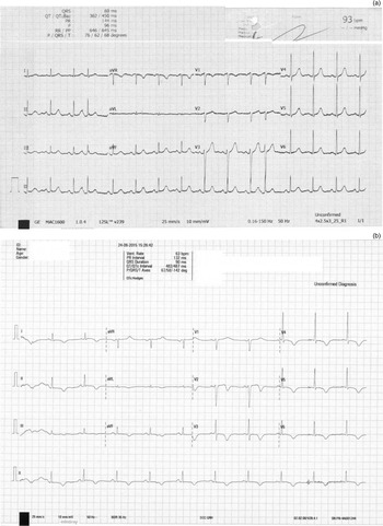
After a period of 24 hours, cardiac rhythm changes were noted on telemetry. A 12-lead electrocardiogram (ECG) showed widespread T-wave inversion and QT prolongation (Figure 1). Throughout this episode, the patient reported no chest pain or other symptoms of an acute coronary syndrome. Analyses of serum cardiac enzymes conducted 6 and 12 hours after ECG changes demonstrated high levels of troponin T. The ECG changes and raised cardiac enzyme levels indicated possible myocardial ischaemia.
(a) Electrocardiogram on admission to the emergency department showing tachycardia and no evidence of ischaemic heart disease. (b) Electrocardiogram 24 hours after admission (following administration of nebulised adrenaline) showing widespread T-wave inversion and QT prolongation. V1–V6 represent pre-cordial electrode placements. Bpm = beats per minute; aVR = augmented vector right; aVL = augmented vector left; aVF = augmented vector foot; vent = ventricular; deg = degrees

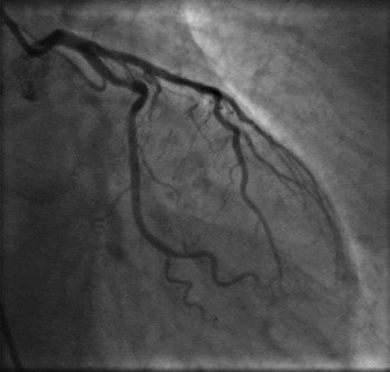
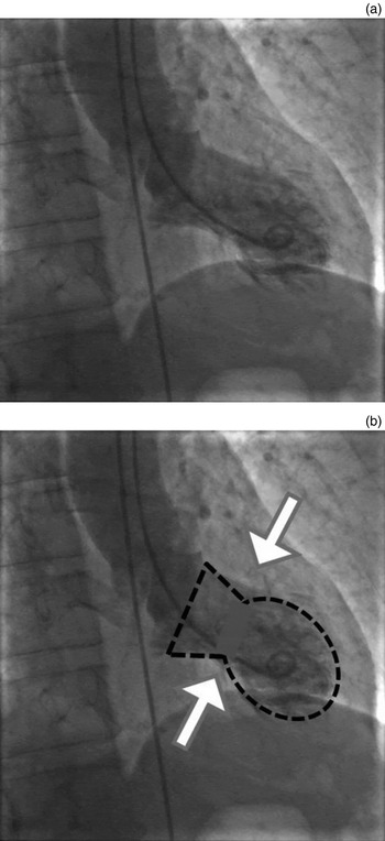
Urgent coronary angiography was performed. This demonstrated no coronary artery disease (Figure 2). However, left ventricular angiography showed marked apical ballooning and apical wall akinesia (Figure 3). A subsequent transthoracic echocardiogram confirmed a hypokinetic apical segment of the left ventricle and a left ventricular ejection fraction by Simpson's method of 42 per cent. These findings are consistent with a diagnosis of takotsubo cardiomyopathy. The patient subsequently underwent laryngectomy for his laryngeal carcinoma.
Angiography showing normal coronary artery, with no defect in filling.

(a) Left ventricular angiography shows apical ballooning and mid-cavity ventricular abnormality. (b) Vase-like appearance of the cavity.

Discussion
Takotsubo cardiomyopathy is also referred to as apical ballooning syndrome, broken heart syndrome, stress cardiomyopathy and Japanese octopus syndrome.Reference Dote, Sato, Tateishi, Huchida and Ishihara 1 – Reference Tsuchihashi, Ueshima, Uchida, Oh-mura, Kimura and Owa 3 It describes the transient regional left ventricular dysfunction manifesting in global ECG changes and raised cardiac biomarkers that mimic myocardial infarction. It is distinguishable from myocardial infarction based on angiographic evidence suggesting no coronary artery disease or plaque rupture.Reference Tsuchihashi, Ueshima, Uchida, Oh-mura, Kimura and Owa 3 – Reference Prasad, Lerman and Rihal 10 Approximately 15 per cent of those with takotsubo cardiomyopathy also have some level of coronary artery disease. If coronary artery disease is found, takotsubo cardiomyopathy can be diagnosed if the changes in ECG are beyond the territory perfused by a single epicardial coronary artery and the wall motion abnormalities are not in the distribution of the coronary disease.Reference Templin, Ghadri, Diekmann, Napp, Bataiosu and Jaguszewski 11
The condition was first described in 1990 in Japan. The term ‘takotsubo’ means octopus trap, which is a vase that has a similar shape to the systolic apical ballooning appearance of the left ventricle, created as a result of depression of the mid and apical segments and hypokinesis of the basal walls.Reference Kurowski, Kaiser, von Hof, Killermann, Mayer and Hartmann 12 – Reference Park, Kang, Song, Kim, Lim and Kang 15
Patients often present with chest pain, retrosternal pain or dyspnoea, but a significant number are asymptomatic and the condition is found on routine investigation.Reference Templin, Ghadri, Diekmann, Napp, Bataiosu and Jaguszewski 11 It affects women more than men and 80 per cent of those diagnosed are post-menopausal. The prevalence is not known; however, 1–2 per cent of patients presenting with troponin-positive suspected acute coronary syndrome are thought to have takotsubo cardiomyopathy.Reference Kurowski, Kaiser, von Hof, Killermann, Mayer and Hartmann 12 – Reference Prasad, Dangas, Srinivasan, Yu, Gersh and Mehran 14
-
• Takotsubo cardiomyopathy following the use of nebulised adrenaline for airway obstruction has not previously been reported
-
• It may mimic true ischaemic heart disease; the possibility of takotsubo cardiomyopathy should be considered in this context
-
• It can mimic myocardial infarction, with electrocardiogram (ECG) and biochemical evidence of ischaemic heart disease
-
• It is distinguished from a myocardial infarct based on angiographic and ECG evidence
-
• There is systolic apical ballooning of the left ventricle resulting from depression of mid and apical segments and hypokinesis of basal walls
The exact pathogenesis of the disease is not known, particularly the reason for the primary involvement of the mid and apical segments of the left ventricle.Reference Park, Kang, Song, Kim, Lim and Kang 15 – Reference Park, Prasad, Rihal, Bell and Oh 17 Takotsubo cardiomyopathy is often but not always precipitated by extreme physical or emotional distress, such as the death of a relative, natural disasters or after sepsis.Reference Bybee, Kara, Prasad, Lerman, Barsness and Wright 2 , Reference Abe, Kondo, Matsuoka, Araki, Dohyama and Tanio 4 , Reference Watanabe, Kodama, Okura, Aizawa, Tanabe and Chinushi 18 This has led to the hypothesis that excess catecholamine release leads to coronary artery spasm and microvascular abnormalities, causing myocardial stunning.Reference Paur, Wright, Sikkel, Tranter, Mansfield and O'Gara 19 This idea has been reinforced by studies that have demonstrated high levels of plasma adrenaline in patients found to have takotsubo cardiomyopathy in the intensive care setting.Reference Park, Kang, Song, Kim, Lim and Kang 15 This has not been replicated in all subsequent studies, with some authors demonstrating no difference in the plasma adrenaline levels found in those with takotsubo cardiomyopathy and those who had a myocardial infarct.Reference Heubach, Ravens and Kaumann 20 However, the findings of vasospasm on angiography and hypoperfusion of the myocardium which resolves following improvement in myopathy strengthen the argument for catecholamine-induced takotsubo cardiomyopathy.Reference Abe, Kondo, Matsuoka, Araki, Dohyama and Tanio 4 , Reference Dec 7 , Reference Lyon, Rees, Prasad, Poole-Wilson and Harding 21 , Reference Kurisu, Sato, Kawagoe, Ishihara, Shimatani and Nishioka 22
Conclusion
Takotsubo cardiomyopathy has not previously been reported following the use of nebulised adrenaline for airway obstruction. It may mimic true ischaemic heart disease and it is important to consider the possibility of takotsubo cardiomyopathy in this context.
The diagnosis requires a high index of suspicion in patients with ECG and biochemical evidence of ischaemic heart disease in the absence of physical symptoms.
Takotsubo cardiomyopathy is confirmed by ECG, with or without angiography, demonstrating normal coronary vessels. Treatment involves removing the causative agent. Left ventricular hypokinesis is transient and often resolves.