Introduction
The large majority of retinal ganglion cells in primate including human comprise midget and parasol cells but in addition to date at least thirteen additional types of ganglion cells have been identified based on their morphology (Dacey et al., Reference Dacey, Peterson, Robinson and Gamlin2003; Kolb et al., Reference Kolb, Linberg and Fisher1992; Masri et al., Reference Masri, Percival, Koizumi, Martin and Grünert2019; Yamada et al., Reference Yamada, Bordt and Marshak2005). Owing to their relative low density and the lack of specific markers, these ganglion cells have been not been studied systematically. Moreover, it is assumed that there are a total of 30 functional ganglion cell types in all mammalian retinas (Baden et al., Reference Baden, Berens, Franke, Roman Roson, Bethge and Euler2016; Dacey, Reference Dacey and Gazzaniga2004; Dacey et al., Reference Dacey, Peterson, Robinson and Gamlin2003; Masri et al., Reference Masri, Percival, Koizumi, Martin and Grünert2019; Sanes & Masland, Reference Sanes and Masland2015); thus, there is still room for more ganglion cell types to be discovered in primates.
Calbindin, like calretinin, parvalbumin, and secretagogin, belongs to the family of EF-hand proteins, which bind calcium with high affinity and play a role in calcium buffering (Baimbridge et al., Reference Baimbridge, Celio and Rogers1992; Heizmann & Hunziker, Reference Heizmann and Hunziker1991). Calbindin is expressed in a variety of neurons in the retina of many species of mammals (Pasteels et al., Reference Pasteels, Rogers, Blachier and Pochet1990; Pochet et al., Reference Pochet, Pasteels, Seto-Ohshima, Bastianelli, Kitajima and Van Eldik1991) but its functional role in the nervous system is not well understood (Schwaller et al., Reference Schwaller, Meyer and Schiffmann2002). Understanding which retinal cell types express calbindin may contribute to a better understanding of the functional role of calbindin (Kántor et al., Reference Kántor, Mezey, Adeghate, Naumann, Nitschke, Enzsoly, Szabó, Lukats, Nemeth, Somogyvari and Völgyi2016).
Calbindin immunoreactivity is widely used in the retina and other brain regions as marker for cell populations. In primate retina, calbindin is present in cones, cells in the inner nuclear layer, and cells in the ganglion cell layer (Chiquet et al., Reference Chiquet, Dkhissi-Benyahya and Cooper2005; Pasteels et al., Reference Pasteels, Rogers, Blachier and Pochet1990; Röhrenbeck et al., Reference Röhrenbeck, Wässle and Boycott1989). The calbindin-labeled cell types in the inner nuclear layer are known to include H2 horizontal cells (Wässle et al., Reference Wässle, Dacey, Haun, Haverkamp, Grünert and Boycott2000), diffuse bipolar-type DB3a cells (Haverkamp et al., Reference Haverkamp, Haeseleer and Hendrickson2003; Jacoby et al., Reference Jacoby, Wiechmann, Amara, Leighton and Marshak2000; Jacoby & Marshak, Reference Jacoby and Marshak2000; Kántor et al., Reference Kántor, Mezey, Adeghate, Naumann, Nitschke, Enzsoly, Szabó, Lukats, Nemeth, Somogyvari and Völgyi2016; Luo et al., Reference Luo, Ghosh, Martin and Grünert1999; Martin & Grünert, Reference Martin and Grünert1992), and several populations of amacrine including the AII cells (Kolb et al., Reference Kolb, Zhang, Dekorver and Cuenca2002; Weltzien et al., Reference Weltzien, Percival, Martin and Grünert2015). The calbindin-positive cells in the ganglion cell layer in fetal human retina were recently identified to include ON ChAT cells (Zhang et al., Reference Zhang, Yu, Hoshino, Huang, Rieke, Reh and Wong2019). Although calbindin-immunoreactive cells in the ganglion cell layer were reported for a variety of primate species (Chiquet et al., Reference Chiquet, Dkhissi-Benyahya and Cooper2005; de Souza et al., Reference de Souza, Nivison-Smith, Christie, Polkinghorne, McGhee, Kalloniatis and Acosta2016; Hamano et al., Reference Hamano, Kiyama, Emson, Manabe, Nakauchi and Tohyama1990; Kántor et al., Reference Kántor, Mezey, Adeghate, Naumann, Nitschke, Enzsoly, Szabó, Lukats, Nemeth, Somogyvari and Völgyi2016), none of these studies attempted to identify the labeled type(s).
In mouse retina, calbindin has been localized in about 18% of the ganglion cell population (Gu et al., Reference Gu, Lee and Jeon2016) including a subpopulation of intrinsically photosensitive (melanopsin-expressing) retinal ganglion cells but which specific types of melanopsin-expressing cells has not yet been revealed (Kriegsfeld et al., Reference Kriegsfeld, Mei, Yan, Witkovsky, Lesauter, Hamada and Silver2008). Primates have two major types of melanopsin-expressing cells. Outer stratifying cells stratify close to the inner nuclear layer and have a soma located either in the inner nuclear layer (INL outer cells) or in the ganglion layer (GCL outer cells). Inner stratifying and bistratified cells (GCL inner cells) have a soma in the ganglion cell layer and mainly stratify close to the ganglion cell layer, with occasional processes extending toward the border with the inner nuclear layer (Dacey et al., Reference Dacey, Liao, Peterson, Robinson, Smith, Pokorny, Yau and Gamlin2005; Hannibal et al., Reference Hannibal, Hindersson, Østergaard, Georg, Heegaard, Larsen and Fahrenkrug2004; Jusuf et al., Reference Jusuf, Lee, Hannibal and Grünert2007; Liao et al., Reference Liao, Ren, Peterson, Marshak, Yau, Gamlin and Dacey2016; Nasir-Ahmad et al., Reference Nasir-Ahmad, Lee, Martin and Grünert2019). To date, no molecular differences between melanopsin cell types have been reported in primates although such differences have been reported for melanopsin-expressing cells in mouse (Chen et al., Reference Chen, Badea and Hattar2011; Li & Schmidt, Reference Li and Schmidt2018) and tree shrew (Johnson et al., Reference Johnson, Westbrook, Shayesteh, Chen, Schumacher, Fitzpatrick and Field2019) retinas.
In the present study, we show that calbindin is present in the two types of melanopsin-expressing ganglion cells in human but not in marmoset retina. In both species, calbindin is expressed by ON ChAT cells but only in a subpopulation of OFF ChAT cells. In addition, some antibody-related differences were observed.
Materials and methods
Human retinas
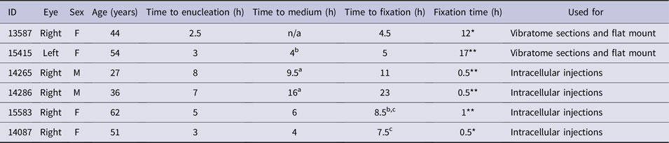
Postmortem human eyes from donors with no known history of eye disease were obtained from the Lions NSW Eye Bank at the Sydney Eye Hospital with donor consent and ethical approval from the University of Sydney Human Research Ethics Committee. Details about the eyes are summarized in Table 1. The posterior eyecups were fixed in 2% or 4% paraformaldehyde in 0.1 M phosphate buffer (PB) when the tissue was intended for standard immunohistochemistry. No obvious staining differences were seen with respect to the concentration of fixative used. The eyes intended for prelabeling and DiI injections were kept in Hibernate-A medium (Gibco, Life Technologies, Mulgrave, Australia) or CO2 independent medium (Gibco) for 1 to 9 h before retina dissection. Some eyecups were placed into carboxygenated (95% O2/5% CO2) Ames’ medium before dissection. The retinas intended for DiI injections were first dissected and then fixed in 4% paraformaldehyde in 0.1 M PB for 30 or 60 min.
Subjects
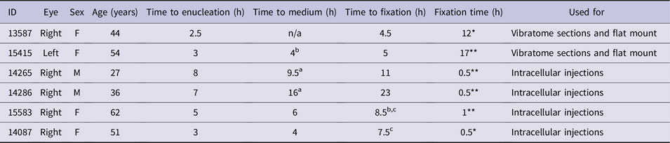
* Fixed in 2% paraformaldehyde in PB.
** Fixed in 4% paraformaldehyde in PB.
a Eyes were kept in Hibernate medium before retina dissection.
b Eyes were kept in CO2 independent medium before retina dissection.
c Eyes were placed into oxygenated Ames’ medium before fixation.
Marmoset retinas
Eyes were obtained from one male and three female adult marmosets (Callithrix jacchus, aged between 1.8 and 4 years) at the end of unrelated electrophysiological experiments. The animals were obtained from the Australian National Health and Medical Research Councils (NHMRC) combined breeding facility. All procedures were conducted according to the provisions of the NHMRC code of practice for the care and use of animals and were approved by the University of Sydney Animal Ethics Committee or by the Monash University Animal Ethics Committee, which also monitored the welfare of the animals. The protocols were in compliance with the principles regarding the care and use of animals adopted by the Society for Neuroscience. At the end of the experiments, animals were overdosed with sodium pentobarbitone (Lethobarb, Virbac, Milperra, NSW, Australia). Three animals were perfused with cold saline; the eyes were enucleated, cut open; the cornea, lens, and vitreous were removed; and subsequently the posterior eyecup was immersion fixed in 4% paraformaldehyde in 0.1 M PB for one hour. One animal was perfused with 4% paraformaldehyde in 0.1 M PB after saline perfusion. Two retinal quadrants from one animal were used for prelabeling and subsequent intracellular DiI injections. Two quadrants were used for vibratome sectioning and one quadrant was processed as a flat mount.
Immunohistochemistry
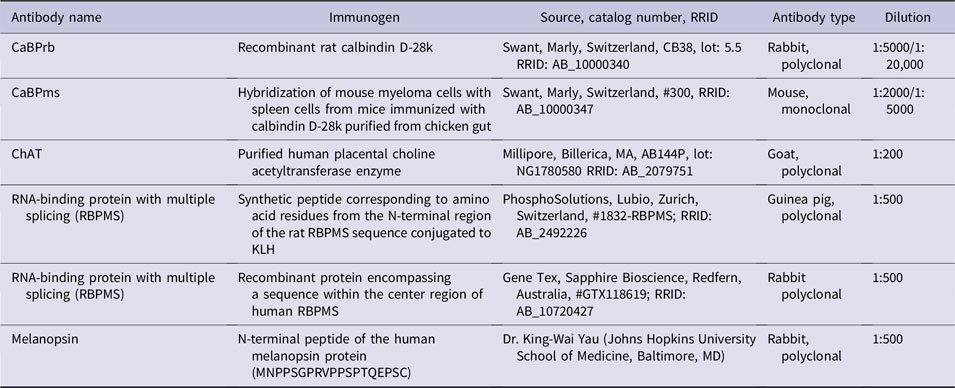
The primary antibodies used are summarized in Table 2. The two antibodies against calbindin D-28 used in the present study were obtained from Swant (Marly, Switzerland). According to the manufacturer, the rabbit calbindin antibody shows some cross-reactivity to the calcium-binding protein calretinin when used at higher concentrations, whereas the mouse calbindin antibody does not cross react with calretinin or other known calcium-binding proteins. Hereinafter, we refer to the polyclonal antiserum against calbindin as calbindin-rabbit and the monoclonal mouse antibody as calbindin-mouse.
Antibodies

Retinal quadrants and 100-µm thick vibratome sections were preincubated for 60 min in 5% normal donkey serum (Jackson Immuno Research Laboratories Inc., Westgrove, PA) in phosphate buffered saline (PBS) containing 0.5% Triton X-100 (BDH Chemicals, Kilsyth, Australia). Subsequently, the tissue was incubated in a mixture of primary antibodies for up to 7 days at 4°C; the tissue was rinsed and then incubated in a mixture of secondary antibodies made in donkey and coupled to Alexa 488 or Alexa 594 (Jackson Immuno Research Laboratories) diluted 1:250 and 1:500, respectively, in PBS containing 3% normal donkey serum and 0.5% Triton X-100 overnight. Retinal sections and pieces were rinsed in PBS (3 × 10 min), mounted onto microscope slides and coverslipped with Vectashield Antifade Mounting Medium (Vector Laboratories Inc., Burlingame, CA). Secure seal adhesive spacers (Invitrogen, Mulgrave, Australia) were used to prevent distortion of the sections.
Prelabeling and DiI injection
Immunolabeling before DiI injection was conducted as described previously (Kao & Sterling, Reference Kao and Sterling2003; Lee et al., Reference Lee, Weltzien, Madigan, Martin and Grünert2016; Weltzien et al., Reference Weltzien, Dimarco, Protti, Daraio, Martin and Grünert2014). Briefly, after fixation, retinal quadrants were rinsed in 0.1 M PB and then incubated with primary antibodies against calbindin-rabbit diluted 1:20,000 (human retina) or 1:5000 (marmoset retina) in PB for 3 to 4 days at 4°C. Tissue was rinsed in PB and then incubated with secondary antibodies coupled to Alexa 488 diluted at 1:250 in PB for 2 to 4 h at room temperature. Triton was omitted from all incubation solutions. Retinal tissue was placed into a superfusion chamber mounted on a fixed stage microscope (Axioskop, Zeiss). Glass electrodes with tip diameter of ∼1 µm were filled with 1% DiI (Invitrogen, Mulgrave, Australia) in 100% ethanol and prelabeled cells were injected with DiI by applying a 10 nA–positive current for 1 to 10 s. After injection, the retinal quadrants were incubated in a solution of 10 µl/ml DAPI (4′,6-diamidino-2-phenylindole dihydrochloride, D9542, Sigma, Sydney, Australia) in PBS at room temperature overnight. The tissue was rinsed in PBS, mounted onto glass slides, and coverslipped with VECTASHIELD mounting medium (H-100, Vector Laboratories, Burlingame, CA) and imaged with a confocal microscope within a week.
Microscopy
Images were acquired using a confocal microscope (Zeiss LSM 700, Carl Zeiss, Jena, Germany) equipped with 405, 488, 555, and 635 nm lasers. Overview images were obtained for each retinal quadrant using a 10×/0.45 Plan-Apochromat objective. Subsequently, stacks of images were taken of individual injected cells using a 20× objective (Plan Apochromat, 20 × 0.8, #420650-9901) at a resolution of 2048 by 2048 pixels, from the ganglion cell layer to the inner nuclear layer (step size 1 µm). The contrast and brightness of the images were adjusted using Zen Black or Zen Blue (Zeiss), Adobe Photoshop CS6, or Imaris (Bitplane, Zurich, Switzerland) software.
Analysis
The dendritic field and soma diameters of DiI-injected cells were measured from stacks of images using Fiji (Schindelin et al., Reference Schindelin, Arganda-Carreras, Frise, Kaynig, Longair, Pietzsch, Preibisch, Rueden, Saalfeld, Schmid, Tinevez, White, Hartenstein, Eliceiri, Tomancak and Cardona2012). The tips of the outermost dendrites of each cell were connected to form a convex polygon. The average of the minimum and maximum diameter of the polygon was taken as the dendritic field diameter (Szmajda et al., Reference Szmajda, Grünert and Martin2008). Cell stratification was determined from the image stacks using orthogonal projections in Zen Blue (Zeiss) or Imaris software (Bitplane, Zurich, Switzerland). DiI-injected cells were reconstructed in 3D from confocal image stacks using the filament tracer in Imaris software. The labeling intensity of calbindin immunofluorescence was determined using the spot tool in Imaris software and normalizing against a white background point.
Results
Calbindin immunoreactive cells in human retina
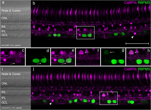
Consistent with previous studies (reviewed in Kántor et al., Reference Kántor, Mezey, Adeghate, Naumann, Nitschke, Enzsoly, Szabó, Lukats, Nemeth, Somogyvari and Völgyi2016), in human retina, calbindin is expressed by cones as well as subpopulations of cells in the inner nuclear layer and in the ganglion cell layer (Fig. 1). To determine whether calbindin expressing cells in the ganglion cell layer are ganglion cells, sections and flat mount preparations were double labeled with antibodies against calbindin-rabbit and antibodies against RBPMS (Kwong et al., Reference Kwong, Caprioli and Piri2010; Rodriguez et al., Reference Rodriguez, Pérez de Sevilla Müller and Brecha2014). As shown in Fig. 1, some RBPMS-positive cells were also calbindin-positive. The soma of these double-labeled cells were either located in the ganglion cell layer (Fig. 1a–1e) or in the inner nuclear layer (Fig. 1f–1i). Strongly calbindin-positive cells in the ganglion cell layer had a smaller soma diameter and were not double labeled (arrowheads in Fig. 1b and 1j), indicating that they are displaced amacrine cells.
Calbindin expression in human retina. Confocal micrographs of vertical sections through peripheral retina from two human donors, processed with antibodies against calbindin (magenta) and the ganglion cell marker RBPMS (green). (a, i) Nomarski optics to reveal the retinal layers. (b, j) Calbindin is expressed by cone photoreceptors, subpopulations of horizontal, bipolar, and amacrine cells and a small population of cells in the ganglion cell layer (GCL). The arrowheads point to calbindin-positive displaced amacrine cells. The rectangles in b and j indicate the region shown at higher resolution in c–e and f–h, respectively. (c) A strongly calbindin positive displaced amacrine cell and a weakly calbindin positive ganglion cell (arrow) are visible in the GCL. (d, e) The same region as in c is shown revealing two RBPMS positive ganglion cells. The double labeled cell is indicated by an arrow. (f–h) A calbindin-labeled cell in the inner nuclear layer is RBPMS-positive (arrow) indicating that it is a displaced ganglion cell. Scale bar shown in b = 100 µm, applies to all. ONL, outer nuclear layer; INL, inner nuclear layer; IPL, inner plexiform layer; GCL, ganglion cell layer.

The proportion of calbindin-positive/RBPMS-positive cells was determined in stacks of images taken from such vibratome sections of peripheral retina (5 to 10 mm eccentricity) in two preparations. In the ganglion cell layer, a total of 545 RBPMS-positive ganglion cells and 157 calbindin-positive cells were counted. Five of the RBPMS-positive cells (0.9%) were also positive for calbindin, whereas in the amacrine cell layer, 4/13 RBPMS-positive cells were calbindin-positive. This result suggests that calbindin-expressing ganglion cells in the inner nuclear layer may form a higher proportion than those located in the ganglion cell layer.
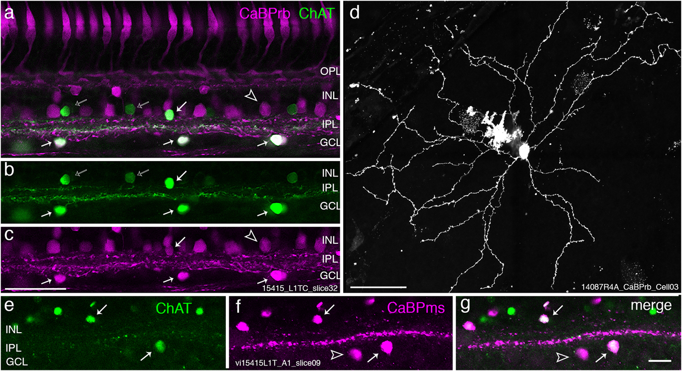
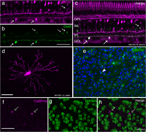
Other vibratome sections (eccentricity range 1 to 8.5 mm) were double-labeled with antibodies to calbindin-rabbit and choline acetyltransferase (ChAT, Fig. 2a–2c) or with calbindin-mouse antibodies and ChAT (Fig. 2e and 2f). There were no obvious differences between the two antibodies with respect to the double-labeling pattern; thus, the quantitative results are presented together. All ON ChAT cells (n = 447) were calbindin-positive, but some of the calbindin-positive cells (16/463 cells or 3%) were ChAT-negative. This result is consistent with the idea that some calbindin-positive cells are ganglion cells.
Calbindin is expressed by starburst amacrine cells in human retina. (a–c) Confocal images of a vertical section through human donor (54 years, female) retina, which was processed with antibodies against calbindin made in rabbit (CABPrb, magenta) and ChAT (green). All ON ChAT cells but not all OFF ChAT cells are double labeled (white arrows). Singly labeled OFF ChAT cells are indicated by open arrows; singly labeled calbindin-positive cells are indicated by arrowheads. (d) Micrograph of a calbindin-positive cell that was injected with DiI after immunolabeling with calbindin. The tissue is from a 51-year-old female donor. The cell has the morphology of a starburst amacrine cell. (e–g) Confocal images of a section double labeled for ChAT and calbindin-mouse (CaBPms). Results are comparable to a–c. Scale bar shown in c = 50 µm, applies to a–c; scale bar shown in d = 50 µm; scale bar shown in g = 20 µm applies to e–g. OPL, outer plexiform layer; INL, inner nuclear layer; IPL, inner plexiform layer; GCL, ganglion cell layer.

In the inner nuclear layer, calbindin-positive amacrine cells comprise multiple populations (Kántor et al., Reference Kántor, Mezey, Adeghate, Naumann, Nitschke, Enzsoly, Szabó, Lukats, Nemeth, Somogyvari and Völgyi2016). Here, we found that calbindin-positive amacrine cells made up 24% (39/164) of the OFF ChAT cells. We also estimated the ratio between ON and OFF ChAT cells at eccentricities between 1 and 8 mm and found at all regions ON ChAT cells (n = 449 cells) outnumbered OFF ChAT cells (n = 164 cells) at a ratio of about 70 to 30. This ratio is comparable to that reported in previous studies of human and macaque retinas (Rodieck & Marshak, Reference Rodieck and Marshak1992).
Morphology of calbindin-immunoreactive cells in human retina
The findings reported above show that in human retina the majority of calbindin-positive cells in the ganglion cell layer are ON ChAT cells, but a small population of ganglion cells is also calbindin-positive. This result was further substantiated by intracellular injections, as follows.
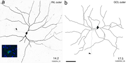
When small calbindin-positive somas were targeted, the injected cells had the morphology of starburst amacrine cells (n = 2 cells, Fig. 2d) comparable to those previously identified in Golgi impregnations in human retina (Rodieck, Reference Rodieck1989; Kolb et al., Reference Kolb, Linberg and Fisher1992). When large calbindin-positive somas were targeted, injected cells (n = 25) usually had a very large and sparse dendritic tree (Fig. 3a and 3b). The dendritic field diameter of these cells ranged between 450 and 760 µm (eccentricities between 8 and 19 mm). The dendritic stratification was analyzed for 21 cells. Nearly all (n = 19) cells had their soma in the ganglion cell layer and stratified close to the inner nuclear layer (Fig. 3b) but one cell stratified close to the ganglion cell layer. One cell had its soma in the inner nuclear layer and stratified close to the inner nuclear layer (Fig. 3a). Thus, based on morphology, dendritic field size, and stratification, calbindin-positive ganglion cells in human retina resemble giant sparse cells (Dacey et al., Reference Dacey, Peterson, Robinson and Gamlin2003; Peterson & Dacey, Reference Peterson and Dacey1999; Rodieck & Watanabe, Reference Rodieck and Watanabe1993), which more recently have been identified as melanopsin-expressing cells (Dacey et al., Reference Dacey, Liao, Peterson, Robinson, Smith, Pokorny, Yau and Gamlin2005; Liao et al., Reference Liao, Ren, Peterson, Marshak, Yau, Gamlin and Dacey2016; Nasir-Ahmad et al., Reference Nasir-Ahmad, Lee, Martin and Grünert2019). In addition in far peripheral retina close to the edge of the retina, some calbindin-positive cells with a relatively large soma diameter were injected (not shown). These cells had a relatively small dendritic tree and may not be fully differentiated. The far peripheral retina has been implicated to be involved with neurogenesis and undifferentiated calbindin-positive cells have been reported in the periphery of human and macaque retina (Martínez-Navarrete et al., Reference Martínez-Navarrete, Angulo, Martín-Nieto and Cuenca2008).
Calbindin is expressed by giant sparse ganglion cells in human retina. (a) Confocal image of a DiI-injected ganglion cell that was prelabeled with antibodies against calbindin (green, inset on the left). The soma of the cell was located in the inner nuclear layer. Blue indicates DAPI-labeled nuclei. (b) Drawing of a DiI-injected calbindin-positive ganglion cell, which had its soma in the ganglion cell layer. The eccentricity (in mm) of the cells is indicated in the lower right corner. The arrowhead indicates the axon. Scale bar shown in b = 100 µm, applies to a and b.

Calbindin expression in ganglion cells of human retina
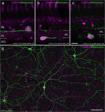
To confirm that calbindin-positive ganglion cells in human retina are indeed melanopsin-expressing ganglion cells, we double-labeled vertical sections and flat mount preparations with antibodies against melanopsin and calbindin-mouse. As shown in Fig. 4a–4c, melanopsin-expressing cells in both the ganglion cell layer and the inner nuclear layer were calbindin-positive. They stratified either close to the inner nuclear layer (outer cells) or close to the ganglion cell layer (inner cells) and thus included the two previously described types of melanopsin-expressing cell (Liao et al., Reference Liao, Ren, Peterson, Marshak, Yau, Gamlin and Dacey2016; Nasir-Ahmad et al., Reference Nasir-Ahmad, Lee, Martin and Grünert2019).
Calbindin is present in melanopsin-expressing ganglion cells in human retina. (a–c) Confocal micrographs of vertical sections through human retina, which were processed with antibodies against calbindin (CaBP, magenta) and melanopsin (green). Double-labeled cells are located in the inner nuclear layer (INL, a) or the ganglion cell layer (GCL, b and c) and stratify either close to the INL or close to the GCL. (d) Maximum intensity projection of a stack of images from a double-labeled flat mount preparation. The somas of all four melanopsin-expressing ganglion cells (green) are also positive for calbindin (magenta). Scale bar shown in c = 20 µm applies to a–c; scale bar in d = 100 µm. INL, inner nuclear layer; IPL, inner plexiform layer; GCL, ganglion cell layer.

In total, 22 melanopsin-expressing cells were analyzed in vibratome sections through central retina (0.91 to 2.3 mm). Eighteen of these cells (82%) were calbindin-positive and comprised GCL inner (n = 9) and INL outer (n = 9) cells. The four calbindin-negative cells included one GCL outer cell and three GCL inner cells. In peripheral retina (5 to 10 mm), 33 melanopsin-expressing cells were analyzed. Nearly all (32/33) were calbindin-positive, comprising GCL inner (n = 4), GCL outer (n = 4), and INL outer (n = 24) cells. The only calbindin-negative melanopsin-expressing cell was a GCL outer stratifying cell.
Consistent results were obtained from regions of interest at eccentricities between 5 and 12 mm in double-labeled flat mounts from two other preparations (Fig. 4d). Nearly all (50/54) melanopsin-expressing cells were calbindin-positive and included GCL inner (n = 12), GCL outer (n = 15), and INL outer (n = 27) cells. The four calbindin-negative cells had their soma in the ganglion cell layer and were GCL outer stratifying cells. Taken together, nearly all melanopsin-expressing ganglion cells in human retina are calbindin-positive and include GCL inner, GCL outer, and INL outer cells.
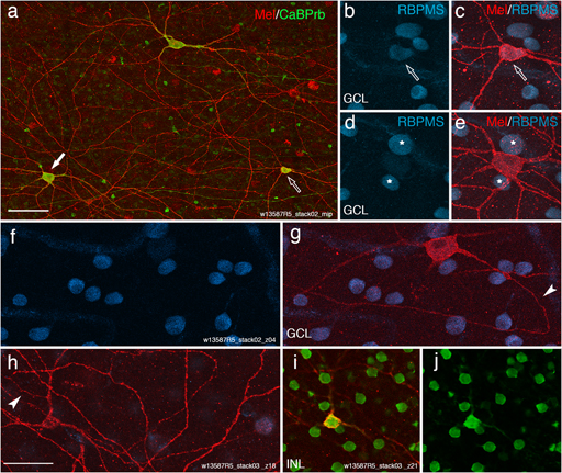
To determine whether all calbindin-positive ganglion cells are melanopsin-expressing cells, a flat mount preparation was triple-labeled using antibodies against calbindin-mouse, melanopsin, and RBPMS (Fig. 5). Five regions of interest at eccentricities between 10 and 13 mm were analyzed. In total, 1434 RBPMS-positive cells, 182 calbindin-positive cells, and thirteen melanopsin-positive cells were counted in the ganglion cell layer. Ten of 182 (5.5%) calbindin-positive cells in the ganglion cell layer were classified as ganglion cells based on their immunoreactivity for RBPMS; all of these cells were also melanopsin-positive (Fig. 5a–5c). Three melanopsin cells were calbindin-negative, which is consistent with the results reported above. In the inner nuclear layer, 45 RBPMS-positive cells including nine calbindin-positive ganglion cells were counted all of which were also melanopsin-positive. Taken together, these results show that in human retina, all calbindin-positive ganglion cells are melanopsin-expressing ganglion cells.
Some melanopsin-expressing cells show only weak RBPMS immunoreactivity. (a) Maximum intensity projection from a stack of images from a double-labeled flat mount preparation showing three melanopsin-expressing ganglion cells (red), which are located in the ganglion cell layer (GCL) and are also calbindin-positive (CaBP, green). The open arrow points to the cell shown in (b, c), which is melanopsin (red) and RBPMS (blue) positive; the white arrow points to the cell shown in (d, e), which has only very weak RBPMS immunoreactivity. The asterisks indicate RBPMS-positive cells located adjacent to the weakly labeled cell. (f, g) A single confocal plane of the same melanopsin-expressing cell as shown in e. The arrowhead points to the axon of the cell. (h–j) Melanopsin-expressing cell located in the inner nuclear layer (INL). This cell was only weakly labeled for RBPMS (not shown) but was strongly calbindin-positive (green). The arrowhead in h indicates the axon. Scale bar in a = 100 µm; scale bar in h = 50 µm valid for b–j.

It is noteworthy that similar to the findings reported above, the proportion of calbindin-positive ganglion cells among the ganglion cells in the ganglion cell layer was lower (10/1434, 0.7%) than that among the ganglion cells located in the inner nuclear layer (9/45, 20%).
The RPBMS counts from the flat mount also enabled us to calculate directly the ganglion cell density and the proportion of melanopsin cells in peripheral retina. The ganglion cell density decreased from 440 cells/mm2 at 10 mm to 200 cells/mm2 at 13 mm. These values are comparable to previous data of ganglion cell density in human retina (Curcio & Allen, Reference Curcio and Allen1990) and indicate that RBPMS is a valid ganglion cell marker in human retina. Melanopsin-expressing ganglion cells in peripheral retina made up about 1% (13/1434) of the RBPMS-positive ganglion cell population in the ganglion cell layer. Previous estimates of the proportion of melanopsin-expressing ganglion cells ranged from 0.2 to 1.5% (Dacey et al., Reference Dacey, Liao, Peterson, Robinson, Smith, Pokorny, Yau and Gamlin2005; Hannibal et al., Reference Hannibal, Hindersson, Østergaard, Georg, Heegaard, Larsen and Fahrenkrug2004; Liao et al., Reference Liao, Ren, Peterson, Marshak, Yau, Gamlin and Dacey2016; Nasir-Ahmad et al., Reference Nasir-Ahmad, Lee, Martin and Grünert2019) but these studies did not measure the proportion directly.
About half (6/13) of melanopsin-expressing cells located in the ganglion cell layer and about two thirds (9/13) of those located in the inner nuclear layer showed very weak or no RBPMS immunoreactivity (Fig. 5d, 5e) suggesting that they may not be ganglion cells. However, close inspection of these cells showed that these cells had a large soma size and distinct axons originating from their primary dendrite (Fig. 5g, 5h). Thus, they resembled melanopsin-expressing ganglion cells described previously (Liao et al., Reference Liao, Ren, Peterson, Marshak, Yau, Gamlin and Dacey2016; Nasir-Ahmad et al., Reference Nasir-Ahmad, Lee, Martin and Grünert2019). All but three of the melanopsin-positive/RBPMS-negative cells expressed calbindin. Weak RBPMS immunoreactivity maybe related to the fact that there was a time delay of 4.5 h between death and fixation in the triple-labeled retina. Previous studies have reported that RBPMS immunoreactivity is weakened after optic nerve injury (Rodriguez et al., Reference Rodriguez, Pérez de Sevilla Müller and Brecha2014). Thus, our result may indicate that the weakly labeled RBPMS cells are degenerating. However, this possibility will need to be studied more systematically. Future studies will also need to determine whether other than melanopsin-expressing ganglion cell types are affected by weak or absence of RBPMS labeling.
For mouse retina, it was reported that all melanopsin-expressing ganglion cells (n = 34) investigated were also immunoreactive to RBPMS (Rodriguez et al., Reference Rodriguez, Pérez de Sevilla Müller and Brecha2014). Taken together, these results show that in human retina all calbindin-positive ganglion cells express melanopsin. They also show that displaced ganglion cells include calbindin-positive cells but are not exclusively made up of calbindin-positive cells. Finally the results indicate that a low percentage of melanopsin-expressing cells are calbindin-negative.
Soma measurements in human retina
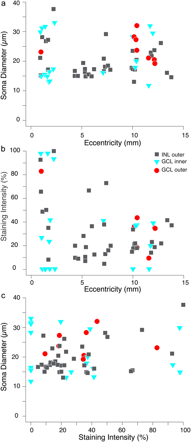
To determine whether different melanopsin-expressing cell types can be distinguished with respect to their calbindin immunoreactivity, the diameter and labeling intensity of melanopsin-positive/calbindin-positive somas was determined for 61 cells in two preparations (eight GCL outer, 35 INL outer, and 18 GCL inner cells). The soma diameters ranged between 11.6 and 37.6 µm but did not change with eccentricity (Fig. 6a). The figure also shows that GCL inner cells are rare at eccentricities between 4 and 12 mm. Similar observations were made previously (Nasir-Ahmad et al., Reference Nasir-Ahmad, Lee, Martin and Grünert2019) but the reason remains unknown. Seven melanopsin-expressing but calbindin-negative cells had soma diameters between 21 and 40 µm, which is in the same range as that of the double-labeled cells.
Scatter plots showing that soma diameter and staining intensity of calbindin-positive melanopsin-expressing ganglion cells in human retina do not change with eccentricity.

Fig. 6b compares the staining intensity for calbindin in the same cells and shows that both inner as well as outer stratifying cells comprise strongly and weakly calbindin-positive cells. Fig. 6c shows that the soma diameter of calbindin-positive melanopsin-expressing cells is not correlated to the staining intensity.
Taken together, different types of melanopsin-expressing ganglion cells in human retina cannot be distinguished based on their calbindin immunoreactivity.
Calbindin-immunoreactive cells in marmoset retina
In marmoset retina, calbindin-rabbit immunoreactivity is found in cones and subpopulations of cells in the inner nuclear and ganglion cell layer (Fig. 7a and 7c). The section shown was also processed with antibodies against ChAT and shows double-labeled cells in the amacrine and ganglion cell layer (Fig. 7b, c). In two regions of interest, 28 ON ChAT cells and 191 OFF ChAT cells were counted. All ON ChAT cells were calbindin-positive, and consistently, intracellular injections showed that calbindin-rabbit labeled cells in the ganglion cell layer had starburst morphology (n = 5, Fig. 7d and 7e). Only 8 (4%) of the OFF ChAT cells were double labeled; thus, in marmoset retina, only a small percentage of OFF ChAT cells is calbindin-positive.
Calbindin is expressed by starburst amacrine cells in marmoset retina. (a–c) Confocal images of a vertical section through marmoset retina, which was processed with antibodies against calbindin-rabbit (magenta) and choline acetyl transferase (ChAT, green). Double-labeled cells are indicated by white arrows. Most ChAT cells in the inner nuclear layer (INL) are not double labeled (open arrows). (d) Micrograph of a calbindin-positive cell that was injected with DiI after prelabeling with calbindin. (e) Same preparation as in d showing calbindin immunoreactivity in the soma of the injected cell (arrowhead). (f–h) Confocal images of a flat mount preparation processed with the calbindin-mouse antibody (magenta) and antibodies against RBPMS (green). Ganglion cells are not double labeled. Open arrows indicate calbindin-positive displaced amacrine cells. Scale bar in c = 50 µm applies to a–c; scale bar in d = 50 µm valid for d and e; scale bar in f = 50 µm applies to f–h. OPL, outer plexiform layer; INL, inner nuclear layer; IPL, inner plexiform layer; GCL, ganglion cell layer.

We compared the number of ChAT cells in regions of interest of mid-peripheral (3.5 mm eccentricity) and central (1 mm eccentricity) retina and found that the ratio between ON (n = 102 cells) and OFF (n = 371 cells) ChAT cells is about 30 to 70. Similar ratios were reported previously for marmoset retina (Moritoh et al., Reference Moritoh, Komatsu, Yamamori and Koizumi2013).
To determine whether calbindin-positive cells in marmoset retina include ganglion cells, vibratome sections were processed with antibodies against calbindin-rabbit and RBPMS (guinea pig). At eccentricities between 1.8 and 3.5 mm, a total 535 RBPMS-positive cells and 82 calbindin-positive cells were counted in the ganglion cell layer. None of the RBPMS cells were double-labeled for calbindin, indicating that there are no calbindin-positive ganglion cells in marmoset retina. Similarly, in the inner nuclear layer, none of the three RBPMS-positive cells were calbindin-positive.
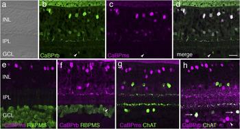
In another set of experiments, we combined the mouse monoclonal calbindin antibody with the RBPMS antibody in a retinal flat mount preparation (eccentricity range of 2.6 to 6.7 mm) and again found that none of the RBPMS-positive cells (n = 1430) was calbindin-positive. In contrast to the staining pattern obtained with the rabbit calbindin antibody, however, only a low number of calbindin-positive cells (n = 19) was found in the ganglion cell layer (Fig. 7f–7h) suggesting that the mouse calbindin antibody does not label ON ChAT cells.
Although the double-labeling experiments including RBPMS antibodies indicated that ganglion cells in marmoset retina do not express calbindin, some of the DiI-injected cells were classified as ganglion cells based on the presence of an axon (data not shown). These cells, however, had only weakly calbindin-positive somas and thus these cells may be subject of some cross-reactivity of the calbindin antibody with calretinin. Calretinin is known to be present in broad and narrow ganglion cell types of marmoset retina (Chandra et al., Reference Chandra, Lee and Grünert2017).
To test directly whether the two antibodies against calbindin show differences in their labeling pattern in marmoset retina, some sections were doubled with the two calbindin antibodies. Other double-labeling experiments included the following combinations: calbindin-mouse and RBPMS; calbindin-rabbit and RBPMS; calbindin-mouse and ChAT; calbindin-rabbit and ChAT. The results show that the two antibodies have similar staining patterns in the inner nuclear layer (Fig. 8a–8d), but cells in the ganglion cell layer are more strongly labeled with the calbindin-rabbit antiserum (arrows). As reported above, RBPMS-positive ganglion cells are labeled neither with the mouse nor with the rabbit calbindin antibody (Fig. 8e and 8f), whereas ON ChAT cells were double labeled with the rabbit but not the mouse calbindin antibody (Fig. 8g and 8h). Taken together, our results suggest that in marmoset retina, RBPMS-expressing ganglion cells do not include calbindin-labeled cells. We could not determine whether there are melanopsin-expressing ganglion cells lacking RBPMS immunoreactivity, because the antiserum against melanopsin used in the present study does label any cells in marmoset retina.
Comparison of the staining pattern obtained with the two calbindin (CaBP) antibodies in marmoset retina. (a–d) Confocal micrographs of vibratome sections through marmoset retina processed with calbindin antibodies made in rabbit (rb, green) and made in mouse (ms, magenta). The two antibodies have comparable staining patterns but the cells in the ganglion cell layer are more strongly labeled by the rabbit antiserum (arrowheads). (e, f) Confocal images of vibratome sections processed with antibodies against RBPMS and antibodies against mouse-calbindin (e) and rabbit-calbindin (f). Neither of the calbindin antibodies label ganglion cells. (g, h) Confocal images of vibratome sections processed with antibodies against choline acetyltransferase (ChAT), mouse-calbindin (g), and rabbit-calbindin (h). ON ChAT cells are only double labeled in the rabbit-calbindin processed section (arrows in h). In addition, some cells that are only calbindin-positive but ChAT-negative cells can be seen (arrowheads in h). Scale bar in d = 20 µm, applies to all. INL, inner nuclear layer; IPL, inner plexiform layer; GCL, ganglion cell layer.

Discussion
Previous studies have shown that the staining pattern obtained with antibodies against the calcium-binding protein calbindin in the retina of different primate species is fairly consistent and includes cone photoreceptors, and subpopulations of horizontal, bipolar, and amacrine cells (Chan et al., Reference Chan, Martin, Clunas and Grünert2001; Chiquet et al., Reference Chiquet, Dkhissi-Benyahya and Cooper2005; de Souza et al., Reference de Souza, Nivison-Smith, Christie, Polkinghorne, McGhee, Kalloniatis and Acosta2016; Haverkamp et al., Reference Haverkamp, Haeseleer and Hendrickson2003; Jacoby & Marshak, Reference Jacoby and Marshak2000; Kántor et al., Reference Kántor, Mezey, Adeghate, Naumann, Nitschke, Enzsoly, Szabó, Lukats, Nemeth, Somogyvari and Völgyi2016; Luo et al., Reference Luo, Ghosh, Martin and Grünert1999; Martin & Grünert, Reference Martin and Grünert1992; Pasteels et al., Reference Pasteels, Rogers, Blachier and Pochet1990; Röhrenbeck et al., Reference Röhrenbeck, Wässle and Boycott1989; Roski et al., Reference Roski, Langrock, Körber, Habermann, Buse, Reichenbach, Pannicke and Francke2018; Wässle et al., Reference Wässle, Dacey, Haun, Haverkamp, Grünert and Boycott2000). On the other hand, eccentricity and species-related differences with respect to calbindin expression have been reported. For example, calbindin expression is absent from foveal cones in macaque and human retina (Haley et al., Reference Haley, Pochet, Baizer, Burton, Crabb, Parmentier and Polans1995; Pasteels et al., Reference Pasteels, Rogers, Blachier and Pochet1990) and some differences between macaque and human have been found with respect calbindin-expressing bipolar types (Haverkamp et al., Reference Haverkamp, Haeseleer and Hendrickson2003). Variability in the expression of other calcium-binding proteins including recoverin (Chan et al., Reference Chan, Martin, Clunas and Grünert2001; Milam et al., Reference Milam, Dacey and Dizhoor1993) and secretagogin (Puthussery et al., Reference Puthussery, Gayet-Primo, Taylor and Haverkamp2011; Weltzien et al., Reference Weltzien, Dimarco, Protti, Daraio, Martin and Grünert2014) in the retina of different primate species has also been noted.
The present study shows that calbindin immunoreactivity is present in melanopsin-expressing ganglion cells of human but not marmoset retina. It further shows that calbindin is localized to ON ChAT cells but only to a subpopulation of OFF ChAT cells in both human and marmoset retina. In addition, we observed differences related to the calbindin antibody used in marmoset retina.
Antibody comparison
Previous studies have indicated that some antibodies against calbindin D28k show cross-reactivity with calretinin depending on the antibody concentration (Pasteels et al., Reference Pasteels, Rogers, Blachier and Pochet1990; Pinol et al., Reference Pinol, Kägi, Heizmann, Vogel, Séquier, Haas and Hunziker1990). This applies to the rabbit antiserum of the present study, which according to the manufacturer (Swant) may also detect calretinin when used highly concentrated whereas the mouse calbindin antibody does not to cross react with calretinin (Swant).
The potential cross-reactivity of the rabbit calbindin antiserum with calretinin could explain the fact that in marmoset we found ganglion cells among the injected cells when the antiserum was used at a higher concentration. In favor of this hypothesis, the injected ganglion cells resembled previously identified calretinin expressing thorny ganglion cells (Chandra et al., Reference Chandra, Lee and Grünert2017). Likewise, the fact that in marmoset ON ChAT cells were labeled with the rabbit calbindin antiserum but not the mouse calbindin antibody may be explained by cross-reactivity, if one assumes that marmoset ON ChAT cells express calretinin but not calbindin. Calretinin-positive ChAT cells have been reported for rodent retina (Gábriel & Witkovsky, Reference Gábriel and Witkovsky1998; Haverkamp & Wässle, Reference Haverkamp and Wässle2000).
In human retina, no obvious differences in the staining pattern between the two antibodies were seen (data not shown). Double-labeling experiments using a ganglion cell marker established that calbindin-rabbit positive cells form a small subpopulation of the ganglion cell population and that the majority of calbindin labeled cells in the ganglion cell layer are displaced amacrine cells. Separate double-labeling experiments showed that the calbindin-mouse positive cells included melanopsin-expressing ganglion cells and that all calbindin-positive displaced amacrine cells are ON ChAT cells. Consistently, intracellular injection of calbindin-rabbit positive cells in the ganglion cell layer of human retina revealed cells with the morphology of melanopsin-expressing and ON starburst amacrine cells.
The reason why the rabbit calbindin antiserum cross reacts with calretinin in marmoset but not in human tissue needs further investigation. Nonetheless the cross-reactivity found in marmoset does not affect the fundamental conclusions of the study that there are differences between marmoset and human retina with respect to calbindin expression in the ganglion cell layer.
Calbindin immunoreactivity in ChAT cells
Consistent with previous studies (Moritoh et al., Reference Moritoh, Komatsu, Yamamori and Koizumi2013; Rodieck & Marshak, Reference Rodieck and Marshak1992; Weltzien et al., Reference Weltzien, Percival, Martin and Grünert2015), we found that the ratio of ON ChAT to OFF ChAT cells is 70/30 in human and 30/70 in marmoset (30/70). The higher number of ON ChAT cells in human retina can be explained by a loss of OFF but not ON ChAT cells during development (Zhang et al., Reference Zhang, Yu, Hoshino, Huang, Rieke, Reh and Wong2019). However, as these authors discuss the loss of ON ChAT cells during development does not seem to be a characteristic of primate retina.
In both human and marmoset retina, calbindin is expressed in a subpopulation of OFF ChAT cells (Figs. 2a–2c; 7a–7c). Similar findings were obtained in fetal human fovea by Zhang et al. (Reference Zhang, Yu, Hoshino, Huang, Rieke, Reh and Wong2019) who used the same calbindin-rabbit antibody as the present study. This result suggests that there are molecular differences within the OFF ChAT population but further work (for example, intracellular injections or labeling with other markers) will be needed to investigate this hypothesis.
In mouse and rabbit retina, OFF ChAT cells costratify with the OFF arbor of direction-selective ganglion cells and play a role in shaping their response to a moving stimulus (Vaney et al., Reference Vaney, Sivyer and Taylor2012). In primate retina, direction-selective ganglion cells are yet to be described and thus the role of ChAT cells is not yet clear.
Cholinergic amacrine cells also play a role during retinal development in mouse (Ford & Feller, Reference Ford and Feller2012) as well as human retina (Zhang et al., Reference Zhang, Yu, Hoshino, Huang, Rieke, Reh and Wong2019) but the role of calbindin in ChAT cells is not understood. There are considerable differences in the calbindin expression of ChAT cells between mammalian species. In mouse retina, calbindin is strongly expressed by all ON and all OFF ChAT cells (Zhang et al., Reference Zhang, Yu, Hoshino, Huang, Rieke, Reh and Wong2019). In rabbit retina, calbindin is not expressed in any ChAT cells (Lee & Jeon, Reference Lee and Jeon2013) and in ground squirrel retina, calbindin is expressed by all ON ChAT and a subpopulation of OFF ChAT cells (Cuenca et al., Reference Cuenca, Deng, Linberg, Fisher and Kolb2003). Thus, in this respect, the ground squirrel retina resembles the human and marmoset retina. But the ground squirrel in addition has a third type of ChAT cell, which is calbindin-negative and stratifies in the middle of the inner plexiform layer (Cuenca et al., Reference Cuenca, Deng, Linberg, Fisher and Kolb2003). A third ChAT cell type has not been described in any other species.
Taken together, there is a confusing variety in the expression of calbindin by ChAT cells in different species, and it is not yet clear to which extent this variety is accompanied by functional differences. Moreover, Wässle et al. (Reference Wässle, Peichl, Airaksinen and Meyer1998) showed that in calbindin null mutant mice the same cell types expressing calbindin as in wild type were still present and appeared normal suggesting that calbindin is not required to maintain retinal morphology.
Calbindin immunoreactivity in ganglion cells
The present study shows that in human retina, calbindin-positive cells make up only a small proportion of the ganglion cell population and mainly comprise melanopsin-expressing cells. We did not see any calbindin-positive ganglion cells in marmoset retina. In contrast, in mouse retina, calbindin-positive cells include a variety of ganglion cell types and make up about 20% of the ganglion cell population (Gu et al., Reference Gu, Lee and Jeon2016). Species-related differences in the expression of calcium-binding proteins by ganglion cells have been observed previously, e.g., in human retina, calretinin is expressed by two populations of bistratified ganglion cells (Lee et al., Reference Lee, Weltzien, Madigan, Martin and Grünert2016), whereas in marmoset calretinin is expressed by thorny ganglion cells (Chandra et al., Reference Chandra, Lee and Grünert2017). Whether these differences are correlated with functional differences will need to be determined.
Calbindin-positive ganglion cells in human included all known types of melanopsin-expressing cell, i.e., inner and outer stratifying cells (Liao et al., Reference Liao, Ren, Peterson, Marshak, Yau, Gamlin and Dacey2016; Nasir-Ahmad et al., Reference Nasir-Ahmad, Lee, Martin and Grünert2019) as well as the giant M1 cell type (Hannibal et al., Reference Hannibal, Christiansen, Heegaard, Fahrenkrug and Kiilgaard2017). This result is based on intracellular injections after prelabeling (calbindin-rabbit, Fig. 3) as well as double-label immunohistochemistry with antibodies against melanopsin and calbindin-mouse (Fig. 4). The labeling intensity and soma size varied significantly between different cells but no obvious differences were detected between the known morphological types (Fig. 6). Thus in human retina, calbindin immunoreactivity cannot be used to distinguish different types of melanopsin-expressing ganglion cells.
Calbindin is known to play a role in calcium buffering and also has been suggested to have a function as a calcium sensor and transporter (Schmidt, Reference Schmidt2012; Schwaller et al., Reference Schwaller, Meyer and Schiffmann2002) but its role in the retina is not well understood (Kántor et al., Reference Kántor, Mezey, Adeghate, Naumann, Nitschke, Enzsoly, Szabó, Lukats, Nemeth, Somogyvari and Völgyi2016). Calbindin appears to be absent from melanopsin-expressing ganglion cells in some mammals including rabbit (Hoshi et al., Reference Hoshi, Liu, Massey and Mills2009), gerbil (Jeong & Jeon, Reference Jeong and Jeon2015), and marmoset (this study) but it is present in mouse (Gu et al., Reference Gu, Lee and Jeon2016; Kriegsfeld et al., Reference Kriegsfeld, Mei, Yan, Witkovsky, Lesauter, Hamada and Silver2008) and human retina (this study). This species-related discrepancy may indicate that calbindin expression in retinal ganglion cells is labile but further studies will be needed to prove this hypothesis.
Acknowledgments
We thank Paul Martin for critical reading of the manuscript. We are grateful to Arzu Demir for providing excellent technical assistance and Rania Masri for help with cutting sections and the analysis. We thank M. Rosa and J. Bourne of the Monash University for providing some of the marmoset retinas. Grant support: National Health and Medical Research Council (NHMRC) Project grant (#1123418 to UG); Sydney Medical School Foundation fellowship (to UG); Claffy Foundation fellowship (SCSL); Australian Research Council Center of Excellence for Integrative Brain Function (CE140100007 to UG).