When considering the embryology of congenital heart disease, it is not surprising that the associated coronary artery patterns can have great variability. It is now understood that coronary arteries originate distally and grow proximally to insert into the aortic sinuses, following the septation of the great arteries.Reference Anderson, Chiu, Spicer and Hlavacek1 When rotation and septation of the great arteries are abnormal, the coronary anatomy can be inconsistent. Recognising this variability is critical when considering surgical or interventional therapies in these patients. This review will discuss the echocardiographic evaluation of the coronary arteries in transposition of the great arteries, congenitally corrected transposition of the great arteries, double-inlet left ventricle, and the conotruncal lesions of the common arterial trunk, tetralogy of Fallot, and double-outlet right ventricle. The patterns of coronary anatomy and clinically relevant variants are described.
Transposition of the great arteries
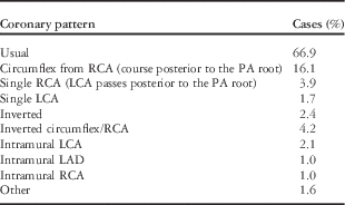
The Leiden classification is the most common nomenclature used to describe coronary artery patterns in D-transposition of the great arteries. This classification describes nine major patterns based on two sinuses of origin, three major coronary branches, and the possibility of an intramural course.2 In the majority of cases, the coronary arteries arise from the sinuses facing the pulmonary valve with the aorta in an anterior and rightward position to the pulmonary artery. The frequency of these coronary artery patterns is shown in Table 1. Coronary artery abnormalities are more common when the great vessels are oriented side-by-side or anterior–posterior.Reference Chiu, Sh and Wang3
Frequency of coronary artery patterns in D-transposition of the great arteries.2
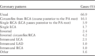
LAD=left anterior descending; LCA=left coronary artery; PA=pulmonary artery; RCA=right coronary artery
In the most common pattern seen in 66.9% of cases, the left anterior descending coronary artery and the circumflex coronary artery arise from the left-facing sinus of Valsalva and the right coronary artery arises from the right-facing sinus of ValsalvaReference Wernovsky and Sanders4 (Fig 1). In 30% of cases, the coronary arteries take a looping course. When the circumflex coronary artery and the right coronary artery arise from the right-facing sinus, as seen in 16.1% of cases, the circumflex coronary artery will take a retro-pulmonary course (Fig 2). When the left coronary artery arises from the right-facing sinus, as seen in 3.9% of cases, a posterior loop is seen, where the left anterior descending coronary artery passes posterior to the pulmonary root before bifurcating.Reference Wernovsky and Sanders4, Reference Gremmels, Tacy, Brook and Silverman5 An anterior course of the coronary artery or a combined anterior and posterior course relative to the aortic and pulmonary roots is present in cases of single coronary arteries (Fig 3). Inverted coronary artery origins are seen in 2.4% of cases, where the right coronary artery arises from the left anterior-facing sinus of Valsalva passing in front of the aorta and the left anterior descending coronary artery and circumflex coronary artery arising from the right posterior-facing sinus looping behind the pulmonary trunk (double loop) (Fig 4).
Usual coronary artery pattern in D-transposition of the great arteries (D-TGA). The left coronary artery (LCA) originates from the leftward-facing sinus. The RCA originates from the rightward-facing sinus. AO=aorta; LMCA=left main coronary artery; PA=pulmonary artery; RCA=right coronary artery.

Second most common coronary artery pattern: the right coronary artery (RCA) arises from the right posterior-facing sinus and gives rise to the circumflex coronary artery. The circumflex coronary artery takes a retro-pulmonary course. AO=aorta; LCx=left circumflex artery; PA=pulmonary artery.
A single coronary artery arising from the left-facing sinus and giving rise to the RCA, the left coronary artery (LCA), and the circumflex coronary arteries. The coronary arteries take an anterior and posterior course relative to the aortic and pulmonary trunks. AO=aorta; LAD=left anterior descending; LCx=left circumflex artery; PA=pulmonary artery; RCA=right coronary artery.
Inverted coronary artery pattern: the RCA arises from the left-facing sinus of Valsalva and passes in front of the aorta, while the LAD coronary artery and the circumflex coronary artery arise from the right posterior-facing sinus looping behind the pulmonary trunk. AO=aorta; LAD=left anterior descending; LMCA=left main coronary artery; PA=pulmonary artery; RCA=right coronary artery.
Intramural coronary arteries are rare, and they typically arise from the opposite-facing sinus and course within the wall of the aorta. They are usually described as having a high take-off from the sinotubular junctionReference Wyman, Mertens and Cohen6 (Fig 5).
Abnormal origin of the left coronary artery (LCA) from the right posterior sinus of Valsalva from a separate ostium than the RCA, taking an intramural course between the great arteries. AO=aorta; LAD=left anterior descending; RCA=right coronary artery.
Echocardiographic evaluation
Echocardiography has proven to be a highly reliable imaging technique to determine the proximal coronary artery anatomy in D-transposition of the great arteries.Reference Gremmels, Tacy, Brook and Silverman5, Reference Pasquini, Sanders and Parness7 Multiple imaging planes are required to diagnose the coronary artery pattern in D-transposition of the great arteries.
The parasternal short-axis view at the level of the aortic root will display the origins of the coronary arteries. With slight clockwise rotation of the probe, the left coronary artery can be seen arising from the left-facing sinus of Valsalva. With slight counterclockwise rotation of the probe, the right coronary artery can be viewed. The usual pattern of distribution of the coronary arteries is depicted in Figure 1.
The apical four-chamber view and left oblique subxiphoid – subcostal frontal sweep – view are useful for locating the left circumflex coronary artery or the left coronary artery when they arise from the right-facing sinus of Valsalva, which are the second and the third most common patterns. In such cases, the coronary artery usually passes posterior to the pulmonary artery (Fig 2). Subcostal and apical views are helpful to delineate an anterior loop or combined anterior and posterior loops present when there is a single coronary artery or inverted coronary artery origins (Figs 3 and 4).
Features that suggest an intramural course include a coronary vessel originating from the opposite sinus of Valsalva with a course that passes between the semilunar valves, and it may also have a high take-off above the sinotubular junction. This can be evaluated in the parasternal short-axis view, which shows an intramural coronary artery travelling between the aorta and the pulmonary arteryReference Wyman, Mertens and Cohen6, Reference Eidem, O’Leary and Cetta8 (Fig 5). The high origin can also be evaluated in the parasternal long-axis view.
Congenitally corrected transposition of the great arteries
In congenitally corrected transposition of the great arteries, the aortic valve has a leftward anterior position relative to the pulmonary valve. In general, the coronary arteries arise from the posterior-facing sinus of the aortic valve, with a mirror-image distribution, and travel with their respective ventricles (Fig 6). The right-sided coronary artery has the epicardial distribution of a morphological left coronary artery, dividing into circumflex and left anterior descending branches. The left-sided coronary artery has the epicardial distribution of a morphological right coronary artery and follows the left-sided atrioventricular groove, giving rise to infundibular and marginal branches.2 The coronary arteries arise in a normal pattern usually (76% of cases).Reference Chiu, Sh and Wang3 Coronary artery abnormalities seen in D-transposition of the great arteries and tetralogy of Fallot can also be seen in congenitally corrected transposition of the great arteries, including single coronary arteries – a main coronary branch travelling anterior to the pulmonary trunk or the right ventricular outflow tract.2, Reference Wyman, Mertens and Cohen6
Usual coronary artery pattern in L-transposition of the great arteries (L-TGA). The coronary arteries have a mirror-image distribution and travel with their respective ventricles. AO=aorta; LAD=left anterior descending; LCx=left circumflex artery; PA=pulmonary artery; RCA=right coronary artery.
Echocardiographic evaluation
The echocardiographic evaluation of the coronary arteries must include the short-axis view with slight superior angulation at the level of the semilunar valves. In the majority of cases, the coronary arteries are inverted, mirror-imaged, with the right coronary artery arising from the left posterior-facing sinus and the left coronary artery arising from the right posterior-facing sinus and dividing into the anterior descending branch and the posterior circumflex.Reference Eidem, O’Leary and Cetta8
Coronary arteries in double-inlet left ventricle
In double-inlet left ventricle, the coronary arteries arise from the two facing aortic sinuses, with a pattern of distribution that depends on the underlying ventricular anatomy. Anterior and posterior delimiting arteries mark the position of the septum and indicate the size of the outflow chamber.Reference Becker, Penkoske and Zuberbuhler9
Common arterial trunk
Common arterial trunk represents embryological failure of the great arteries to septate. The resultant coronary patterns are the attempts to grow into the aortic portion of the truncal valve. The ventricular musculature of the pulmonary root is felt to be impervious to embryological coronary growth. Therefore, the left anterior aspect of the truncal root is typically devoid of coronary arteries.Reference Anderson, Chiu, Spicer and Hlavacek1 There is a higher incidence of coronary anomalies with quadrifoliate truncal valves than with tri-leaflet valves, where coronary arteries typically arise from opposite, non-adjacent leaflets. There is an 18% incidence of single coronary ostia with a posterior origin. The circumflex coronary artery arises from the right coronary artery in 3.6% of patients, and the left coronary artery arises from the right coronary artery in 2.4% of patients. In 13% of patients, a major coronary artery will cross the anterior surface of the right ventricle – an important consideration when placing or intervening on a pulmonary artery conduit. Another important coronary variant is the coronary ostia origin above the sinotubular junction. High left coronary ostia are seen in 32% of patients and high right coronary ostia are seen in 20% of patients.Reference Baraona, Valente and Porayette10 This finding can increase the risk of surgical correction when the ostia are adjacent to the origins of the pulmonary arteries. The coronary ostia can arise from a zone of apposition of the valve leaflets, with ostial stenosis or slit-shape origin.Reference Anderson, Baker and Penny11 This increases the risk of ischaemic changes secondary to the associated physiological issues of low diastolic pressures of pulmonary run-off and truncal valve insufficiency.
Echocardiographic evaluation
The echocardiographic assessment of the coronary arteries in common arterial trunk begins with the parasternal short-axis view at the level of the valve leaflets. Subtle clockwise and counterclockwise rotations of the probe will assist in identification of the coronary origins. Colour Doppler at Nyquist limits of 20–40 cm/second confirms the direction of flow. Visualisation of a distal portion of the coronary artery will allow the reader to trace the coronary artery proximally to the insertion site in the truncal valve. The echocardiogram must include a careful interrogation for coronary ostia in the posterior portion of the truncal valve or close to leaflet hinge points.
Assessment of the area superior to the sinotubular junction must be performed to exclude a high take-off of a coronary artery. Parasternal long-axis, apical four-chamber view, and subcostal long-axis view at the level of the aortic root will all visualise a high origin.Reference Wyman, Mertens and Cohen6
Tetralogy of Fallot
The majority of patients with tetralogy of Fallot have normal coronary anatomy; however, 5% of patients have a coronary pattern that results in a major coronary artery crossing the right ventricular outflow tract.Reference Baraona, Valente and Porayette10 When the echocardiogram assessment of the coronary arteries shows a coronary artery crossing the right ventricular outflow tract or when the echocardiogram is equivocal, the coronary concerns must be communicated to the surgeon so that direct inspection of the coronary anatomy will be performed. Alternative surgical interventions must be considered in these cases in order to avoid transection of the coronary artery. Coronary artery compression is also a concern in these patients following right ventricle-to-pulmonary artery conduit stenting and right pulmonary artery stenting.
Coronary patterns in tetralogy of Fallot that result in major coronary branches crossing the right ventricular outflow tract include the left anterior descending coronary artery from the right coronary artery in 3% of patients and dual left anterior descending coronary arteries in 1.8% of patients. Distinguishing between a large conal branch-off of the right coronary artery (6.4% of patients) versus dual left anterior descending coronary arteries can be challenging by echocardiogram alone, and is based on whether or not the branch reaches the anterior interventricular grove. Additional coronary anomalies include a single right coronary artery (0.3% of patients) and a single left coronary artery (0.2% of patients), both of which result in a coronary artery crossing the right ventricular outflow tractReference Need, Powell, Del Nido and Geva12 (Fig 7). The aortic annulus in tetralogy of Fallot rotates abnormally during embryonic septation of the great arteries. This results in the aortic sinuses having a clockwise rotation, with the left coronary artery origin located more posteriorly and the right coronary artery origin located more anteriorly. Of interest, the left coronary artery is dominant in 28% of patients with tetralogy of Fallot as opposed to 10% in the normal population.Reference Baraona, Valente and Porayette10
Patterns of coronary artery anatomy in tetralogy of Fallot. This figure is re-printed with permission from Elsevier.Reference Need, Powell, Del Nido and Geva12 Cx=circumflex artery; LAD=left anterior descending; LCA=left coronary artery; RCA=right coronary artery.
Echocardiographic evaluation
The coronary anatomy in tetralogy of Fallot is examined primarily from the parasternal short-axis view at the level of the aortic root. Colour Doppler at Nyquist limits of 20–40 cm/second confirms the direction of flow. The left coronary artery is interrogated with special attention to the left anterior descending coronary artery by rotating the transducer clockwise and slightly apical. The circumflex coronary artery should also be visualised branching from the left coronary artery. All coronary branches-off of the right coronary artery must be interrogated with care to exclude a left anterior descending coronary artery off of the right coronary artery, especially when the right coronary artery appears to be generous in size (Fig 8a). Any branching-off of the right coronary artery should be followed from the short-axis view into the parasternal long-axis view, with angulations up towards the right ventricular outflow tract. An anterior descending coronary artery can be seen in cross-section on the epicardial surface of the right ventricular free wall (Fig 8b). Additional assessment can be performed in the apical four-chamber sweep angled to visualise the anterior wall of the right ventricular outflow tract.Reference Wyman, Mertens and Cohen6
(a) Parasternal short-axis view in tetralogy of Fallot, with the origin of the LAD from the RCA. (b) Parasternal long-axis view angled anteriorly to show the LAD crossing the epicardial surface of the right ventricular outflow tract. LAD=left anterior descending; MPA=main pulmonary artery; RCA=right coronary artery; RVOT=right ventricular outflow tract.
Double-outlet right ventricle
The abnormal rotation and septation of the great arteries in double-outlet right ventricle result in great variability in the relationship of the aorta with the pulmonary artery. It is, therefore, not surprising that there is great variability in the coronary anatomy associated with this diagnosis. The coronary pattern tends to follow the position of the great arteries. As such, a rightward and posterior aorta is typically associated with the coronary patterns seen in tetralogy of Fallot (Fig 9). A rightward and anterior aorta is associated with the coronary patterns seen in D-transposition of the great arteries. When the aorta is leftward and anterior, the right coronary artery will always cross in front of the right ventricular outflow tract.Reference Baraona, Valente and Porayette10 The coronary patterns of tetralogy of Fallot and D-transposition of the great arteries have already been reviewed.
Parasternal short-axis view of double-outlet right ventricle. The aorta is rightward and posterior with a dual LAD shown off of the RCA. LAD=left anterior descending; RCA=right coronary artery.
The surgical and interventional considerations from a coronary anatomy standpoint are also similar to those of tetralogy of Fallot and D-transposition of the great arteries. Some coronary artery patterns will exclude otherwise feasible surgical options, making the pre-operative assessment extremely important.
Echocardiographic evaluation
Echocardiographic assessment of the coronary arteries in double-outlet right ventricle is primarily from the parasternal short-axis view at the level of the aortic root. Colour Doppler at Nyquist limits of 20–40 cm/second confirms the direction of flow. The echocardiographic evaluation of the various coronary patterns of tetralogy of Fallot and D-transposition of the great arteries has already been reviewed.
Conclusion
The coronary patterns of congenital heart disease offer great insight into the embryological development of the coronary arteries. This includes the way in which the major coronary arteries grow into the aortic sinuses adjacent to the pulmonary artery, while avoiding the impervious subpulmonary musculature.Reference Anderson, Chiu, Spicer and Hlavacek1 Knowing this allows for anticipation of possible treacherous coronary artery patterns before surgery. Accurately diagnosing the coronary anatomy in congenital heart disease helps guide the re-implantation of coronary artery origins and avoid transection of the coronary artery that crosses the outflow tract. The improving echocardiography technology, with its emphasis on coronary anatomy, continues to improve the sensitivity, specificity, and accuracy in diagnosing coronary artery patterns.Reference Gremmels, Tacy, Brook and Silverman5, Reference Need, Powell, Del Nido and Geva12 As a result, pre-operative cardiac catheterisation and CT angiography are no longer the norm. We must now consider coronary anatomy for future interventional procedures such as stenting of conduits, transcatheter valve replacement, and transcatheter coronary artery disease therapy. As patients with congenital heart disease are living longer lives, understanding the variants in coronary anatomy is now critical, not only for the initial surgical success but also for the success of future procedures. The role of coronary echocardiography will continue to grow in importance.
Acknowledgements
None.
Financial Support
This research received no specific grant from any funding agency, commercial, or not-for-profit sectors.
Conflicts of Interest
None.
Ethical Standards
The authors assert that all the procedures contributing to this work comply with the ethical standards of the relevant national guidelines on human experimentation and with the Helsinki Declaration of 1975, as revised in 2008.