INTRODUCTION
Echinococcosis (hydatidosis; hydatid disease) is caused mainly by infection with the larvae of the dog/fox tapeworms Echinococcus granulosus and E. multilocularis. This brief review reflects on several of my papers published on the biochemistry of these fascinating parasites in Parasitology in the 1970s and 1980s, and briefly discusses where the field has advanced since then, and where it might lead to.
Two of the publications (McManus and Smyth, Reference McManus and Smyth1978, Reference McManus and Smyth1982) discuss strain variation and metabolic differences in Echinococcus, and are some of the most detailed studies carried out on the biochemistry and intermediary metabolism of these parasites. The third paper (McManus and Barrett, Reference McManus and Barrett1985) discusses detailed biochemical analysis of the tegumental surface of the protoscolex of E. granulosus. It provided the first insight into the range of proteins and enzymes present in this complex structure, and revealed novel information on the membrane-bound proteins, many exhibiting enzyme activity associated with the tegumental brush border of this larval stage. The final two publications arose from my earlier report of the first successful isolation of functional nucleic acids from Echinococcus spp. (McManus et al. Reference McManus, Knight and Simpson1985). One describes the first genomic cloning of Echinococcus DNA fragments (Rishi and McManus, Reference Rishi and McManus1987) and the other their use, along with other DNA markers, in molecular identification of E. granulosus isolates collected worldwide from areas endemic for hydatid disease (McManus and Rishi, Reference McManus and Rishi1989).
STRAIN VARIATION AND METABOLISM IN ECHINOCOCCUS GRANULOSUS
A key feature of E. granulosus that fascinated me early in my career, and still does, is the fact that this parasite comprises a number of forms that exhibit considerable genetic variation. This extensive strain variation appears to influence life-cycle patterns, host specificity, development rate, antigenicity, transmission dynamics, sensitivity to chemotherapeutic agents and pathology of E. granulosus (Thompson and McManus, Reference McManus2002; McManus and Thompson, Reference McManus and Thompson2003). It is now recognized that this variability has potentially important implications for the design and development of vaccines, diagnostic reagents and drugs impacting on the epidemiology and control of echinococcosis (McManus et al. Reference McManus, Zhang, Li and Bartley2003; Craig et al. Reference Craig, McManus, Lightowlers, Chabalogoity, Garcia, Gavidia, Gilman, Gonzalez, Lorca, Naquira, Nieto and Schantz2007).
In a paper published in Parasitology, Des and Mim Smyth (Smyth and Smyth, Reference Smyth and Smyth1964), first pointed out that the Echinococcus organisms have a mode of reproduction which favours the expression of mutants, with the result that new parasite variants or strains can readily arise. However, the key in our understanding of this phenomenon of strain variation came in the early 1970s with the first demonstration of physiological differences between E. granulosus from British sheep and horses in another paper co-authored by Des Smyth (Smyth and Davies, Reference Smyth and Davies1974); in identical culture media, the parasite of sheep origin developed in vitro to sexual maturity whereas that from horses did not. In addition to these differences in in vitro growth characteristics, epidemiological data published at the time (Hatch, Reference Hatch1975) suggested that the horse-dog form may be a different strain with possibly no, or only low infectivity to humans. Indeed, all subsequent studies have reinforced this view and, indeed, separate species status for the two strains has been proposed with important consequences for the epidemiology of hydatid disease and public health in the UK and elsewhere (Thompson and McManus, Reference McManus2002; McManus and Thompson, Reference McManus and Thompson2003).
Although minor morphological differences had long been recognized between the two forms of E. granulosus, possible biochemical differences had not been investigated. In my first paper published in Parasitology, Des Smyth and I (McManus and Smyth, Reference McManus and Smyth1978) compared the basic biochemical composition and carbohydrate metabolism of protoscolex larvae of the two strains of E. granulosus and between E. multilocularis. The biochemical differences were marked and provided a striking example of a phenomenon described for other helminths such as Hymenolepis diminuta and Haemonchus contortus that also exhibit intra-species variation in metabolism (Bryant, Reference Bryant1983). Protoscoleces of the horse strain contained less protein and RNA and more lipid than those of the sheep strain. E. multilocularis had more lipid and DNA than either, but otherwise it occupied an intermediate position. Of perhaps greater interest were the differences in respiratory parameters. Under aerobic conditions, the sheep strain used more oxygen and glycogen and produced more succinic and acetic acids but less lactic acid than the horse strain. Anaerobically, there was little difference in glycogen utilization, but the sheep strain produced less succinic and lactic acids, and more acetic acid and ethanol. In other words, both aerobically and anaerobically, the major end-products of the sheep strain were mainly acetic acid with some succinic acid, whereas those of the horse strain were mainly lactic acid with some succinic acid. E. multilocularis showed an entirely different pattern in each case which was not surprising in view of its separate species status.
A subsequent, wider ranging study I carried out on E. granulosus from Kenya involving protoscoleces from 5 different host species (sheep, goats, camels, cattle and humans) and adults, also indicated metabolic variability (McManus, Reference McManus1981) although the significance of the variability was, at the time, unclear. Subsequent isoenzymatic (Macpherson and McManus, Reference Macpherson and McManus1983) and DNA (Wachira et al. Reference Wachira, Bowles, Zehyle and McManus1993) studies of Kenyan isolates indicated the presence of 2 distinct strains; a sheep-dog strain, infective to humans and a camel-dog strain which is not infective to humans. This work led to later, more extensive studies by my group and others investigating further the molecular basis for this variation.
REGULATION OF RESPIRATORY PATHWAYS IN ECHINOCOCCUS
In the 1980s the emphasis in studies of parasite biochemistry changed from the elucidation of pathways, typified by the classic studies of Agosin (Reference Agosin1968) on E. granulosus, to an understanding of how they are regulated. Somewhat surprisingly, although there are many examples of such regulatory studies in the parasite literature, to date, only one, published in Parasitology (McManus and Smyth, Reference McManus and Smyth1982) has been carried out with E. granulosus, where the activities of glycolytic and associated enzymes, and the concentrations of their substrates in the protoscoleces of the horse and sheep strains and in E. multilocularis were measured. By assaying each of the component enzymes, the presence of a complete glycolytic pathway was demonstrated. Furthermore, by comparing the observed mass action ratios of these enzymes with their theoretical equilibrium constants, phosphorylase, hexokinase, phosphofructokinase and pyruvate kinase were shown to be regulators of carbon flow in glycolysis, an observation similar to other organisms.
Pyruvate kinases from the two species of Echinococcus were stimulated by fructose bisphosphate but were not inhibited by alanine, a common invertebrate allosteric effector. Phosphoenolpyruvate carboxykinase was more specific for inosine diphosphate (IDP) than adenosine diphosphate (ADP) but was activated to a greater extent by Mg2+ than Mn2+. This conflicted with observations made earlier on E. granulosus protoscoleces from South American sheep by Agosin and Repetto (Reference Agosin and Repetto1965) – possibly reflecting a strain difference –, and in other parasites (Bryant, Reference Bryant1975), where the enzyme was shown to be more sensitive to Mn2+.
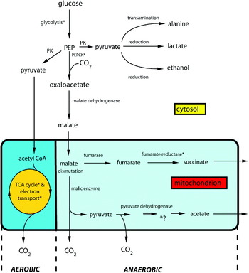
The horse and sheep strains of E. granulosus and E. multilocularis were shown to possess the complete suite of tricarboxylic acid cycle enzymes with relatively high activities and the levels of intermediates were of the same order as those reported in tissues with an active cycle (McManus and Smyth, Reference McManus and Smyth1982). On the other hand, fumarate reductase, which catalyses the reverse reaction to succinic dehydrogenase, had a low activity, although the assay method used would have seriously under-estimated the activity, because of its instability and because it is notoriously difficult to measure. The precise role of the tricarboxylic acid cycle is difficult to assess in parasitic helminths generally because a respiratory end product, succinate, is also an intermediate in the cycle (Fig. 1) and, along with partly oxidized metabolites, is produced even under aerobic conditions. Nevertheless, in our earlier study (McManus and Smyth, Reference McManus and Smyth1978) we had shown marked differences in oxygen uptake between the sheep and horse strains of E. granulosus, again reinforcing the existence of biochemical variants with greater or lesser dependence of tricarboxylic acid cycle activity (McManus and Bryant, Reference McManus, Bryant, Thompson and Lymbery1995).
Respiratory metabolism in Echinococcus granulosus (after McManus and Bryant, Reference McManus, Bryant, Thompson and Lymbery1995). *Presumed sites of ATP synthesis; PK, pyruvate kinase; PEPCK, phosphoenolpyruvate carboxykinase; TCA, tricarboxylic acid. The reactions which take place outside the box occur in the cytosol; those inside the box occur in the mitochondria. Almost certainly, aerobic and anaerobic reactions do not occur simultaneously. Further, it is probable that 2 distinct types of mitochondria exist.

Our end-product analysis showed that, while the tricarboxylic acid cycle is functional at least in part in E. granulosus, fermentative pathways yielding acetic, succinic and lactic acids occur even under aerobic conditions (McManus and Smyth, Reference McManus and Smyth1982). We (McManus and Bryant, Reference McManus, Bryant, Thompson and Lymbery1995) presumed that succinic and acetic acids are formed in the mitochondria, as is the case with other helminths, but whether this occurs in the same mitochondria which house the tricarboxylic acid cycle is unknown. In the pathway illustrated in Fig. 1, NAD(P)H, produced from the glyceraldehyde-3-phosphate dehydrogenase step in glycolysis and by the activity of cytoplasmic malic enzyme, can be re-oxidized by the reduction of pyruvate to lactate or ethanol and of oxaloacetate to malate. In the mitochondrion, reducing equivalents generated on the acetate arm are used to drive the fumarate reductase reaction in the generation of succinate. The adenine nucleotide contents of the protoscoleces of E. granulosus and E. multilocularis were comparable with those of other parasites; the adenylate energy charge: ((ATP)+(ADP))/((ATP)+(ADP)+(AMP)) in the range 0·86–0·92 was, however, the highest recorded for a parasite. ATP: AMP ratios were also very high and probably reflect the low demand for ATP by a fully developed protoscolex in a hydatid cyst.
BIOCHEMICAL CHARACTERIZATION OF THE ECHINOCOCCUS GRANULOSUS TEGUMENT
Cestodes lack a gut and the host/parasite interface is represented by the tegument that is bounded on the external surface by a series of spine-like processes, the microtriches. These structures morphologically resemble the brush border microvilli of many vertebrate and invertebrate cells and they are covered with a single outer plasma membrane overlain by a glycocalyx composed of mucopolysaccharide and glycoproteins (Smyth and McManus, Reference Smyth and McManus1989). The tegument plays a vital role in the physiology of cestodes, being involved in nutrient uptake, defence against enzyme and immunological attack by the host, in excretion and ionic exchange, and in metabolic transfer (Smyth and McManus, Reference Smyth and McManus1989).
In the 1970s and 1980s, cognisant of the obvious importance of the cestode tegument, a series of studies were aimed at achieving a greater understanding of its biochemical nature. The majority of these considered the adult stage of Hymenolepis microstoma or H. diminuta (e.g. Oaks et al. Reference Oaks, Knowles and Cain1977; Pappas and Narcisi, Reference Pappas and Narcisi1982). Our study published in Parasitology was different in that it involved the protoscolex of E. granulosus (McManus and Barrett, Reference McManus and Barrett1985). Being a tissue-dwelling larval stage enclosed within several discrete coverings (brood capsule wall and the germinal, laminated and pericyst layers of the hydatid cyst), the protoscolex encounters different physiological challenges than those of an adult tapeworm. As with others at the time, we utilized the unusual syncytial nature of the cestode tegument to lift off the distal cytoplasm and brush border by physical and/or chemical means, thereby leaving the remainder of the syncytium including the basement membrane. Several approaches were adopted for the disruption and removal of the tegumental surface from protoscoleces. The effectiveness of each method and the purity of subsequent microthrix-enriched fractions obtained by differential centrifugation were evaluated by electron microscopy, by the amount of protein released and by the degree of enrichment of surface plasma membrane marker enzymes. Incubation in saponin for 10 min produced the purest microtriche preparation, but in low yield; freeze/thawing, incubation in Triton X-100 for 10 min or in saponin for 20 min produced fractions containing significant amounts of relatively pure microtriches, but mild homogenization was a poor method for surface disruption and subsequent isolation of microtriches. Phosphodiesterase, adenosine triphosphatase (ATPase) (total and ouabain-inhibited), leucine aminopeptidase and glutamyltransferase were active in the protoscoleces but none were enriched in any of the microthrix fractions. In contrast, alkaline phosphatase, acid phosphatase, 5′ nucleotidase and maltase were enriched significantly in all of the isolated microtriche preparations, which suggested that these enzymes are predominantly surface-membrane bound. The protein profiles of the microthrix-enriched fractions, following SDS-PAGE, were basically similar, although there were some qualitative and quantitative differences in the proteins released by each isolation procedure. Three major PAS-staining components were present in all the preparations and these probably originated from the glycocalyx. One of these PAS-positive components, with an approximate molecular weight of 110 kDa was a glycoprotein, likely of glycocalyx origin, and putatively specific to the horse strain of E. granulosus. Its identity has still not been established.
CLONING OF ECHINOCOCCUS GRANULOSUS DNA
In the 1980's it had become apparent that the new techniques in molecular biology offered great potential as tools for parasite detection and identification, and would provide a new approach to studies on the taxonomy, genetics and population biology of parasites. The sensitivity and specificity of DNA analysis was ideal for addressing important problems in diagnostic parasitology and achieving two of my research objectives of producing a simple field test for distinguishing Echinococcus eggs from those of other taeniid cestodes, and ascertaining the extent of strain variation in E. granulosus.
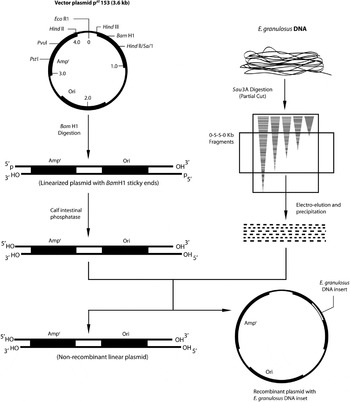
Andrew Simpson and I had used cloned DNA fragments of the ribosomal RNA gene of Schistosoma mansoni to discriminate between isolates of E. granulosus from UK horses and sheep as well as E. granulosus from E. multilocularis (McManus and Simpson, Reference McManus and Simpson1985). However, I reasoned it would be necessary to use cloned DNA markers, constructed from E. granulosus DNA, to provide increased powers of resolution for identification purposes. Accordingly, Arun Rishi and I described in Parasitology the construction of a small, size selected (0·5–5·0 kbp) genomic DNA library in a plasmid vector (pAT153) of Escherichia coli using total DNA isolated from E. granulosus (Fig. 2) (Rishi and McManus, Reference Rishi and McManus1987). A panel of taeniid cestode DNAs was then used in conjunction with hybridization and restriction-enzyme analysis to identify in the library 2 recombinant plasmids with Echinococcus-specific inserts and a single recombinant plasmid (coded pEG18) with a DNA fragment unique for E. granulosus. These, and other recombinant plasmids with E. granulosus DNA inserts, were then used in restriction endonuclease, Southern transfer and hybridization analysis to independently and reproducibly discriminate between the UK horse and sheep strains of E. granulosus; these probes had the potential for characterizing isolates from other endemic areas. The recombinant insert (approximately 2·3 kbp in size) of pEG18 had a low copy number – estimated at approximately 26 – and it proved later to be insufficiently sensitive for practical use as a DNA probe in the identification of small numbers (ideally, a single egg) of E. granulosus eggs by molecular hybridization. Success in this area was only achieved later after the advent of the polymerase chain reaction (PCR) and its application in research on Echinococcus.
Cloning of Echinococcus granulosus genomic DNA using the plasmid vector, PAT153. kb, 103 base pairs (after Rishi and McManus, Reference Rishi and McManus1987).

We used a similar procedure, described in a paper published the following year in Parasitology to clone specific DNA fragments from Taenia solium that we used to detect intra-specific genetic variation in cysticerci of T. solium from India, Mexico and Zimbabwe, and to distinguish T. solium from T. saginata in a simple dot-blot assay (Rishi and McManus, Reference Rishi and McManus1988).
GENETIC HETEROGENEITY WITHIN ECHINOCOCCUS GRANULOSUS CHARACTERIZED WITH DNA PROBES
In a paper published in Parasitology in 1989, we used the E. granulosus- and S. mansoni-derived DNA probes in restriction endonuclease fragment length (RFLP) and Southern blot analysis to distinguish between isolates of larval and adult E. granulosus from different host species (sheep, goats, cattle, camels, humans, buffalo, horses, donkeys, pigs and dogs from various geographical areas (United Kingdom, Kenya–Masailand and Turkana-, Sudan, Somalia, Uruguay, Argentina, Italy, Turkey, Spain, Lebanon, Jordan, Holland, India, Ireland, Yugosalvia and Poland); a total of 251 larval isolates, 16 individual adult worms and 3 pools of adult worms were analysed (McManus and Rishi, Reference McManus and Rishi1989). The study did not demonstrate any significant genetic variation within the United Kingdom horse/dog or sheep/dog strains but confirmed the distinctiveness of the two strains shown in previous studies. The sheep/dog strain was shown to be cosmopolitan in its distribution and fertile bovine material originating from the United Kingdom, Kenya, Spain and India conformed to this strain by DNA hybridization. In contrast, cattle isolates from Holland produced markedly different DNA hybridization banding profiles indicating that cattle can harbour more than 1 strain of E. granulosus. Similarly, it was shown that goats can harbour 2 different strains of E. granulosus, the sheep/dog strain and a form which infects camels. The analysis showed that the strain of E. granulosus infecting equines in Spain and Ireland was genetically identical to that infecting horses in the United Kingdom. There was also a different strain infecting pigs in Poland and Yugoslavia. This pig/dog strain appeared to be very similar genetically to the forms of E. granulosus which use camels and goats as intermediate hosts and was similar, though not identical, to the variant infecting Dutch cattle. The study also showed that E. granulosus material, fixed for a prolonged period in ethanol, or lyophilized, was amenable to DNA analysis and that it was possible to characterize the DNA of a single adult worm.
QUO VADIT?
A number of years have elapsed since these early studies on the biochemistry of Echinococcus published in Parasitology and, in this Centenary Issue article, it is timely to reflect briefly on some of the subsequent work that has been undertaken in this area, highlighting some of the significant contributions made, and providing some insight into possible future research.
Metabolism of Echinococcus larvae
There have been, surprisingly, few recent studies investigating the basic metabolism or the regulation of respiratory pathways in Echinococcus. One area of interest that has been pursued has involved determining the effects of anti-hydatid drugs on the activity of enzymes of carbohydrate metabolism in the cyst wall of E. granulosus, where glycolysis was shown to be the main respiratory pathway operating (Xiao et al. Reference Xiao, Feng, Guo, Jiao, Yao and Jiao1994, Reference Xiao, Feng and Yao1995). These studies suggested that pyruvate kinase, phosphoenolpyruvate carboxykinase and ATPase might be key targets for mebendazole and albenzdazole but not praziquantel, which appears to act by interfering with calcium flux across membranes. On this theme, Bonilla et al. (Reference Bonilla, Denicola, Novoselov, Turanov, Protasio, Izmendi, Gladyshev and Salinas2008) demonstrated the existence of functional linked thioredoxin-glutathione systems in the cytosolic and mitochondrial compartments of E. granulosus through a single thioredoxin-glutathione reductase (TGR) enzyme, a promising drug target, and indeed, auranofin, a known TGR inhibitor, killed larval worms in vitro.
Studies by Matsumoto et al. (Reference Matsumoto, Sakamoto, Shinjyo, Kido, Yamamoto, Yagi, Miyoshi, Nonaka, Katakura, Kita and Oku2008) suggest that the mitochondrial respiratory system of E. multilocularis is also a good target for the development of novel selective anti-echinococcal compounds, as demonstrated previously for other helminths. Their most notable finding was that E. multilocularis protoscoleces possess a unique mitochondrial respiratory system that is highly adapted to anaerobic conditions. Specifically, the predominant enzymatic activity in an enriched mitochondrial fraction prepared from the protoscoleces was NADH-fumarate reductase, which does not normally function in the aerobic respiratory chain of mammals, compared with that of NADH oxidase, an enzyme involved in aerobic respiration in aerobic organisms. The high-performance liquid chromatography-mass spectrometry that they carried out revealed that the primary quinone of the parasite mitochondria was rhodoquinone-10, which is commonly used as an electron mediator in anaerobic respiration by the NADH-fumarate reductase system of other eukaryotes. This also suggested that the mitochondria of E. multilocularis protoscoleces possess an anaerobic respiratory chain in which complex II of the parasite functions as a rhodoquinol-fumarate reductase. Furthermore, in vitro treatment assays using respiratory chain inhibitors against the NADH-quinone reductase activity of mitochondrial complex I demonstrated that they had a potent ability to kill protoscoleces, emphasizing the promise of the mitochondrial respiratory chain of the parasite as a target for chemotherapy of alveolar echinococcosis.
In our earlier study, we had observed that E. granulosus protoscoleces cultured under anaerobic conditions produced more succinate than parasites kept under aerobic conditions, suggesting their survival under anaerobic conditions by utilizing the NADH-fumarate reductase system (McManus and Smyth, Reference McManus and Smyth1978). We further reported that the specific activity of fumarate reductase in Echinococcus protoscoleces was lower than those of enzymes involved in the tricarboxylic acid cycle (McManus and Smyth, Reference McManus and Smyth1982). These results, however, did not establish the importance of NADH-fumarate reductase activity in the mitochondrial respiratory system of the parasites because the other enzyme activities were not analysed. Clearly, experiments on E. granulosus, along the lines undertaken by Matsumoto et al. (Reference Matsumoto, Sakamoto, Shinjyo, Kido, Yamamoto, Yagi, Miyoshi, Nonaka, Katakura, Kita and Oku2008) on E. multilocularis would be highly informative and indicate the potential for developing specific inhibitors of the mitochondrial respiratory chain as lead compounds for the chemotherapy of cystic echinococcosis.
Proteins of the Echinococcus granulosus tegument
Some recent proteomics analysis of E. granulosus has been undertaken with 15 prominent proteins including actins, tropomyosin, paramyosin, thioredoxin reductase, antigen P-29, cyclophilin, and the heat shock proteins hsp70 and hsp20 shown to be present in protoscoleces (Chemale et al. Reference Chemale, van Rossum, Jefferies, Barrett, Brophy, Ferreira and Zaha2003). As with studies on schistosomes, a combination of proteomics with differential extraction procedures (Braschi et al. Reference Braschi, Curwen, Ashton, Verjovski-Almeida and Wilson2006) or biotinylation (Braschi and Wilson, Reference Braschi and Wilson2006) of the tegument would likely reveal very useful information on the nature of the echinococcal parasite surface and its membrane-associated proteins. Notwithstanding, there are some recent reports on tegumental enzymes and proteins from larval E. granulosus. A fatty acid-binding protein was shown to be present in the tegument of protoscoleces (Esteves et al. Reference Esteves, Dallagiovanna and Ehrlich1993; Jakobsson et al. Reference Jakobsson, Alvite, Bergfors, Esteves and Kleywegt2003), paramyosin (Mühlschlegel et al. Reference Mühlschlegel, Sygulla, Frosch, Massetti and Frosch1993) and molecules with both Fc-binding and proteolytic activity are present on the surface, likely playing a role in the host-parasite interaction (Baz et al. Reference Baz, Carol, Marco, Casabó, Jones, Dunne and Nieto1998). A novel 29 kDa antigen (immunologically related to antigen 5, one of the 2 major antigenic components of hydatid cyst fluid) has been shown to be distributed in the tegument and rostellum (González et al. Reference González, Spinelli, Lorenzo, Hellman, Nieto, Willis and Salinas2000), and the antioxidant protein thioredoxin peroxidase is expressed in the tegument and subtegument as well as calcareous corpuscle forming cells in protoscoleces of E. granulosus (Hou et al. Reference Hou, Wang, Zhang, Cao, Zhang and Zhang2007).
Following a study on schistosomes (Smyth et al. Reference Smyth, McManus, Smout, Laha, Zhang and Loukas2003), we used the signal sequence trap technique to identify genes coding for secreted and membrane, including tegumental surface membrane, bound proteins from E. granulosus (Rosenzvit et al. Reference Rosenzvit, Zhang, Motazedian, Smyth, Pearson, Loukas, Jones and McManus2006b). Many of these proteins are likely to be involved in host-parasite interactions, and because of their potential exposure to the host immune system, such components are candidates for improved diagnostic tests, as well as being new putative drug and vaccine targets. Several of the clones obtained shared between 30 and 40% amino acid identity with amino acid transporters belonging to members of the neutral and excitatory amino acid transporters of the Dicarboxylate/Amino Acid Na+ and/or H+Cation Symporter family (DAACS) (TC 2.A.23).
In a subsequent study, Camicia et al. (Reference Camicia, Paredes, Chalar, Galanti, Kamenetzky, Gutierrez and Rosenzvit2008) confirmed one of these clones (termed egat1) as a DAACS member and bioinformatics analysis showed the presence of 9–10 transmembrane domains, consensus sequences for N-glycosylation between the third and fourth transmembrane domain, a highly similar hydropathy profile with ASCT1 (a known member of DAACS family), high score with SDF (sodium dicarboxylate family) and similar motifs with EDTRANSPORT, a fingerprint of excitatory amino acid transporters. In situ hybridization indicated distribution of egat1 mRNA throughout the tegument and immunofluorescence showed localization of the EgAT1 protein in the subtegument of protoscoleces particularly around suckers and rostellum. The close proximity to the surface of the larva, its expression in all life-cycle stages (Rosenzvit et al. 2006 a), and particularly the intimate association with the suckers and rostellum, which are of major importance for the survival in the final host, could make EgAT1 a particularly amenable target for drug development. The presence of a dicarboxylate amino acid (Na+ or H+) symporter (DAACS) member in protoscoleces of E. granulosus is supported by the concomitant presence of the active Na+/K+-ATPase that we had described earlier (McManus and Barrett, Reference McManus and Barrett1985).
Genetic variation in Echinococcus
In the 1990s, it had become clear that application of the powerful polymerase chain reaction (PCR) would increase considerably our capabilities for studying the molecular genetics of Echinococcus. In combination with direct sequencing approaches and PCR, we were able to sequence and compare 2 regions (cytochrome oxidase subunit 1 (cox1) and NADH dehydrogenase (nad1)) of the mitochondrial genome of a large number of hydatid isolates (Bowles et al. Reference Bowles, Blair and McManus1992; Bowles and McManus, Reference Bowles and McManus1993). As a result, we were able to distinguish the 4 recognized species of Echinococcus and group E. granulosus into 7 genotypically different forms of genoptypes. To date, 10 distinct genotypes within E. granulosus have now been identified, and this categorization follows very closely the pattern of strain variation emerging based on published biological characteristics (McManus, Reference McManus2002, Reference McManus2006).
Complete sequences for the mitochondrial genomes of E. granulosus (horse and sheep strains) and E. multilocularis have been obtained as well as sequences for variable mitochondrial genes from additional genotypes of E. granulosus, from E. vogeli and E. oligarthrus (Le et al. Reference Le, Pearson, Blair, Dai, Zhang and McManus2002). Based on this and considerable biological information, a re-evaluation of the taxonomy and phylogeny of E. granulosus has been advocated (Thompson and McManus, Reference McManus2002; McManus and Thompson, Reference McManus and Thompson2003; McManus, Reference McManus2006, Nakao et al. Reference Nakao, McManus, Schantz, Craig and Ito2007). Indeed, mitochondrial DNA-based detection of Echinococcus species has been shown generally to be an excellent tool for analysis of strain/genotypic variation in the genus, determining phylogenetic relationships, and informing taxonomic species questions (McManus and Thompson, Reference McManus and Thompson2003; McManus, Reference McManus2006, Nakao et al. Reference Nakao, McManus, Schantz, Craig and Ito2007; Xiao et al. Reference Xiao, Qiu, Nakao, Li, Yang, Chen, Schantz, Craig and Ito2005). As well, several stool-based PCR (copro-PCR) tests have been developed that allow rapid identification of Echinococcus DNA from eggs or from adult parasites, thereby providing a highly complementary approach for positive and highly specific diagnosis of canines infected with E. granulosus and E. multilocularis and environmental detection of Echinococcus eggs in soil samples (Abbasi et al. Reference Abbasi, Branzburg, Campos-Ponce, Abdel Hafez, Raoul, Craig and Hamburger2003; Deplazes et al. Reference Deplazes, Dinkel and Mathis2003; Eckert and Deplazes, Reference Eckert and Deplazes2004; Shaikenov et al. Reference Shaikenov, Rysmukhambetova, Massenov, Deplazes, Mathis and Torgerson2004; Stefanic et al. Reference Stefanic, Shaikenov, Deplazes, Dinkel, Torgerson and Mathis2004; Reiterova et al. Reference Reiterova, Miterpakova, Turcekova, Antolova and Dubinsky2005; Naidich et al. Reference Naidich, McManus, Canova, Gutierrez, Zhang, Guarnera and Rosenzvit2006; Boufana et al. Reference Boufana, Campos-Ponce, Naidich, Buishi, Lahmar, Zeyhle, Jenkins, Combes, Wen, Xiao, Nakao, Ito, Qiu and Craig2008). None of these tests, however, have been shown to have the sensitivity required for identification of a single egg, and this is clearly an area for future research.
Concluding comment
Since the 1970s, when I first began my research career, considerable advances have been made in the area of parasite biochemistry and physiology. Molecular biology provided a huge armamentarium of new and sophisticated approaches for the study of Echinococcus that firstly my group and then others have used to progress the field. For the future, one can predict complete genome sequencing of several Echinococcus taxa, the generation of a large Echinococcus transcriptome database and the application of post-genomic technologies, including DNA microarray profiling, advanced proteomics, glycomics, immunomics and RNA interference (RNAi), leading to a more complete understanding of the biochemistry, physiology and general biology of these important parasites, and to the development of effective interventions for hydatid disease, including new drugs and vaccines.
I would like to gratefully acknowledge my co-authors of the early papers that I published in Parasitology and which form the basis of this retrospective – Nicky Barrett, Arun Rishi and, of course, the late Des Smyth, who was such a guiding light and mentor to me in the years following my Ph.D. and beyond. I would also like to thank all my staff, students and numerous colleagues and collaborators with whom I have worked over the past 35 years and the support of the various funding bodies that have allowed me to undertake my research. Mimi Kersting's help with the figures is also much appreciated.