


Introduction
Animals such as parasites are often overlooked in biodiversity research and conservation efforts (Gómez and Nichols Reference Gómez and Nichols2013). This is perhaps unsurprising, considering that most parasites are small, and many live inside their hosts, hidden from the naked eye. However, documenting and characterising parasite biodiversity is essential for several reasons. First, it is thought that parasites constitute up to 50% of metazoan biodiversity, are ubiquitous across the world’s ecosystems and play multiple functional roles in natural systems (Dobson et al Reference Dobson, Lafferty, Kuris, Hechinger and Jetz2008; Marcogliese Reference Marcogliese2004; Poulin and Morand Reference Poulin and Morand2004). Consequently, a comprehensive understanding of ecosystem structure and functioning is not possible without accounting for the parasites present. Second, many parasites are more vulnerable to climate change than their hosts as they rely entirely on their host’s survival (sometimes multiple host species) for their own persistence (Cizauskas et al Reference Cizauskas, Carlson, Burgio, Clements, Dougherty, Harris and Phillips2017). For parasites with complex life cycles, the extinction of any single host species across two-, three-, or four-stage life cycles may result in parasite extinction (Lafferty Reference Lafferty2012). Third, parasites can cause or be associated with disease in their hosts, meaning that their documentation is essential for the safeguarding and management of host wildlife. For these reasons, and others, it is critical that parasite biodiversity is documented and characterised before it is lost, or emerges as a threat to host health in future.
Bennett et al. (Reference Bennett, Presswell and Poulin2022) demonstrated that fewer than 10% of marine species in Aotearoa New Zealand had ever been characterised for their parasitic fauna. However, in recent years a significant effort has been made to address this shortfall, including the publication of updated checklists of parasites infecting marine invertebrates (Bennett et al Reference Bennett, Poulin and Presswell2022a) and marine mammals (Lehnert et al Reference Lehnert, Poulin and Presswell2019); biodiversity studies targeting specific parasite groups (acanthocephalans, Bennett, Poulin and Presswell (Reference Bennett, Poulin and Presswell2023); nematodes, Bennett et al (Reference Bennett, Poulin and Presswell2022b)) and papers on parasite assemblages within specific host taxa (e.g. shags, Presswell and Bennett (Reference Presswell and Bennett2021); penguins, Bennett et al (Reference Bennett, Presswell, Little, Lagrue, Webster, Dutoit, Poulin, Davidson, Cahill and Hutson2025)), as well as individual new species accounts. Collectively, these works have resulted in the description of several species new to science, and identified a number of species with potential to cause or be associated with disease. This paper further contributes to this work by documenting a comparatively high diversity of trematode parasite species infecting the two marine gulls of New Zealand: red-billed gull Chroicocephalus scopulinus (Forster) and black-backed gull Larus dominicanus Lichtenstein.
The red-billed gull (known as tarāpunga to indigenous Māori) is endemic to New Zealand and its outlying islands, having colonised from Australia approximately 130,000 years ago (Given et al Reference Given, Mills and Baker2005). Although estimated to comprise greater than 100,000 birds, the population is listed ‘at risk and declining’ according to the New Zealand Threat Classification criteria (Robertson et al Reference Robertson, Baird, Elliott, Hitchmough, McArthur, Makan, Miskelly, O’Donnell, Sagar, Scofield and Michel2021). On the other hand, the Southern black-backed gull (karoro, referred to as black-backed gull herein) is a recently arrived native to New Zealand and its outlying islands, but has a broad distribution in the Southern Hemisphere. This species is much more abundant on New Zealand’s mainland than the red-billed gull, and is listed as ‘not threatened’ (Robertson et al Reference Robertson, Baird, Elliott, Hitchmough, McArthur, Makan, Miskelly, O’Donnell, Sagar, Scofield and Michel2021). Both are highly opportunistic feeders in New Zealand, frequently scavenging human refuse at landfills, ports and urban areas as well as natural food sources along coasts and estuaries. Given their wide distribution across New Zealand and their broad, opportunistic diets, parasite communities in these species are expected to be highly variable and closely linked to dietary breadth. Although a number of studies have investigated selected parasite species in red-billed and black-backed gulls (e.g. Bennett and Presswell Reference Bennett and Presswell2019; Bowie Reference Bowie1981; Clark Reference Clark1978a, Reference Clarkb; Fredensborg et al Reference Fredensborg, Latham and Poulin2004; Latham and Poulin Reference Latham and Poulin2002; Presswell Reference Presswell2022; Presswell and Bennett Reference Presswell, Bennett and Smales2020; Presswell et al Reference Presswell, Bennett and Smales2020; Rind Reference Rind1984), the lack of sufficiently large sample sizes of host individuals has limited comprehensive, systematic assessments of their parasite faunas at a population or species level. Previously reported helminth parasites are listed in Table 1.
Previous records of trematodes from red-billed and black-backed gulls in New Zealand: hosts, references and how they are interpreted in this study. BBG = black-backed gull; RBG = red-billed gull

Table 1. Long description
The table consists of four columns: Trematode, Gull host, Reference, and In this study. B B G refers to black-backed gull and R B G refers to red-billed gull.
* Apatemon jamiesoni: B B G; Presswell 2022; Apatemon jamiesoni.
* Ornithobilharzia canaliculata: B B G and R B G; Rind 1984 and this study; Ornithobilharzia canaliculata.
* Schistosomatidae gen. sp. 1: B B G; Bennett et al. 2023; Gigantobilharzia cf. patagonensis.
* Curtuteria australis: R B G; Fredensborg et al. 2004; N O T F O U N D.
* Himasthla sp. 1: B B G and R B G; Bennett et al. 2023; Himasthla davisi n. sp.
* Parorchis acanthus: B B G and R B G; Latham and Poulin 2002 and Wisely 1977; N O T F O U N D.
* Philophthalmus attenuatus: B B G and R B G; Bennett and Presswell 2019; Philophthalmus attenuatus.
* Galactosomum otepotiense: B B G and R B G; Presswell and Bennett 2020; Galactosomum otepotiense.
* Levinseniella sp. 1: B B G and R B G; Bennett et al. 2023; Levinseniella (Monorrhenos) trisepsilon n. sp.
* Maritrema novaezealandensis: B B G and R B G; Martorelli et al. 2004; Maritrema novaezealandense.
* Microphallus sp.: B B G and R B G; Leung et al. 2009; Microphallus lagruei n. sp.
* Microphallus sp. 1: B B G and R B G; Bennett et al. 2023; Microphallus lagruei n. sp.
* Echinoparyphium recurvatum: B B G and R B G; Latham and Poulin 2002 and Fredensborg et al. 2004; N O T F O U N D.
* Echinostoma revolutum: B B G; Latham and Poulin 2002; N O T F O U N D.
* Acanthoparyphium sp. A: B B G and R B G; Leung et al. 2009; Acanthoparyphium argillamartelliarum n. sp.
* Acanthoparyphium sp. 1 A: B B G and R B G; Bennett et al. 2023; Acanthoparyphium argillamartelliarum n. sp.
* Cryptocotyle micromorpha: B B G; Presswell and Bennett 2022; Cryptocotyle micromorpha.
This study characterises the diversity of trematode parasites infecting red-billed and black-backed gulls from southern New Zealand. We describe 6 species new to science, and provide ecological data and remarks on all 15 species recovered (including new genetic data for 12 species). Together, these data establish a baseline survey of trematode biodiversity against which future studies can be compared, and demonstrate that although parasites are frequently underrepresented or excluded from biodiversity assessments, their substantial contribution to overall biodiversity means they should not be overlooked.
Methods
Gull collection and trematode sampling
Between 2018 and 2025, we examined the parasites of 113 red-billed and 114 black-backed gulls from various locations along the Otago coast. The specimens were obtained deceased from the Wildlife Hospital, Dunedin, and the Department of Conservation. Gull specimens were received frozen and subsequently thawed for dissection, when gastrointestinal tracts were removed and examined under a dissecting scope for the presence of helminth parasites. Nematodes, cestodes and acanthocephalans were kept aside and will be presented in subsequent publications. Trematodes were preserved in 70% ethanol for whole-mounting or 10% buffered formalin for scanning electron microscope (SEM) photography and 70–99% ethanol for genetic analysis.
Parasite identification
Parasite species were identified to the lowest possible taxonomic level using a combination of morphological and molecular data. For morphological analyses, trematodes were stained using iron acetocarmine, dehydrated through a graded ethanol series, cleared in clove oil and mounted in Canada balsam. Measurements were made using ImageJ software (Wayne Rasband, NIH, USA) from photographs taken on an Olympus BX51 compound microscope mounted with a DP25 camera attachment. All measurements are in micrometres given as ranges with the mean in parentheses, unless otherwise specified. Drawings were made with the aid of a drawing tube mounted to an Olympus compound microscope, or from light micrographs using a light box. For SEM, specimens were transferred to 2.5% glutaraldehyde in 0.1 M phosphate buffer, then post-fixed in 1% osmium tetroxide and dehydrated through a gradient series of ethanols, critical-point dried in a CPD030 BalTec critical-point dryer (BalTec AG, Balzers, Liechtenstein) using carbon dioxide, mounted on aluminium stubs, and sputter coated with gold/palladium (60:40) to a thickness of 10 nm in an Emitech K575X Peltier cooled high-resolution sputter coater (EM Technologies, Ashford, Kent, UK). The specimens were viewed with a JEOL 6700F field-emission scanning electron microscope (JEOL Ltd., Tokyo, Japan) at the Otago Centre for Electron Microscopy (OCEM, University of Otago, New Zealand). Taxa unfamiliar to the authors were identified using morphological keys (Bray et al Reference Bray, Gibson and Jones2008; Gibson et al Reference Gibson, Jones and Bray2002; Jones et al Reference Jones, Bray and Gibson2005), and original species descriptions.
When condition and numbers of specimens allowed, molecular data were also obtained to support taxonomic identifications. Partial 28S gene sequences were targeted in the first instance. DNA was extracted using DNeasy® Blood and Tissue kit (Qiagen, Hilden, Germany) and amplified using primers T16/T30 (Harper and Saunders Reference Harper and Saunders2001) with polymerase chain reactions (PCR) of 94 °C for 5 min, 38 cycles of 94 °C for 30 s, 45 °C for 30 s and 72 °C for 2 min, and 72 °C for 7 min. The partial cox1 gene was also targeted for comparison with closely related sequences on GenBank and previously published data. Cox1 PCRs involved primers JB3 (Bowles et al Reference Bowles, Hope, Tiu, Xushian and McManus1993)/Trem.cox1.rrnl (Králová-Hromadová Reference Králová-Hromadová, Špakulová, Horáčková, Turčeková, Novobilský, Beck, Koudela, Marinculić, Rajský and Pybus2008) with conditions of 95 °C for 2 min, 40 cycles of 95 °C for 30 s, 48 °C for 40 s and 72 °C for 1 min, and 72 °C for 10 min. All positive sequences were cleaned using EXOSAP™ Express PCR Product Cleanup Reagent (USB Corporation, Cleveland, OH, USA), and Sanger sequencing by capillary electrophoresis was performed by the Genetic Analysis Service, Department of Anatomy, University of Otago (Dunedin, New Zealand). Sequences were viewed and manually edited on Geneious Prime® v11.0.20.1. To support morphological identifications, we used BLASTn searches (https://blast.ncbi.nlm.nih.gov/) to compare relatives on GenBank by percentage identity. Voucher specimens (whole-mount slides) of parasites are deposited at the Museum of New Zealand Te Papa Tongarewa under accession W.004100–W.004112 and genetic sequences have been deposited to NCBI GenBank under accessions PZ622610–20, PZ656439 and PZ669438–51 (Table 2). When comparing closely related species, uncorrected pairwise genetic distances were used to obtain intra- and interspecific genetic variation, estimated in MEGA v11 (Tamura et al Reference Tamura, Stecher and Kumar2021).
Accession numbers of trematode species recovered in this study submitted to GenBank. BBG = black-backed gull; RBG = red-billed gull

Table 2. Long description
The table consists of four columns: Trematode species, I D, Gene, and Accession.
* Gigantobilharzia cf. patagonensis: I D BBG42tre1, Gene 28 S, Accession PZ622610.
* Ornithobilharzia canaliculata: I D BBG26tre1, Gene 28 S, Accession PZ656439; also Gene cox 1, Accession PZ669348.
* Diplostomum spathaceum: I D BBG96tre2, Gene 28 S, Accession PZ622611.
* Mesorchis carsonae n. sp.: I D BBG51tre1, Gene 28 S, Accession PZ622612; also Gene cox 1, Accession XXXXXX.
* Acanthoparyphium argillamartelliarum n. sp.: I D BBG64tre1, Gene 28 S, Accession PZ622613; also Gene cox 1, Accession PZ669350.
* Himasthla davisi n. sp.: I D BBG35tre1, Gene 28 S, Accession PZ622614.
* Echinostomatoidea gen. sp.: I D BBG23tre2, Gene 28 S, Accession PZ622615; also Gene cox 1, Accession PZ669349.
* Gymnophalloides hutsonae n. sp.: I D BBG112tre1A, Gene 28 S, Accession PZ622616.
* Cryptocotyle micromorpha: I D BBG28tre1, Gene 28 S, Accession PZ622617.
* Levinseniella trisepsilon n. sp.: I D BBG8tre1, Gene 28 S, Accession PZ622618.
* Microphallus lagruei n. sp.: I D RGB26tre1, Gene 28 S, Accession PZ622619; also I D BBG36tre1, Gene cox 1, Accession PZ669351.
* Maritrema novaezealandense: I D BBG29tre1, Gene 28 S, Accession PZ622620.
Results
General results
A total of 15 trematode species were recovered from 227 gulls sampled across southern New Zealand. Nine of these species were found in both host species; all 15 species were recorded from black-backed gulls, and no trematodes were detected in red-billed gulls that were not also present in black-backed gulls. Most gulls harboured a single trematode species, and the maximum number of species infecting a bird was two (in red-billed gulls) and three (in black-backed gulls). Infection intensities were generally low, with most infected individuals hosting only one or two trematode individuals, although microphalloids occurred in the hundreds in five instances. Accounts for each species are given below, including full descriptions of new species and taxonomic remarks for all taxa. Prevalence and intensities (following Bush et al Reference Bush, Lafferty, Lotz and Shostak1997) are given for each parasite under the species account.
Diplostomida Olson, Cribb, Tkach, Bray & Littlewood, 2003
Schistosomatoidea Stiles & Hassall, 1898
Schistosomatidae Stiles & Hassall, 1898
Gigantobilharzia cf. patagonensis (Lorenti, Brant, Gilardoni, Diaz & Cremonte, Reference Lorenti, Brant, Gilardoni, Diaz and Cremonte2022) Dutton & Bullard in Dutton, Du Preez, Netherlands, Jordaan & Bullard, 2024; (Figure 1a)
Diplostomida species recovered from gulls in New Zealand: (a) Gigantobilharzia cf. patagonensis anterior half of worm, composite light microphotograph. (b) Ornithobilharzia canaliculata paired male and female, composite light microphotograph. (c) Ornithobilharzia canaliculata scanning electron micrograph of forebody showing oral and ventral suckers, and lack of body spines. (d) Diplostomum spathaceum whole worm. Dotted line indicates fragment removed for DNA extraction. (e) Apatemon jamesoni whole worm. Scale bars: (a) 500 μm; (b) 1mm; (c), (d), (e) 200 μm.

Figure 1. Long description
A five-panel collection of micrographs labeled a through e.
* Panel a. A composite light microphotograph of the anterior half of Gigantobilharzia cf. patagonensis. The worm is elongated and translucent with internal structures visible. A scale bar at the bottom right represents 500 micrometers.
* Panel b. A composite light microphotograph of Ornithobilharzia canaliculata showing a paired male and female. The male is larger and thicker, while the female is thinner and partially enclosed within the male. A scale bar at the bottom right represents 1 millimeter.
* Panel c. An S E M micrograph of the forebody of Ornithobilharzia canaliculata. It shows a smooth body surface without spines, featuring a distinct oral sucker at the tip and a protruding ventral sucker on the side. A scale bar at the bottom right represents 200 micrometers.
* Panel d. A light microphotograph of a whole Diplostomum spathaceum worm. It has a widened anterior end and a narrower posterior. A dotted line on the right side of the anterior section indicates where a fragment was removed for D N A extraction. A scale bar at the bottom right represents 200 micrometers.
* Panel e. A light microphotograph of a whole Apatemon jamesoni worm. It features a bulbous, cup-shaped anterior segment and a thicker posterior body. A scale bar at the bottom right represents 200 micrometers.
Synonyms: Gigantobilharzia patagonense (Lorenti, Brant, Gilardoni, Diaz & Cremonte, Reference Lorenti, Brant, Gilardoni, Diaz and Cremonte2022) Dutton & Bullard in Dutton, Du Preez, Netherlands, Jordaan & Bullard [ill-formed ending]; Marinabilharzia patagonense Lorenti, Brant, Gilardoni, Diaz & Cremonte, Reference Lorenti, Brant, Gilardoni, Diaz and Cremonte2022; Schistosomatidae gen. sp. 1 of Bennett, Presswell & Poulin (Reference Bennett, Presswell and Poulin2023).
Single specimens of this trematode were found in the intestines of two black-backed gulls. The mounted specimen was identified as Marinabilharzia patagonense (now regarded as Gigantobilharzia patagonensis (Dutton et al Reference Dutton, DuPreez, Netherlands, Jordaan and Bullard2024; WoRMS)) based on the following diagnostic features: an elongate body; a short gynaecophoric canal in the male; absence of both oral and ventral suckers; caeca bifurcating near the anterior end; a long common caecum without diverticula; and a genital pore located at the anterior end of the gynaecophoric canal (Khalil Reference Khalil, Jones, Bray and Gibson2002).
Of the two specimens available, one was used for DNA extraction, while the other (a not fully mature male) was mounted. This specimen was incomplete, as a small piece of the posterior end had been removed for confirmatory DNA analysis. Briefly, the main measurements were: body length >8.08 mm; maximum body width at the gynaecophoric canal 270 μm; oesophagus 632 μm long; gynaecophoric canal 1.2 mm long; and testes more than 100 in number.
Remarks
Specimens recovered from black-backed gulls were morphologically consistent with the original description of Marinabilharzia patagonense as described by Lorenti et al. (Reference Lorenti, Brant, Gilardoni, Diaz and Cremonte2022), later synonymised with Gigantobilharzia by Dutton et al. (Reference Dutton, DuPreez, Netherlands, Jordaan and Bullard2024). This identification was further supported by molecular evidence, with our 28S sequence (PZ622610) exhibiting 99.7% similarity (2 bp difference across 594bp) to M. patagonense from gulls from Argentina (OK338636; Lorenti et al Reference Lorenti, Brant, Gilardoni, Diaz and Cremonte2022). Although low, this distance falls within the range of interspecific divergence reported by other authors (e.g. Lawton et al Reference Lawton, Lim, Dukes, Cook, Walker and Kirk2014). In view of this, we conservatively identify these specimens as Gigantobilharzia cf. patagonensis. DNA sequence was identical to larval specimens from a limpet Patelloida corticata (Hutton) (Bennett et al Reference Bennett, Poulin and Presswell2022a) and false-limpet Siphonaria australis Quoy & Gaimard (Li et al Reference Li, Bennett and Poulin2026), establishing these gastropods as first intermediate hosts. This is the first species of Gigantobilharzia to be recorded from New Zealand.
Ornithobilharzia canaliculata (Rudolphi, Reference Rudolphi1819) Odhner, Reference Odhner1912; (Figure 1b–c)
Synonyms: Bilharzia kowalewskii Parona & Ariola, 1896; Bilharziella canaliculata (Rudolphi Reference Rudolphi1819) Braun, 1902; Distoma canaliculatum Rudolphi, 1819; Ornithobilharzia intermedia Odhner, 1912; Ornithobilharzia kowalewskii (Parona & Ariola, 1896).
This trematode was found in the mesenteric blood vessels of 2 red-billed (1.8%) gulls and 16 black-backed (14.0%). Intensity was greater in black-backed (range 1–50, average 7.35) than in red-billed gulls (range 1–6). The specimens were identified as belonging to genus Ornithobilharzia by the following features: elongate body; male with long gynaecophoric canal; oral and ventral suckers present; common caecum short; caeca anastomose before uniting; >100 testes, intercaecal in anterior half of body; cirrus sac small or absent; genital pore opens immediately posterior to ventral sucker; ovary in anterior half of body; in mesenteric veins of birds (Khalil Reference Khalil, Jones, Bray and Gibson2002).
Comparison with the original description (Rudolphi Reference Rudolphi1819) and redescriptions (Rind Reference Rind1984; Witenberg and Lengy Reference Witenberg and Lengy1967) suggests the specimens belong to O. canaliculata, described from a tern (Sterna sp.). All specimens were adults, many found paired, with the female located in the gynaecophoric canal of the male. Descriptive details of male specimens were as follows: body length 11.1–14.2 (12.4) mm, body width 590–684 (631), oral sucker 278–318 (300) wide, pedunculate ventral sucker, diameter 417–455 (434), and testes c.112–114 in number. Females: body length 6.3–6.6 (6.4) mm, body width 185–258 (210), oral sucker 54–64 (59) wide, ventral sucker 55–58 (56) wide.
Remarks
Specimens recovered were morphologically consistent with the original description of Ornithobilharzia canaliculata, as well as with the descriptions of its synonyms and subsequent redescriptions. Although the literature records a range of body sizes, males are generally reported to reach lengths of up to 16 mm and females around 6 mm (Braun Reference Braun1902; Macko Reference Macko1963; Odhner Reference Odhner1912; Price Reference Price1929; Witenberg and Lengy Reference Witenberg and Lengy1967; Rind Reference Rind1984). The early dates of many of these sources complicate direct comparison, as interpretations have varied over time. The main point of variation among previous accounts concerns the presence or absence of spines. Odhner (Reference Odhner1912) reported ‘blunt spines’ on the dorsal tegument and ‘small sharp spines’ lining the gynaecophoric canal. Rind (Reference Rind1984) described the ventral sucker as ‘spinous’, while Witenberg and Lengy (Reference Witenberg and Lengy1967) noted that the oral sucker was ‘beset with rows of minute spines’. In contrast, Price (Reference Price1929) stated explicitly that there were no tubercles or spines present. In SEM photographs of the present material, there is no evidence of spines or spine scars on any external surface, including the suckers and gynaecophoric canal (Figure 1c). Whether this reflects true taxonomic variation or an artefact of preservation remains unclear and may warrant further investigation.
Identification of the specimens as Ornithobilharzia canaliculata is strongly supported by molecular evidence. The 28S rDNA sequence generated in this study showed 100% identity with published O. canaliculata sequences obtained from Larus delawarensis Ord in the USA (Lockyer et al Reference Lockyer, Olson, Ostergaard, Rollinson, Johnston, Attwood, Southgate, Horak, Snyder, Le, Agatsuma, McManus, Carmichael, Naem and Littlewood2003; AY157194). In addition, both the 28S rDNA (PZ656439) and Cox1 sequences (PZ669348) were identical (100%) to sequences reported from the gastropod Pirenella cingulata (Gm.) in Iran (Khosravi et al. Reference Khosravi, Thieltges, Shamseddin and Georgieva2022; ON928982/3).
Since its original description from a tern in Brazil (Rudolphi Reference Rudolphi1819), O. canaliculata has been widely reported from larid and procellariid seabirds across the Holarctic and Neotropical regions. Witenberg and Lengy (Reference Witenberg and Lengy1967) synonymised O. aviani McLeod, 1940, O. filamenta McLeod, 1940, O. intermedia Odhner, Reference Odhner1912 and O. kowalewskii (Parona & Ariola, 1896) with this species. However, the extensive host range and reported morphological variation presented in the literature suggest it may represent a complex of closely related taxa rather than a single, highly successful and variable species.
Despite its apparent ubiquity in birds, only two intermediate hosts are known: the related marine gastropods, Lampanella minima (Gm.) from the Americas and Pirenella cingulata from the Persian Gulf. In New Zealand, O. canaliculata has been recorded only twice previously, from a black-backed gull in Lyttelton (Rind Reference Rind1984), and from a Caspian tern Hydroprogne caspia (Pallas) (Presswell and Bennett Reference Presswell, Bennett, Kelly, Mills, Terezow, Sim-Smith and Nelson2023). No larval stages have yet been identified in molluscan hosts in New Zealand.
Diplostomoidea Poirier, 1886
Diplostomidae Poirier, 1886
Diplostomum spathaceum (Rudolphi, Reference Rudolphi1819) Olsson, 1876 (Figure 1d)
Synonyms [as given in WoRMS]: Cercaria flexicauda Cort & Brooks, 1928, C. paracaudum Iles, 1959, Diplostomulum flexicaudum (Cort & Brooks, 1928), Diplostomulum spathaceum (Rudolphi Reference Rudolphi1819), Diplostomum erythrophthalmi Shigin, 1969, D. flexicaudum (Cort & Brooks, 1929) Van Haitsma, 1931, D. macrostomum Shigin, 1965, D. paracaudum (Iles, 1959) Shigin, 1977, Distoma spathaceum Rudolphi, 1819, Hemistomum spathaceum (Rudolphi Reference Rudolphi1819) Diesing, 1850.
One black-backed gull was infected with a single specimen of this trematode. The worm conformed to the genus Diplostomum, based on its bipartite body; trilobate anterior extremity; subround holdfast organ; ovary in anterior of opisthoma; asymmetrical anterior testis; vitellarium extending into the prosoma; and absent genital cone (Niewiadomska Reference Niewiadomska, Gibson, Jones and Bray2002). The specimen was an adult gravid worm, measuring; 1840 µm total length, prosoma 804 µm long by 506 µm wide, opisthoma 1102 µm long by 344 µm wide, oral sucker 57 µm long by 68 µm wide, pharynx 92 µm long by 45 µm wide, pseudosuckers 65 µm long by 41 µm wide, ventral sucker 85 µm long by 79 µm wide, holdfast 243 µm long by 150 µm wide, average egg size 101 µm long by 56 µm wide.
Remarks
Comparison with the original description confirmed that the specimens belonged to Diplostomum spathaceum, a species redescribed by Dubois (Reference Dubois1968) and widely reported in the literature, particularly at the larval stages. The usual hosts are gulls and terns. Our 28S rDNA sequence (PZ622611) showed 100% identity with various representative sequences of D. spathaceum, including from Larus ridibundus (L.) from the Czech Republic (Brabec et al. Reference Brabec, Kostadinova, Scholz and Littlewood2015; KR269765), L. cachinnans Pallas, and L. argentatus Pontoppidan from Ukraine (Achatz et al Reference Achatz, Martens, Kostadinova, Pulis, Orlofske, Bell, Fecchio, Oyarzún-Ruiz, Syrota and Tkach2022; MZ314171–2).
Another single specimen of this species was discovered by us previously in the Caspian tern (Presswell and Bennett Reference Presswell, Bennett, Kelly, Mills, Terezow, Sim-Smith and Nelson2023). Although genetically identical, the tern specimen was only 75% the total length of the gull specimen, with all organs concomitantly smaller. This is the first record of a named Diplostomum species in New Zealand. Two unnamed metacercariae were reported from the eyes of common bullies Gobiomorphus cotidianus McDowall, one located in the lens and the other in the vitreous humour (Ruehle et al. Reference Ruehle, Presswell and Bennett2021). The former was found to be closely related to Diplostomum spathaceum, although not conspecific, while the latter clustered with D. baeri Dubois, 1937.
As the type species of the genus, Diplostomum spathaceum has historically served as a catch-all for countless Diplostomum specimens, rendering virtually all pre-molecular records ambiguous. Dubois’ redescription (1968) exacerbates this issue: his morphometric ranges are so expansive that nearly any specimen could be shoe-horned into D. spathaceum. Such extreme variability is unlikely to reflect genuine phenotypic plasticity in a single taxon and instead strongly suggests that multiple, distinct species have been conflated under the name D. spathaceum.
Strigeidae Railliet, 1919
Apatemon jamiesoni Presswell, 2022 (Figure 1e)
A single black-backed gull was found to be infected with 32 specimens of this trematode. The worms conformed to the genus Apatemon, based on their bipartite body; cup-shaped prosoma; subcylindrical opisthoma; vitellarium confined to the opisthoma; tandem testes; small genital cone poorly delimited from the parenchyma; and absence of a ringnapf (Niewiadomska Reference Niewiadomska, Gibson, Jones and Bray2002). All specimens from black-backed gulls were either gravid adults (1500–1776 μm long) or subadults (1054–1609 μm long).
Remarks
The original description of A. jamiesoni (based on these specimens) was comprehensive, so no additions are needed here (Presswell Reference Presswell2022). DNA sequence from the gull material was submitted to GenBank (28S OM949809). Further sequences are available from larval stages and other definitive hosts (Blasco-Costa et al. Reference Blasco-Costa, Poulin and Presswell2016; 28S OM949807–8 and KT334166–9; ITS1-5.8S-ITS2, KT334170–2; COI, KT334181–2). The species was also recorded in mallard Anas platyrhynchos L., spotted shag Phalacrocorax punctatus (Sparmann), and as larval stages in bullies Gobiomorphus cotidianus and G. breviceps (Stokell), Galaxias anomalus (Stokell), and the snail Potamopyrgus antipodarum (Gray).
Plagiorchiida La Rue, 1957
Echinostomatoidea Looss, 1902
Echinochasmidae Odhner, 1910
Mesorchis Dietz, 1909
Mesorchis carsonae n. sp. (Figure 2a–d)
Mesorchis carsonae n. sp. ex red-billed gull Chroicocephalus scopulinus: (a) Whole body. (b) En-face view of collar. (c) Male genitalia. (d) Female genitalia. Scale bars: (a) 1 mm; (b) 50 μm; (c), (d) 100 μm.

Figure 2. Long description
A four-panel scientific illustration of the trematode Mesorchis carsonae n. sp.
Panel a: A vertical line drawing of the whole body. At the anterior end is a small oral sucker surrounded by a collar. Moving down the elongated body, there is a larger ventral sucker, followed by a series of internal organs including the ovary and two large tandem testes. The posterior half is filled with numerous small vitelline follicles. A vertical scale bar on the left represents 1 mm.
Panel b: A detailed en-face view of the anterior collar. It features a central oral sucker surrounded by a ring of approximately 27 sharp, spine-like structures. A horizontal scale bar below represents 50 micrometers.
Panel c: A drawing of the male genitalia, showing a large, muscular cirrus sac containing a seminal vesicle and a protrusible cirrus. A horizontal scale bar below represents 100 micrometers.
Panel d: A drawing of the female genitalia, illustrating the ovary at the top, followed by the Mehlis gland and the beginning of the uterus containing several large, oval eggs. A horizontal scale bar below represents 100 micrometers.
Specimens of Mesorchis were found in one red-billed gull (n = 1) and two black-backed gulls (n = 9 and 25). They conformed to the generic diagnosis of Mesorchis (Cajiao-Mora et al Reference Cajiao-Mora, Dutton, Jacobs, Beytell, Netherlands, DuPreez and Bullard2025) in possessing an elongate body; head collar with 22 spines, dorsal and lateral spines forming a single interrupted row, with 2 corner spines each side of the collar; pharynx at level of corner spines; testes equatorial, rounded or ovoid in shape, with smooth margins; and the vitellaria extending from posterior body end to level of anterior testis. They were found in the intestine, and infections consisted of mature adults and immature worms. Based on a literature search and supported by genetic evidence, the specimens were determined to represent a new species of Mesorchis, and we therefore provide a description below.
Description [Based on 12 adult specimens from black-backed gulls, stained and mounted]
Body very elongate, with constriction behind collar and tapering to a rounded point posteriorly; body length 4.8–6.7 (5.8) mm, widest at ventral sucker, 433–587 (518). Tegument spinose, spines extending to uterine region, occasionally claw-like, lost in many specimens. Head collar reniform, 149–226 (178) long by 298–388 (331) wide, with 22 spines arranged in a single row interrupted dorsally, 2 angle spines; spines relatively stout, sometimes ventrally curved (Figure 2b). Terminal spines slightly smaller than others, 42–71 long by 18–23 wide; other spines 51–83 long by 19–25 wide. Oral sucker subround, subterminal; 108–146 (124) long by 109–135 (119.6) wide. Prepharynx 60–105 (95.1) long. Pharynx oval; 120–148 (134.8) long by 89–130 (103.1) wide. Oesophagus 384–850 (578.8) long. Caeca terminate around posterior end of body. Ventral sucker larger than oral sucker, round to oval, anterior edge 866–1290 (1111) from anterior of body, at 17–22 (19)% of body length; 285–380 (338.3) long by 316–390 (341.9) wide. Oral sucker to ventral sucker ratio 1:2.4–3.2 (2.9). Testes entire, tandem, in close proximity; anterior testis subround to oval, 221–655 (475.2) long by 135–348 (287.9) wide, equatorial; posterior testis oval 263–687 (550.9) long by 129–329 (259.7) wide, post-equatorial. Cirrus sac rounded, tapering posteriorly, anterior to ventral sucker, containing seminal vesicle, pars prostatica, prostatic cells, and inconspicuous cirrus. Seminal vesicle bipartite; 160–358 (226.1) long by 95–174 (142.4) wide. Genital pore set in a tegumental fold, posterior to oesophageal bifurcation. Ovary round to oval, pre-equatorial, anterior to testes; 133–189 (159.8) long by 122–163 (144.6) wide. Mehlis’ gland sinistro-dorsal and slightly posterior to ovary; uterine seminal receptacle postero-dextral to ovary. Uterus ventral to ovary, intercaecal, coiling between ventral sucker and anterior edge of anterior testis; distance covered by uterus as a percentage of body length 15–22% (18%). Vitellarium in bilateral fields, sometimes unequal in length, extending between posterior end and ¼ to ¾ length of anterior testis. Post testicular length 34–45 (39)% of body length. Eggs large, numbering up to approximately 50; 80–100 (90) long by 50–60 (56) wide.
Type host. Black-backed gull Larus dominicanus Lichtenstein (Aves: Laridae)
Type locality. Port Chalmers, Dunedin, Otago, New Zealand. 45°49′04″S 170°37′08″E
Other hosts. Red-billed gull Chroicocephalus scopulinus (Forster) (Aves: Laridae)
Other locality: Waihola, Otago, South Island, New Zealand. 46°02′S 170°06′E
Site of infection. Intestine
Prevalence and intensity. One (0.9%) red-billed gull (n = 1) and 2 (1.8%) black-backed gulls (n = 9 and 25)
First intermediate host. Unknown
Second intermediate hosts. Unknown
Type material. Holotype W.004102, Paratypes W.004102–3 deposited with the Museum of New Zealand, Te Papa Tongarewa, Wellington, NZ.
Representative DNA sequences. 28S PZ622612 and cox1 Supplementary material 1
Zoobank reference. urn:lsid:zoobank.org:act:B40C7E9A-B274-4F14-A5DF-38E9C1BE718F
Etymology. The specific epithet carsonae honours Dr Sally Carson, of the Portobello Marine Studies Centre, Department of Marine Science at the University of Otago, whose conservation work through citizen science has significantly aided in advancing our knowledge of gull distribution and population numbers in southern New Zealand. The name is treated as a noun in the genitive singular.
Remarks
The new species can be differentiated from its congeners as follows: Of the 26 Mesorchis species currently recognised as valid (Cajiao-Mora et al Reference Cajiao-Mora, Dutton, Jacobs, Beytell, Netherlands, DuPreez and Bullard2025), six exceed the maximum body length observed in the present material and may be excluded from consideration. A further 15 are notably smaller than the current specimens (the largest of these barely exceeding 4 mm in length), so are equally unlikely to be conspecific. Two species, for which we do not have data, may also be excluded: M. jacaretinga (Teixeira de Freitas & Lent, 1938) is a parasite of crocodilians, and M. pandei (Nath, 1973) originates from fish in India. The remaining species, with a length of over 4 mm, can be differentiated from Mesorchis carsonae n. sp. as follows: M. fusca (Lal, 1939) has smaller collar spines (50 vs. 42–83), a smaller ventral sucker (250 vs. 316–390), a longer prepharynx (150 vs. 60–105), a shorter oesophagus (275 vs. 384–850), and smaller eggs (50 × 25 vs. 90 × 55) (data from Mehra Reference Mehra1980); M. magniovatus (Yamaguti Reference Yamaguti1939) reaches a greater length (>8 mm vs. <7 mm), is much wider (700–800 vs. 433–587), has smaller collar spines (30–50 vs. 42–83), and the eggs reach a larger size (up to 108 × 72 vs. 100 × 60) (Yamaguti Reference Yamaguti1939); M. microtestius Kurova, 1927 is described as smaller (4560 vs. 4796–6656 × 306 vs. 433–587), with a much larger collar (width 478 vs. 298–300), a smaller ventral sucker (265 vs. 316–390), a short prepharynx (48 vs. 60–105), smaller eggs (77–87 × 43–48 vs. 80–100 × 50–60), and the vitellaria reach only the posterior edge of the posterior testis (Sonin Reference Sonin1985).
The species most similar to Mesorchis carsonae n. sp. is the type species, M. pseudoechinatus (Olsson, 1876), originally described from the gull Larus marinus L. in Sweden. However, M. pseudoechinatus differs in having a smaller pharynx (88–110 vs. 120–148 long), very long, thin oval eggs (88–110 × 33–44 vs. 80–100 × 50–60), and vitellaria extending to the anterior margin of the anterior testis. Moreover, although closely related, their hosts are geographically distinct, occupying the northern and southern hemispheres.
These morphological differences, and the geographic isolation of the present specimens in non-migratory gulls in New Zealand, support recognition of Mesorchis carsonae n. sp. as a distinct species.
We obtained mitochondrial cox1 and nuclear 28S rDNA sequences for Mesorchis carsonae n. sp. Both markers exhibited the highest similarity to sequences deposited under Stephanoprora, the former generic name of Mesorchis. The cox1 sequence of M. carsonae n. sp. showed 93.9% similarity to Stephanoprora chasanensis Besprozvannykh, Rozhkovan, & Ermolenko, 2017 from an experimentally infected chicken in Russia (Besprozvannykh et al. 2017: KU757307–8) and 93.3% similarity to Stephanoprora amurensis Tatonova, Izrailskaia & Besprozvannykh, 2020, also from an experimental avian host in Russia (Tatonova et al. Reference Tatonova, Izrailskaia and Besprozvannykh2020; MT444911–4). The 28S rDNA sequence was most similar to those of the same hosts (Besprozvannykh et al. Reference Besprozvannykh, Rozhkovan and Ermolenko2017; KU757308, Tatonova et al. Reference Tatonova, Izrailskaia and Besprozvannykh2020; MT444914), differing by a single base pair (99.82% similarity).
Until recently, echinostomes with 22 collar spines, an anteriorly placed pharynx, and smooth, elongate oval testes were placed in Stephanoprora Odhner, 1902. However, Cajiao-Mora et al. (Reference Cajiao-Mora, Dutton, Jacobs, Beytell, Netherlands, DuPreez and Bullard2025) redesignated Stephanoprora as a monotypic genus containing only the type species, S. ornata Odhner, 1902, with 26 collar spines. The remaining species from this genus were moved to a resurrected Mesorchis, previously subsumed as a subgenus of Stephanoprora (Kostadinova Reference Kostadinova, Jones, Bray and Gibson2005), the type species of which was M. pseudoechinatus, which possesses 22 spines. Our specimens are placed in Mesorchis in compliance with the findings of Cajiao-Mora et al. (Reference Cajiao-Mora, Dutton, Jacobs, Beytell, Netherlands, DuPreez and Bullard2025), and the new species represents the first record of the genus in New Zealand.
Himasthlidae Odhner, 1910
Acanthoparyphium Dietz, 1909
Acanthoparyphium argillamartelliarum n. sp. (Figure 3a–c)
Acanthoparyphium argillamartelliarum n. sp. ex red-billed gull Chroicocephalus scopulinus: (a) Whole body. (b) Ventral view of collar. (c) En face scanning electron micrograph of specimen from a South Island pied oystercatcher Haematopus finschii. Scale bars: (a) 200 μm; (b) 100 μm; (c) 50 μm.

Figure 3. Long description
Panel a is a line drawing of the whole body of the fluke. At the anterior end is a head collar with spines, followed by a long neck leading to a large circular ventral sucker. Below the sucker are coiled reproductive organs and two large tandem testes. Dark, rounded vitelline follicles line the lateral margins of the posterior half of the body. A vertical scale bar at the bottom right represents 200 mu m.
Panel b is a line drawing of the ventral view of the collar. It shows a central oral sucker surrounded by a ring of large, pointed circumoral spines. Below the oral sucker is a small, bipartite pharynx. A horizontal scale bar below the pharynx represents 100 mu m.
Panel c is an en face S E M micrograph showing the three-dimensional structure of the head collar. The oral sucker is at the center, surrounded by a dense, radiating crown of thick, peg-like spines. The surface texture is highly granular. A horizontal white scale bar at the bottom center represents 50 mu m.
Synonyms: Acanthoparyphium sp. A of Leung et al. (Reference Leung, Donald, Keeney, Koehler, Peoples and Poulin2009), Acanthoparyphium sp. 1A of Bennett, Presswell and Poulin (Reference Bennett, Presswell and Poulin2023) (see Bennett et al. Reference Bennett, Poulin and Presswell2022a for additional references to this genetic species.
Specimens of Acanthoparyphium were found in one (0.9%) red-billed gull (n = 11) and 5 (4.6%) black-backed gulls (n = 1–29, average 10.8). They conformed to the generic diagnosis of Acanthoparyphium (Kostadinova Reference Kostadinova, Jones, Bray and Gibson2005) in possessing a well-developed head collar with a single row of 23 spines, with no grouped angle spines; spinose tegument; small oral sucker; large ventral sucker between first and second quarters of body; large, contiguous post-equatorial testes; long cirrus sac reaching into hindbody between ventral sucker and testes; vitelline fields non-confluent, anteriorly level with anterior testis. They were found in the intestine, and infections consisted of mature adults and immature worms in a wide range of body sizes. Based on a search of the relevant literature, supported by genetic evidence, the specimens were determined to represent a new species of Acanthoparyphium, and we therefore provide a description below.
Description [Based on 14 adult specimens stained and mounted]
Body elongate, with slight constriction behind collar and tapering to a rounded posterior; body length 1.4–2.5 (1.9) mm, widest between ventral sucker and anterior testis, 265–417 (330). Tegument spinose, spines extending to about level with ventral sucker. Head collar reniform, 115–192 (161) long by 265–417 (330) wide, with 23 spines arranged in a single row uninterrupted dorsally, no grouped angle spines; terminal spine smaller and pointed, 24–46 (38) long by 6–14 (11) wide, other spines 56–65 (60) long by 11–15 (13) wide. Oral sucker usually funnel-shaped, subterminal; 83–117 (95.4) long by 80–102 (88) wide. Prepharynx 0–59 (33) long. Pharynx oval; 61–93 (79) long by 46–69 (59) wide. Oesophagus 223–433 (353) long. Caeca terminate around posterior end of body. Ventral sucker larger than oral sucker, round to oval, anterior edge 460–650 (570) from anterior of body, at 25–36 (30)% of body length; 224–322 (268) long by 202–327 (261) wide. Oral sucker to ventral sucker ratio 1:2.3–3.6 (3.0). Testes spherical to oval, tandem, in close proximity; anterior testis 155–252 (217) long by 175–251 (216.9) wide, post-equatorial; posterior testis 149–286 (238.0) long by 151–246 (197) wide. Cirrus sac elongate, club-shaped, over one half its length posterior to ventral sucker, reaching to near ovary; approximately 500 long, 83 wide at base. Seminal vesicle oval, in expanded posterior portion of cirrus sac; prostate cells surround sperm duct anterior to seminal vesicle. Cirrus spined. Genital pore immediately anterior to ventral sucker. Ovary oval, equatorial, slightly dextral; 55–108 (85.5) long by 72–155 (121.5) wide. Mehlis’ gland, sinistro-dorsal and slightly posterior to ovary; seminal receptacle dextral to ovary. Uterus ventral to ovary, fills space between ventral sucker and anterior edge of anterior testis; distance covered by uterus as a percentage of body length 9–13%. Vitelline follicles oval, extend from anterior testis to posterior end in two lateral fields; 50–70 in length. Post testicular length 20–27 (25)% of body length. Eggs large and few (maximum n=30); 94–110 (100) long by 55–69 (63) wide.
Type host. Black-backed gull Larus dominicanus Lichtenstein (Aves: Laridae)
Type locality. Andersons Bay, Dunedin, Otago, South Island, New Zealand. 45°53′42″S, 170°31′42″E
Other hosts. Red-billed gull Chroicocephalus scopulinus (Forster) (Aves: Laridae), South Island pied oystercatcher Haematopus finschi Martens (Aves: Haematopodidae)
Other localities: St. Clair Beach, Dunedin, 45°54′S, 170°29′E, Taiaroa Head, Otago 45°46′S, 170°43′E
Site of infection. Intestine
Prevalence and intensity. One (0.9%) red-billed gull (n = 11), 5 (4.6%) black-backed gulls (n = 1–29, average 10.8), and 5 out of 11 birds (45%) South Island pied oystercatcher (n = 50–1000s)
First intermediate host. Mud whelk Zeacumantus subcarinatus (Sowerby)
Second intermediate hosts. Cockle Austrovenus stutchburyi (Wood), green chiton Chiton glaucus Gray, snakeskin chiton Sypharochiton pelliserpentis (Quoy & Gaimard), wedge clam Macomona liliana (Iredale), estuarine limpet Notoacmea scapha (Suter) (see Bennett et al. Reference Bennett, Poulin and Presswell2022a)
Type material. Holotype W.004100, Paratypes W.004101 deposited with the Museum of New Zealand, Te Papa Tongarewa, Wellington, NZ.
Representative DNA sequences. 28S PZ622613 and cox1 PZ669350.
Zoobank reference. urn:lsid:zoobank.org:act:1B2360E9-C4A7-49C3-82AC-1A565B454C26
Etymology. The species is named for Dr Lisa Argilla (Wildlife Hospital Director) and Angelina Martelli (Senior Wildlife Vet Nurse) of the Dunedin Wildlife Hospital (www.wildlifehospitaldunedin.org.nz/), a veterinary facility specialising in the treatment of New Zealand native birds, who have provided countless specimens for parasitological studies when the birds could not be saved. The name is treated as a noun in the genitive plural.
Remarks
The new species can be differentiated from its congeners based on the following features. Of the 14 species currently recognised as valid (WoRMS 2021), six exceed the maximum body length observed in the present material and may be excluded from consideration. Among the remaining species, most can be differentiated from A. argillamartelliarum n. sp. by a combination of collar width relative to body length, dimensions of the collar spines, size of the oral sucker, and the distribution of the vitellaria.
Acanthoparyphium spinulosum Johnston, 1917 was reported in New Zealand from the bar-tailed godwit Limosa lapponica baueri Naumann (Allison Reference Allison2000) and is approximately twice the size of A. argellamartelliarum n. sp., with correspondingly larger organs. The species most similar to A. argillamartelliarum n. sp. is A. kurogamo Yamaguti, 1939, described from ducks Melanitta americana (Swainson) and Aythya marila (L.) in Japan. However, A. kurogamo differs in possessing a larger collar width relative to body length (9.1% vs. <8.4% in A. argillamartelliarum n. sp.), an extended uterine field between the ventral sucker and anterior testis, and a reduced post-testicular region, such that the testes are positioned considerably more posteriorly within the hindbody.
The only species reported from Australia are also clearly distinct: A. spinulosum, by its substantially larger overall body size; A. marilae Yamaguti, 1934, by vitellaria confined to the post-testicular field; and A. squaterolae Yamaguti, 1934, by its diminutive oral sucker and collar spines. In addition to the gulls, we have identified A. argillamartelliarum n. sp. in the South Island pied oystercatcher in New Zealand, confirmed by genetic identity, which probably corresponds to the unnamed specimens found in this host by Allison (Reference Allison1973). The only other species of Acanthoparyphium reported from oystercatchers, A. haematopi Gu & Chiu, 1979, is considerably larger (8.42 mm × 1.2 mm) with proportionally massive organs. Gulls are not a usual host for Acanthoparyphium species. Although A. spinulosum and A. tyosenense Yamaguti, 1939 have been reared experimentally in gulls, both occur naturally in wading birds. Taken together, these morphological differences, the absence of any congeneric species from gulls, and the isolation of the present specimens in non-migratory gulls in New Zealand, support recognition of A. argillamartelliarum n. sp. as a distinct species.
We obtained 28S and cox1 sequences for this species. Our 28S sequence matched representatives of Acanthoparyphium sp. 1A from various intermediate hosts in the same location as this study (Bennett, Presswell and Poulin Reference Bennett, Presswell and Poulin2023; OQ407744–6, OQ407748), and our cox1 data (666 bp) had 100% similarity with cercariae of Acanthoparyphium from the mudsnail Zeacumantus subcarinatus from Otago haplotypes 22 and 26 (Keeney et al. Reference Keeney, Palladino and Poulin2015; KJ956273, KJ956277). This species has been reported many times in studies from the Otago parasitology group under various provisional names; the present description establishes its name and identity. Previous records are summarised in Bennett et al. (Reference Bennett, Poulin and Presswell2022a).
Himasthla Dietz, 1909
Himasthla davisi n. sp. ( Figure 4a – d )
Himasthla davisi n. sp. ex black-backed gull Larus dominicanus: (a) Whole body. (b) Ventral view of collar. (c) Terminal genitalia. (d) Schematic of three stages of growth showing massive range of sizes between immature and fully mature individuals. Scale bars: (a) 200 μm; (b) 100 µm; (c) 200 μm; (d) 1 mm.

Figure 4. Long description
Panel a shows the whole body of the parasite. At the top is the head with a collar of spines, followed by a circular oral sucker. Below this is a larger ventral sucker. The middle and lower body contains a dense arrangement of vitelline follicles along the sides, with larger reproductive organs including testes located near the posterior end. A vertical scale bar representing 200 mu m is at the bottom left.
Panel b provides a detailed ventral view of the head collar. It features a central oral sucker surrounded by a horseshoe-shaped arrangement of large, elongated spines. A horizontal scale bar is positioned below the collar.
Panel c illustrates the terminal genitalia. It shows a large, rounded cirrus sac at the top, connected to a long, winding tubular structure that extends downward. A vertical scale bar representing 200 mu m is to the left.
Panel d is a schematic showing three stages of growth. From left to right, the individuals increase significantly in length and complexity. The first is a small immature stage, the second is an intermediate stage, and the third is a fully mature, elongated individual. A vertical scale bar representing 1 mm is placed next to the largest individual.
Synonym: Himasthla sp.1 of Bennett, Presswell and Poulin (Reference Bennett, Presswell and Poulin2023)
Specimens of Himasthla were found in four red-billed gulls and five black-backed gulls. They conformed to the generic diagnosis of Himasthla (Kostadinova Reference Kostadinova, Jones, Bray and Gibson2005) in possessing an elongate, slender body, with short forebody; reniform collar with 31 or 29 spines, dorsal and lateral spines forming a single interrupted row, with 5 corner spines; testes close to posterior extremity, elongate oval, with smooth margins; and vitellaria extending from posterior body end to mid-level of cirrus sac. They were found in the intestine, and infections consisted of mature adults and immature worms. Based on a literature search, supported by genetic evidence, the specimens were determined to represent a new species of Himasthla, and we therefore provide a description below.
Description [Based on 16 adult specimens stained and mounted]
Body elongate to very elongate, with constriction behind collar and tapering to a rounded point posteriorly; body length 2.83–7.01 (4.29) mm, width at ventral sucker 314–440 (364.3). Tegumental spines from just posterior to collar, extend to just anterior to ovary; lost in most specimens. Forebody (anterior end to anterior edge of ventral sucker) 371–840 (631.0) long. Oral sucker subterminal 79–103 (91.1) long by 77–105 (85.2) wide. Reniform head collar 254–337 (299.4) wide, bearing 31 (occasionally 29) spines arranged in a single continuous row of 21, with 5 angle spines on each side; angle spines smaller, 35–54 (45.7) long by 11–15 (13.2) wide; lateral and dorsal collar spines 44–55 (48.8) long by 12–17 (15.1) wide. Ventral sucker 222–276 (246.9) long by 200–315 (254.6) wide, anterior edge at 11–15% (for specimens <4 mm long), or 15–20% (for specimens >4 mm long) of body length. Sucker width ratio 1:2.2–3.4 (3.0). Prepharynx 36–87 (59.0) long. Pharynx 88–103 (96.4) long by 50–94 (66.3) wide. Oesophagus 205–394 (314.0) long, bifurcating anterior to ventral sucker; caeca long, nearly reaching posterior end. Testes smooth, tandem, contiguous, subround to oval, close to posterior end; anterior testis 209–505 (344.5) long by 156–283 (219.6) wide; posterior testis usually longer, 222–557 (366.8) long by 161–297 (212.6) wide. Post-testicular field 3.3–7.8% body length. Cirrus sac 600–853 (711.3) long, extends into hindbody far beyond posterior margin of ventral sucker. Internal seminal vesicle long, sinuous, occupying most of length of cirrus sac. Prostatic duct surrounded by prostatic cells, ending in armed cirrus; spines only apparent at base of cirrus within cirrus sac, not observed on everted specimens. Genital pore ventral, median, postbifurcal. Ovary smooth, round to oval, 102–161 (128.2) long by 107–148 (121.5) wide; anterior to testes. Oviduct exits posteriorly to large ootype with surrounding Mehlis’ gland. Uterus essentially preovarian, intercaecal, ending in long metraterm, ventral to cirrus sac: occupying 29–47% (for specimens <4 mm long), or 29–56% (for specimens >4 mm long) of body length. Vitellaria in lateral fields, overlapping end of cirrus sac anteriorly, and reaching posterior end of worm, in two bands either side of testes and partially filling area posterior to testes. Anterior ends of vitelline fields usually extend to different levels. Vitelline reservoir level with and ventral to Mehlis’ gland. Uterus containing 15–40 eggs (in specimens <4 mm long) up to c.100 (in specimens >4 mm long). Eggs 85–113 long by 54–75 wide.
Type host. Black-backed gull Larus dominicanus Lichtenstein (Aves: Laridae)
Type locality. Central Dunedin, Otago, South Island, New Zealand, 45°52′27″S, 170°30′13″E
Other hosts. Red-billed gull Chroicocephalus scopulinus (Forster) (Aves: Laridae); South Island pied oystercatcher Haematopus finschi Martens (Aves: Haematopodidae).
Other localities: Otago; Kaikourai Valley 45°52′S, 170°28′E, St. Clair Beach 45°54′S, 170°29′E, Port Chalmers 45°49′S, 170°36′E, Portobello 45°50′S, 170°38′E, Blueskin Bay 45°44′S, 170°37′E
Site of infection. Intestine
Prevalence and intensity. 4 (3.7%) red-billed gulls (n = 2 to 12, average 5.0); 5 (4.6%) black-backed gulls (n = 1 to 23, average 9.4).
First intermediate host. Limpet Sigapatella novaezelandiae (Lesson).
Second intermediate host. Wedge clam Macomona liliana (Iredale), Blue mussel Mytilus edulis L. (see Bennett et al. Reference Bennett, Poulin and Presswell2022a, Reference Bennett, Poulin and Presswell2023)
Type material. Holotype W.004104, Paratypes W.004105 deposited with the Museum of New Zealand, Te Papa Tongarewa, Wellington, NZ.
Representative DNA sequences. 28S PZ622614
Zoobank reference. urn:lsid:zoobank.org:act:F368F562-254A-4316-AFAB-5429DEEF4CB3
Etymology. This species epithet honours Dr. Norman E. Davis, whose passion for science and for parasites has contributed significantly to the understanding of echinostome and schistosome knowledge in New Zealand aquatic ecosystems. The specific epithet davisi is treated as a noun in the genitive singular.
Remarks
The new species can be differentiated from its congeners as follows: Of the 25 species currently recognised as valid (Diaz and Cremonte Reference Diaz and Cremonte2004), five have predominantly 31 collar spines. Of these, H. alincia Dietz, 1909, H. incisa Linton, 1928, H. muehlensi Vogel, 1933, and H. limnodromi Didyk & Burt, 1997 exceed the maximum body length observed in the present material by a considerable margin, which, along with other characters such as gonad size and sucker size, excludes these species from comparison with the new material. The remaining species with 31 collar spines, H. quissetensis (Miller and Northup, 1926), was grown experimentally in Larus dominicanus from the snail Ilyanassa obsoleta (Say), and bears the closest resemblance to H. davisi n. sp. This species, however, is also longer and wider than the new species, as well as having much longer, elongate, slightly lobed testes (anterior testis 500–1000 long, posterior testis 700–1100 long vs. 209–505 and 222–557 long), and vitelline follicles are absent alongside the testes. The natural snail host of H. quissetensis occurs on the Atlantic coast of North America, so these two species are also geographically separated. The only previously published record of a species of this genus in New Zealand is H. leptosoma (Creplin, 1829), reported from the eastern bar-tailed godwit Limosa lapponica baueri (Allison Reference Allison2000). H. leptosoma differs from H. davisi n. sp. in being larger and possessing only 29 collar spines. Additionally, there is a 4.2% divergence between the two species in the 28S rRNA gene, strongly indicating genetic differentiation (Tkach et al. Reference Tkach, Kudlai and Kostadinova2016).
Our 28S sequence of H. davisi n. sp. matches 100% with representatives of larval stages of an unidentified Himasthla sp. 1 from a blue mussel Mytilus edulis L. (ON661322) and wedge clam Macomona liliana (Iredale) (ON661321) (Bennett et al. Reference Bennett, Poulin and Presswell2022a, Reference Bennett, Poulin and Presswell2023). The closest representatives with comparable available sequence is Himasthla limnodromi (KT956943) from a short-billed dowitcher Limnodromus griseus (Gm.) from the USA (Tkach et al. Reference Tkach, Kudlai and Kostadinova2016) with 98.2% similarity across 501 bp of the partial 28S gene.
Specimens examined in this study displayed considerable variation in overall size and body proportions (length:width ratio), making specific identification challenging (Figure 4d). As these worms mature, they become markedly longer and more slender, leading to substantial variation in morphometric features associated with body size and maturity (e.g. forebody length, oesophagus length, testis size). In contrast, characters not correlated with growth—such as sucker sizes and their ratios, egg sizes, and spine measurements—remain consistent across both smaller and larger specimens. Due to this variation, we report some measurements separately for worms under and over 4 mm in length. While it could be argued that such morphological differences may indicate the presence of more than one species (and we acknowledge that genetic corroboration is lacking for many of these specimens), similar variation has been previously reported for members of this genus. Notably, Adams and Martin (Reference Adams and Martin1963), Yamaguti (Reference Yamaguti1971) and Fried (Reference Fried2001) mentioned the variation of body sizes in Himasthla spp. Linton (Reference Linton1928), in reference to H. elongata, remarked that specimens ‘while they vary considerably in size and proportions, appear to belong to the same species’. Based on this precedent and our own morphological comparisons, we consider our specimens to represent a single species. South Island pied oystercatchers examined harboured hundreds of immature worms but no adults, raising the possibility that this host is unsuitable for completion of the parasite’s life cycle.
Philophthalmidae Looss, 1899
Philophthalmus attenuatus Bennett & Presswell, Reference Bennett and Presswell2019 (Figure 5a)
Plagiorchiida species recovered from gulls in New Zealand: Philophthalmus attenuatus: (a) Whole worm. (b) Echinostomoidea gen. and sp. unknown. Middle part of worm. (c) Galactosomum otepotiense whole worm. (d) Cryptocotyle micromorpha whole worm. (e) Maritrema novaezealandense whole worm. Scale bars: (a), (b) 500 μm; (c) 200 μm; (d), (e) 100 μm.

Figure 5. Long description
A multi-panel micrograph containing five vertical panels labeled a through e.
Panel a shows a whole Philophthalmus attenuatus worm. It is elongated and stained red, featuring a distinct oral sucker at the top, a large ventral sucker in the upper third, and dark, coiled internal structures leading to two large, rounded testes at the posterior end. A vertical scale bar representing 500 mu m is positioned to the right.
Panel b shows the middle part of an unknown Echinostomoidea species. It is a light purple, rectangular segment filled with dense, dark granular clusters throughout the central body. A vertical scale bar representing 500 mu m is positioned to the left.
Panel c shows a whole Galactosomum otepotiense worm. It is slender and tapered, with a light-colored anterior and a very dark, opaque posterior half. A vertical scale bar representing 200 mu m is positioned to the left.
Panel d shows a whole Cryptocotyle micromorpha worm. It has a pear-shaped, translucent body. The lower half contains a ring of dark purple vitelline follicles surrounding a central cluster of brown eggs. A vertical scale bar representing 100 mu m is positioned to the left.
Panel e shows a whole Maritrema novaezealandense worm. It is oval-shaped and translucent. It features a visible pharynx near the top and a distinct horizontal arrangement of dark follicles and reproductive organs in the lower third. A vertical scale bar representing 100 mu m is positioned to the left.
This trematode was found beneath the nictitating membrane in the eyes of several red-billed and black-backed gulls, in numbers ranging from 1 to 31 per eye. Not all birds had their eyes examined. The worms were identified as belonging to the genus Philophthalmus, based on the following features: body aspinous, large, elongate and flattened; absence of head collar; pre-equatorial ventral sucker; tandem testes in hindbody; long cirrus sac; and vitelline field consisting of lateral rows of unequal length. Their location in the conjunctival cavity is particular to this genus (Kanev et al Reference Kanev, Radev, Fried, Jones, Bray and Gibson2005). The species was described from these specimens by Bennett and Presswell (Reference Bennett and Presswell2019), with the black-backed gull designated as the type host. Worms recovered from the gulls were either gravid adults (3.0–5.4 mm long) or sub-adults (<3 mm long) with few, undeveloped eggs.
Remarks
The original description of P. attenuatus was comprehensive, and no additions are needed here. DNA sequences from gull material were submitted to GenBank (COI, MK482102–MK482103). Although we ceased collecting Philophthalmus attenuatus specimens following the publication of its description in 2019, informal examination of the eyes continued to reveal that this species is very common in gulls. At the time of writing the original paper (Bennett and Presswell Reference Bennett and Presswell2019), eye flukes had been found in 5 of 14 red-billed gulls (36%) and 6 of 12 black-backed gulls (50%). We suspect, however, based on casual observation, that the true incidence is likely higher.
Echinostomatoidea Looss, 1902 gen. and sp. unknown (Figure 5b)
A fragment of a large echinostomatoid trematode was recovered from a single black-backed gull. The specimen was incomplete, broken below the ventral sucker and between the two testes, and measured 4435 μm in total length and 1042 μm at its widest point. The cirrus sac extended well into the hindbody, the remaining posterior portion of which measured 1518 μm in length. The anterior testis measured 858 μm long by 690 μm wide and was relatively large. The vitelline fields were arranged in a single row of discrete, rounded follicles approximately 80 μm in diameter, extending for 2233 μm. These clearly ended anterior to the level of the anterior testis, although it remained unclear whether they resumed posteriorly. The ovary appeared to be situated just anterior to the anterior testis, though it was largely obscured by eggs. The eggs were elongate-oval, measuring approximately 55–65 × 30–32 μm. They were embryonated but lacked visible eyespots. Unfortunately, the site of infection within the host was not recorded.
Remarks
Morphologically, the specimen most closely resembled a species of Philophthalmus, but it did not appear conspecific with P. attenuatus. The fragment suggested a substantially larger individual, with a significantly larger anterior testis. In addition, the eggs were smaller than those of P. attenuatus and lacked eyespots, further differentiating the two. This fragment could be excluded from Himasthla based on several morphological differences. Known species of Himasthla possess comparatively much larger eggs, and their vitelline fields are broader and more densely follicular. Additionally, in Himasthla, the vitelline fields typically extend lateral to, and beyond the level of the anterior testis, unlike in the present specimen, where they clearly terminate anterior to it.
While morphologically, it appears to be allied to Philophthalmus, molecularly there is considerable divergence from other species of the genus. A BLASTn search of the 28S rDNA sequence (PZ622615) revealed closest affinity to other genera within the Echinostomatoidea, though the closest match, a species of Cloacitrema (OQ448948), was only 88.9% similar over a 614 bp region. Our cox1 sequence (PZ669349) showed the highest similarity with the echinostome Curtuteria australis Allison, 1979 in New Zealand (FJ654455; Leung et al Reference Leung, Donald, Keeney, Koehler, Peoples and Poulin2009) on GenBank, but again with considerable difference (81.2% similarity over 708 bp). Unless more complete specimens are found, the identity and taxonomic placement of this interesting echinostomatoid trematode will remain unresolved. Nonetheless, both morphological and molecular data suggested that it represented a previously uncharacterized species, likely within or closely related to Philophthalmus.
Opisthorchioidea Looss, 1899
Heterophyidae Leiper, 1909
Galactosomum otepotiense Presswell & Bennett, 2020 (Figure 5c)
This trematode was found in the intestine of 9 red-billed (8.3%) gulls and 7 black-backed (6.4%). Mean intensity was greater in black-backed (66, range 2–>230) than in red-billed gulls (4.78, range 1–10). The worms were identified as belonging to the genus Galactosomum by the following features: elongate, spinose body; oval, slightly dextral ventral sucker with spines on external face; bipartite seminal vesicle; expulsor; and permanent ventro-genital sac with unspined gonotyl bearing the genital pore and a tubular excretory vesicle (Pearson Reference Pearson, Bray and Gibson2008). The species was described from some of these specimens by Presswell and Bennett (2020), with the Caspian tern Hydroprogne caspia designated as type host. Worms recovered from the gulls were mostly gravid adults (1.5–2.0 mm long).
Remarks
The original description of G. otepotiense was comprehensive, and no additions are needed here. DNA sequences from gull material were submitted to GenBank (28S, MN227729, ITS2 MN227730). In addition to the two gull species, Galactosomum otepotiense was originally reported from little blue penguins Eudyptula novaehollandiae (Stephens) and Caspian terns. Since its initial description (Presswell and Bennett Reference Presswell and Bennett2020), G. otepotiense has also been identified in the white-fronted tern Sterna striata (Gm.), thereby extending its known host range (unpublished data). Its occurrence in such a diversity of avian hosts suggests a notable degree of host generalism. However, as demonstrated by Presswell and Bennett (Reference Presswell and Bennett2020), considerable morphological variation exists among specimens from different hosts, despite their genetic identity. Such cases of host-induced morphological variability remain relatively uncommon in the parasitological literature. The first intermediate host of Galactosomum otepotiense is the mudsnail Zeacumantus subcarinatus (Presswell and Bennett Reference Presswell and Bennett2020), which is currently recognised as the most heavily parasitised invertebrate in New Zealand waters, harbouring larvae of at least 11 helminth species (Bennett et al. Reference Bennett, Poulin and Presswell2022a). The second intermediate host, presumed to be a fish, has yet to be identified.
Opisthorchiidae Looss, 1899
Cryptocotyle micromorpha Presswell & Bennett, 2022 (Figure 5d)
Two black-backed gulls were infected with this species of trematode. They were assignable to the genus Cryptocotyle Lühe, 1899 based on the following: linguiform body; tegumentary spines; ventrogenital sac with protractile bulge bearing rudimentary ventral sucker; and oblique testes (Pearson Reference Pearson, Bray and Gibson2008). DNA sequence (28S rDNA) and morphology showed them to be the same species as described previously from the Australasian crested grebe Podiceps cristatus australis Gould in New Zealand (Presswell and Bennett Reference Presswell and Bennett2022). Specimens were all adult gravid worms 665–771 μm long by 294–337 μm wide, fully within the range of sizes found in the grebe.
Remarks
The original description of C. microphorpha was comprehensive, and no additions are needed here. The specimens reported here from the black-backed gull were mentioned in the original description (Presswell and Bennett Reference Presswell and Bennett2022), and DNA sequences from the grebe were submitted to GenBank (28S OL470523, cox1 OL504983). To these we add a sequence from the black-backed gull (28S PZ622617), which is identical to the 28S sequence from Australasian Grebe.
Microphalloidea Ward, 1901
Microphallidae Ward, 1901
Maritrema (M.) novaezealandense Martorelli, Fredensborg, Mouritsen & Poulin, 2004; (Figure 5e)
Synonym: Maritrema novaezealandensis Martorelli, Fredensborg, Mouritsen & Poulin, 2004 [ill-formed ending]
One red-billed gull and nine black-backed gulls were infected with specimens of this trematode. They were assignable to the genus Maritrema (Maritrema) Nicoll, 1907 based on a linguiform body; tegumentary spines; suckers subequal; J-shaped cirrus sac; genital sac without spines, cirrus spinous; and metraterm thick-walled and convoluted (Deblock Reference Deblock, Bray, Gibson and Jones2008). Comparison with the original description showed them to belong to M. (M.) novaezealandense, described by Martorelli et al. (Reference Martorelli, Fredensborg, Mouritsen and Poulin2004) from the red-billed gull. Specimens were all adult gravid worms. Specimens from red-billed gulls measured 421–427 μm length, 222 to 230 μm width, with a ventral sucker diameter of 35–37 μm, a size range fitting with the original description. In black-backed gulls, body length was 538–616 μm and width 242–307 μm, oral sucker 44–46 μm and ventral sucker 40–48 μm diameter. In both cases, egg size measured an average of 17 × 11 μm, as in the original description.
Remarks
The specimens recovered from both red-billed and black-backed gulls were morphologically consistent with the original description of M. novaezealandense (Martorelli et al. Reference Martorelli, Fredensborg, Mouritsen and Poulin2004). However, those obtained from the black-backed gull were slightly larger in all measured dimensions, with the exception of egg size. DNA sequences from the black-backed gull specimens were identical to those obtained from multiple crustacean hosts, confirming that the specimens represent the same species in both the gulls and the second intermediate hosts. As our material from both gull species was prepared using identical methods, the difference in size is more plausibly explained by phenotypic plasticity in response to host characteristics. Specifically, it appears that this trematode species attains a larger body size when developing in the larger host.
DNA sequences of this species are available for larval specimens of this species including: cox1, FJ765472–FJ765476 (Leung et al. Reference Leung, Donald, Keeney, Koehler, Peoples and Poulin2009), GQ868102–GQ868242 (Keeney et al. Reference Keeney, King, Rowe and Poulin2009); ITS1, KJ144184 (Presswell et al. Reference Presswell, Blasco-Costa and Kostadinova2014); ITS2, KJ540203 (Born-Torrijos et al. Reference Born-Torrijos, Poulin, Raja and Holzer2014); 28S, KJ144178 (Presswell et al. Reference Presswell, Blasco-Costa and Kostadinova2014), OQ407756–OQ407759 and ON661324–ON661327 (Bennett et al. Reference Bennett, Poulin and Presswell2023). To these we add a 28S sequence from a black-backed gull (PZ622620) with 100% match to previous representatives of M. novaezealandense from first and second intermediate hosts.
Levinseniella (Monarrhenos) Deblock & Pearson, Reference Deblock and Pearson1970
Levinseniella (Monarrhenos) trisepsilon n. sp. ( Figure 6a – b )
Levinseniella trisepsilon n. sp. ex black-backed gull Larus dominicanus: (a) Whole worm. Note only one side of vitellaria is shown, inferred from immature worms. (b) Terminal genitalia. Scale bars: (a) 100 μm; (b) 20 μm.

Figure 6. Long description
The figure consists of two panels labeled a and b.
Panel a: A line drawing of the whole worm, which is pear-shaped and tapers toward the anterior end. At the top is a circular oral sucker. Below it, a short esophagus leads to a small, circular pharynx. The body widens toward the posterior, containing a central ventral sucker. Clustered around the ventral sucker are reproductive organs, including two large, lobed testes at the base and a smaller ovary. A group of vitellaria follicles is visible on the lower left side. A vertical scale bar at the bottom left represents 100 micrometers.
Panel b: A high-magnification drawing of the terminal genitalia. At the center is a large, circular genital atrium with radial striations and a central slit-like opening containing a small, pointed sclerotized structure. Above the atrium is a large, convoluted seminal vesicle. To the right of the atrium, there are several smaller glandular and duct structures, including the pars prostatica and a small, lobed atrial pouch. A horizontal scale bar at the center-left represents 20 micrometers.
Synonyms: Levinseniella sp.1 (Bennett et al. Reference Bennett, Poulin and Presswell2022a; Bennett, Presswell and Poulin Reference Bennett, Presswell and Poulin2023)
Specimens of a species of Levinseniella were found in one of each species of birds examined (<1%), identifiable by a flattened, linguiform body; ventral sucker distinctly postequatorial surrounded with annular, subtegumentary muscle fibres; long oesophagus; postovarian testes, symmetrical and lateral in hindbody; pyriform seminal vesicle, intercaecal in forebody, wrapping around ventral sucker, with prostatic gland enveloping distal part of seminal vesicle and surrounding characteristic cylindrical prostatic chamber; club-shaped fleshy phallus; and a sinistral genital atrium with multiple accessory sacs of Type 1; uterus postcaecal, in hindbody only; vitellaria in hindbody two symmetrical clusters of large follicles (Deblock Reference Deblock, Bray, Gibson and Jones2008). The absence of Type II accessory sacs placed the specimens in subgenus Monarrhenos (Deblock and Pearson Reference Deblock and Pearson1970). The worms were found in the intestine of both red-billed and black-backed gulls, and infections consisted of mature adults, all of a similar size. Based on a literature search, the specimens were adjudged to represent a new species of Levinseniella, and we therefore provide a description below.
Description [Based on 16 adult specimens stained and mounted]
Body linguiform to pyriform, forebody thin and fragile, hindbody thick with eggs, which form a distinctive dark, rounded V-shape; body length 492–655 (580), widest at testes, 207–273 (240). Tegument spinose, scale-like spines extending to about level with ventral sucker. Oral sucker round or funnel-shaped, subterminal, surrounded closely by tegumental spines, without ventrolateral papillae; 55–71 (64) long by 55–73 (64) wide. Prepharyngeal sphincter present but inconspicuous. Prepharynx 74–108 long. Pharynx feeble, rarely visible, round; 26–36 (29) long by 25–36 (28) wide. Oesophagus 77–142 (118) long. Caeca unclear in most specimens, terminating around posterior of ventral sucker. Ventral sucker larger than oral sucker, round to elongate oval, with strongly visible annular muscle fibres, anterior edge at 57–69 (62)% of body length; 72–102 (85) long by 70–85 (78) wide. Oral sucker to ventral sucker ratio 1:1.1–1.4 (1.2). Testes posterior to ventral sucker, laterally or slightly obliquely oval, usually obscured by uterus and eggs; right testis situated posterior to ovary, rarely overlapping posterior fraction of ovary; right testis 41–53 (46) long by 61–75 (65) wide; left testis 32–46 (39) long by 56–67 (61) wide. Seminal vesicle and pars prostatica closely follow anterior curvature of ventral sucker, surrounded by thin membrane, retort-shaped, intercaecal; seminal vesicle 55–86 (66) long by 20–32 (24) wide; pars prostatica thick-walled, 39–52 (45) long by 16–30 (22) wide. Prostatic cells form an undefined mass surrounding pars prostatica and proximal end of seminal vesicle. Ejaculatory duct short, ending in fleshy ovoid male genital papilla (average 34 long), projecting into genital atrium. Genital atrium sinistral to, and smaller than, ventral sucker bearing sinistrally three Type I (‘male’) accessory sacs, without hooks or spines; enveloped by a common peripheral cell mass, well differentiated from the surrounding body parenchyma. Atrial pore slit-like, opening ventrally to genital atrium. Ovary dextral, irregular oval to pyriform, oblique, level with ventral sucker; 47–67 (53) longer axis by 24–45 (32) shorter axis. Vitellaria mainly posterior to testes in about five circular follicles, heavily obscured by uterus and eggs. Uterus fills space posterior to ventral sucker, upper loops level with middle of ventral sucker, usually obscures all hindbody organs including testes, but excluding ovary in part. Metraterm enters genital atrium close to male papilla. Eggs small, tanned, smooth; 17–24 (21) long by 11–14 (12) wide. Excretory bladder V-shaped, opening terminally.
Type host. Black-backed gull Larus dominicanus Lichtenstein (Aves: Laridae)
Type locality. Dunedin, Otago, South Island, New Zealand. 45°52′03″S 170°30′37″E
Other hosts. Red-billed gull Chroicocephalus scopulinus (Forster) (Aves: Laridae)
Site of infection. Intestine
Prevalence and intensity. Two in 1 of 108 red-billed gulls (0.9%), several hundred in 1 of 109 black-billed gulls (0.9%).
First intermediate host. Unknown
Second intermediate host. Purple shore crab Hemigrapsus sexdentatus (see Bennett et al. Reference Bennett, Poulin and Presswell2022a, Reference Bennett, Poulin and Presswell2023)
Type material. Holotype W.004107, Paratypes W.004108 deposited with the Museum of New Zealand, Te Papa Tongarewa, Wellington, NZ.
Representative DNA sequences. 28S PZ622618
Zoobank reference. urn:lsid:zoobank.org:act:564D09B3-E823-44D8-8184-D6FA4296F127
Etymology. The species epithet trisepsilon is a noun in apposition derived from the Greek tri- (three) and epsilon (the letter E), referring to the three individuals, Emily Brewer, Emma Chen and Elizabeth Thomas, in whose honour the species is named, in recognition of their outstanding work in animal rescue and care.
Remarks
A distinctive feature of this new species of Levinseniella is its relatively strong ventral sucker. Species of this genus usually possess a ventral sucker that is smaller and relatively weak compared to the oral sucker. However, the species described herein differs in that the ventral sucker is 10 to 40% larger than the oral sucker. This feature distinguishes it from all other species in the genus except L. (M.) capitanea Overstreet & Perry, 1972, L. (M.) polydactyla Deblock & Rose, 1962, and L. (L.) howensis Johnston, 1917. Of these, L. capitanea and L. polydactyla (both described from metacercariae) possess a higher number of accessory sacs (11–21 and 10–13, respectively, compared to 3 in L. trisepsilon n. sp.). In addition, L. capitanea is considerably larger than L. trisepsilon n. sp. (body length 1871–3633 vs. 492–655 in L. trisepsilon). Morphologically and geographically close to L. trisepsilon n. sp. is L. (L.) howensis, described from a golden plover Pluvialis dominica (Müller) in Australia. This species is reported to have oral and ventral suckers of roughly equal size and possesses two Type I accessory sacs. It is placed in the subgenus Levinseniella as it also includes a Type II accessory sac, and it has a distinctively patterned egg (Pearson and Deblock Reference Pearson and Deblock1979). The only other species reported from Australasia is L. (M.) monodactyla Deblock & Pearson, Reference Deblock and Pearson1970, found in Australia, which grows to a similar size as L. trisepsilon n. sp. and lacks a Type II sac but has a weak ventral sucker about 20% smaller than the oral sucker (Deblock and Pearson Reference Deblock and Pearson1970).
Our 28S sequence showed a 100% match to Levinseniella sp. 1 metacercariae from the purple shore crab Hemigrapsus sexdentatus, from a New Zealand locality close to the origin of the gull specimens (Bennett et al. Reference Bennett, Poulin and Presswell2023; OQ407755). The next closest representatives on GenBank were various species of Microphallus, which differed by at least 7.8% at the 28S gene.
Species of the genus Levinseniella are principally distinguished by features of the complex genital atrium and its accessory sacs, which number from zero to around 20 and may bear spines and show sclerotinisation (Deblock and Pearson Reference Deblock and Pearson1970). As immediate dissection after host death was not feasible in our case, specimens were examined only after a period of freezing. Consequently, the worms were often not in optimal condition, making it difficult to discern the fine details of such intricate structures, particularly in small, fragile individuals. Nevertheless, sufficient detail could be observed in a small number of specimens to enable the above description. An additional complication was the density and abundance of eggs within the uterus, which extended more anteriorly than in other species and obscured all of the hindbody organs. Testes were visible as darker ovals in some whole mounts, but vitellaria were almost entirely invisible, and their extent has been inferred from younger specimens where the vitellaria were immature. This is the first species of Levinseniella to be described from New Zealand.
Microphallus Ward, 1901
Microphallus lagruei n. sp. ( Figure 7a – d )
Microphallus lagruei n. sp. ex red-billed gull Chroicocephalus scopulinus: (a) Whole worm. Note only one side of vitellaria is shown. (b) Terminal genitalia. (c) Scanning electron micrograph of body spines, showing spatulate shape with digitate margins. (d) Scanning electron micrograph showing phallus in everted (left) and inverted (right) positions, alongside ventral sucker. Scale bars: (a) 100 μm; (b) 50 μm; (c) 1 μm; (d) 10 μm.

Figure 7. Long description
A four-panel figure labeled a through d.
Panel a is a line drawing of the whole worm in a pear-shaped profile. At the anterior end is a circular oral sucker, followed by a small pharynx and a long esophagus that bifurcates into two intestinal ceca. The posterior half contains complex reproductive organs including a ventral sucker, testes, and vitellaria. A vertical scale bar on the left represents 100 micrometers.
Panel b is a detailed line drawing of the terminal genitalia located in the mid-body. It shows the cirrus sac, seminal vesicle, and the relationship between the ventral sucker and the genital pore. A horizontal scale bar represents 50 micrometers.
Panel c is an S E M micrograph showing a high-magnification view of the tegumental spines. The spines are spatulate with multiple finger-like digitate margins at their tips. A scale bar in the bottom left represents 1 micrometer.
Panel d consists of two side-by-side S E M micrographs. The left image shows the phallus in an everted, protruding state. The right image shows the phallus in an inverted position within the genital atrium, situated next to the circular ventral sucker. Scale bars in both images represent 10 micrometers.
Synonyms: Microphallus sp. 1 of Bennett, Presswell & Poulin (Reference Bennett, Presswell and Poulin2023); Microphallus sp. of Leung et al. (Reference Leung, Donald, Keeney, Koehler, Peoples and Poulin2009).
Specimens of a species of Microphallus were found in <1% of both bird species examined, identifiable by a flattened, linguiform body; ventral sucker postequatorial; medium-length oesophagus; postovarian testes, symmetrical and lateral; thin vesiculoprostatic pouch; prostatic gland enveloping distal part of seminal vesicle and surrounding short ejaculatory duct; muscular fleshy phallus surrounded closely by phallus pouch; and postcaecal uterus (Deblock Reference Deblock, Bray, Gibson and Jones2008). The worms were found in the intestine of both red-billed and black-backed gulls, and infections consisted of mature adults, all of a similar size. Based on a literature search, the specimens were adjudged to represent a new species of Microphallus, and we therefore provide a description below.
Description [Based on 21 adult specimens stained and mounted]
Body medium-sized linguiform to elongate spatulate, organs restricted to hindbody, visible as a distinctive dark rounded diamond shape; body length 455–561 (495), widest at testes, 195–244 (210). Body length in width, 1:1.95–2.7 (1:2.35). Tegument spinose, spines extending to about level with ventral sucker; spatulate with irregularly digitate margins, 3 μm long (Figure 7c). Oral sucker subterminal, 40–49 (44.5) long by 43–55 (49) wide. Ventral sucker smaller than oral sucker, sub-round, situated at two-thirds of body length; 33–44 (40) long by 30–44 (39) wide. Oral sucker to ventral sucker ratio 1: 0.58–0.91 (0.80). Prepharynx 25–42 (33) long. Pharynx small, oval; 21–28 (24) long by 18–23 (20) wide. Oesophagus 134–199 (156) long. Caeca short, preacetabular, diverging anterior to the seminal vesicle; left 88–113 (99) long, right 84–96 (91). Testes immediately posterior to ventral sucker, laterally or slightly obliquely oval, usually partly obscured by uterus and eggs; right testis situated posterior to ovary; right testis 26–38 (32) long by 43–54 (49) wide; left testis 27–41 (32) long by 44–58 (50) wide. Seminal vesicle and pars prostatica anterior to ventral sucker, forming a large round structure, surrounded by a thin membrane. Seminal vesicle sinuous, variable in size and shape according to degree of sperm accumulation; 52–77 (70) long by 17–28 (23) wide; prostatic cells large, surround tubular pars prostatica and proximal end of seminal vesicle. Ejaculatory duct short, ending in fleshy, muscular male genital papilla, which fills genital atrium, papilla variable in size and shape according to state of eversion (Figure 7d), generally sugarloaf shape or round, somewhat amorphous when fully everted; 51–70 (61) long, 28–43 (36) wide. Ovary dextral, irregular oval to reniform or pyriform, oblique, level with ventral sucker; 37–53 (45.5) longer axis by 22–35 (29) shorter axis. Vitellaria in groups of approximately 5 to 8 large follicles, mainly posterior to testes, heavily obscured by uterus and eggs, and never observed in total. Lateral uterine loops entirely post-caecal. Metraterm enters genital atrium close to male papilla. Eggs small, tanned, smooth; 14–17 (16) long by 7–9 (8) wide. Excretory bladder not observed.
Type host. Red-billed gull Chroicocephalus scopulinus (Forster) (Aves: Laridae)
Type locality. Green Island, Otago, South Island, New Zealand. 45°54′50″S, 170°25′24″E
Other hosts. Black-backed gull Larus dominicanus Lichtenstein (Aves: Laridae)
Other locality: Kaikourai Valley, Dunedin 45°52′S, 170°28′E; Waldronville, Dunedin 45°55′S, 170°25′E
Site of infection. Intestine
Prevalence and intensity. 300 and 60 in 2 of 108 red-billed gulls (1.8%), 120 in one black-billed gull (0.9%).
First intermediate host. Zeacumantus subcarinatus (Sowerby) (Prosobranchia: Batillariidae)
Second intermediate hosts. Crabs, Austrohelice crassa (Dana), Cyclograpsus lavauxi Milne-Edwards, Hemigrapsus crenulatus (Milne-Edwards), Hemigrapsus sexdentatus (Milne-Edwards) (Varunidae), Hemiplax hirtipes (Hombron & Jacquinot) (Macrophthalmidae) (see Bennett et al. Reference Bennett, Poulin and Presswell2023; Leung et al. Reference Leung, Donald, Keeney, Koehler, Peoples and Poulin2009).
Type material. Holotype W.004111, Paratypes W.004112 deposited with the Museum of New Zealand, Te Papa Tongarewa, Wellington, NZ.
Representative DNA sequences. 28S PZ622619 and cox1 PZ669351
Zoobank reference. urn:lsid:zoobank.org:act:A938AA1E-925F-4B41-9460-7D0BF398B63D
Etymology. The species is named for Clément Lagrue, Science Advisor for Ecosystems with the New Zealand Department of Conservation, who has been an enthusiastic advocate for communicating the ecological importance and occasional charm of parasites to a broader audience. The specific epithet is treated as a noun in the genitive singular; the authors trust that its combination with the generic name Microphallus will be received in the good humour intended.
Remarks
The new species can be distinguished from congeners as follows. It is excluded from consideration with species known to have two-host (dixenous) life cycles (the pygmaeus group and related species; see Galaktionov et al. Reference Galaktionov, Blasco-Costa and Olson2012), as we have identified the larval stages from both first and second intermediate hosts (see below), indicating a trixenous life cycle. It differs from all species possessing genital papillae smaller than the ventral sucker (following in part the key of Deblock and Pearson (Reference Deblock and Pearson1969)). It further differs from species whose body length does not exceed 450 μm, as well as from those whose maximum body length exceeds 600 μm. In addition, species for which molecular data place them on separate branches of the phylogenetic tree are excluded (see below; tree not shown). Of the remaining species, M. debuni Biguet, Deblock & Capron, 1958, M. oblonga Ching, 1965 and M. papillorobustus (Rankin, 1940) all possess ventral suckers larger than M. lagruei n. sp. (width 41–82, 53, 50 respectively vs. 30–44 in M. lagruei n. sp.), as well as other distinguishing features (ornamented papilla in M. debuni, seminal vesicle smaller than ventral sucker in M. oblonga, two rows of spines around suckers in M. papillorobustus). M. koreanus Guk, Chai, Sohn, Kim, Sim & Seo, 2008, although a similar length to M. lagruei n. sp., is considerably wider, and has a strongly ‘waisted’ body shape with the organs filling the entire posterior half of the worm. The three species morphologically closest to M. lagruei n. sp. are the Australian species M. paragrapsi Smith, 1983, M. pearsoni Deblock & Canaris, 1997 and M. vaginosus Deblock & Pearson, Reference Deblock and Pearson1969 . M. paragrapsi differs in having a smaller oral sucker (width 36–44 (41) vs. 43–55 (49) in M. lagruei n. sp.), and a proportionally narrower body (width:length 1:2.89 vs. 1:1.95–2.69 in M. lagruei n. sp.), and a short prepharynx (8–23 long vs. 25–42 in M. lagruei n. sp.). M. vaginosus differs by its larger ventral sucker (width 48 vs. 30–44 in M. lagruei n. sp.), and a highly convoluted metraterm. Finally, M. pearsoni is most similar to the new species, but differs in having a slightly larger ventral sucker (width 40–52 vs. 30–44 in M. lagruei n. sp.), resulting in a higher OS:VS ratio (1:0.94 vs. 1:0.58–0.91). The two Australian species M. pearsoni and M. vaginosus have been reported from waders such as turnstones and plovers, while the definitive host of M. paragrapsi remains unknown, though its metacercariae unusually inhabit the limb nerves of crabs. These factors, combined with the geographical separation and the fact that the new species was found in non-migratory gulls in New Zealand, support the recognition of the specimens as a new species.
We obtained 28S sequence from a red-billed gull specimen which matched 100% with cercariae of Microphallus sp. 1 from mudsnail Z. subcarinatus, and metacercariae from purple shore crab H. sexdentatus from the same study location, Otago Harbour (Bennett, Presswell and Poulin Reference Bennett, Presswell and Poulin2023; OQ407760–OQ407761). Our cox1 sequence (from a black-backed gull specimen) matched 100% with an unspecified Microphallus sp. from the same mudsnail Z. subcarinatus (FJ765477, FJ765479–FJ765482) and various crabs (FJ765478, FJ765483–FJ765484) (Leung et al. Reference Leung, Donald, Keeney, Koehler, Peoples and Poulin2009). In a BLAST search, the 28S sequences are closest to (94% similarity) Microphallus ochotensis Galaktionov & Blasco-Costa, 2018 from eider ducks in the Sea of Okhotsk (MG783589).
That Microphallus lagruei n. sp. most closely resembles Australian species suggests a relatively recent divergence, although differences in both morphology and host use are evident. Unlike the new species, which was found in gulls, related Australian species infect waders such as Arenaria interpres (L.), Charadrius ruficapillus Temminck, and Pluvialis fulva (Müller). The original record of Microphallus vaginosus from Charadrius dominicus Müller (now Pluvialis dominica), a species rarely recorded in Australia, likely refers instead to P. fulva, a regular migrant. Given the close similarity between M. vaginosus and M. lagruei, P. fulva may have contributed to the origin or dispersal of the new species. Its long-distance migrations between Arctic breeding grounds and southern regions, including Australia and New Zealand, provide a plausible route for parasite movement. By contrast, other hosts are either resident or involve largely separate migratory populations, making parasite exchange less likely, especially given the involvement of local intermediate hosts. This is the first named species of Microphallus to be recorded from New Zealand.
Gymnophalloidea Odhner, 1905
Gymnophallidae Odhner, 1905
Gymnophalloides Fujita, 1925
Gymnophalloides hutsonae n. sp. ( Figure 8 )
Gymnophalloides hutsonae n. sp. ex black-backed gull Larus dominicanus: Whole worm. Scale bar 100 μm.

Figure 8. Long description
A detailed line drawing of a flatworm specimen.
* At the anterior end, a large, bowl-shaped oral sucker dominates the top, featuring a wide opening and muscular striations.
* Just below the oral sucker is a small, rounded pharynx connected to a short esophagus.
* The esophagus bifurcates into two intestinal caeca that extend down the lateral sides of the body, depicted with wavy, irregular borders.
* In the mid-body region, two elongated, oval-shaped testes are positioned symmetrically.
* The posterior half of the worm contains a cluster of reproductive organs, including a large, rounded ventral sucker with radial markings, a granular ovary, and several dark, textured eggs.
* Small, circular vitelline follicles are scattered along the lateral margins of the mid and posterior sections.
* The body tapers slightly at the posterior end.
* A vertical scale bar is located at the bottom right, representing 100 mu m.
Specimens of a gymnophallid trematode were found in two black-backed gulls; a single worm in one bird and 80 in the other. In their key to the Gymnophallidae, Cremonte et al. (Reference Cremonte, Pina, Gilardoni, Rodrigues, Chai and Ituarte2013) characterised Gymnophalloides by the absence of a pars prostatica, the presence of a well-developed ventral pit comparable in size to the ventral sucker, and a genital pore that is inconspicuous and situated at the anterior margin of the ventral sucker. Although the condition of our specimens was generally poor, each of these diagnostic features was observable in at least one individual. In addition, molecular evidence confirms the placement of these specimens within Gymnophalloides. Based on a literature search, the specimens were adjudged to represent a new species, and we therefore provide a description below.
Description [Based on 17 adult specimens stained and mounted]
Body elongate, straight-sided, slightly narrower behind oral sucker, slightly tapering posteriorly; 429–736 (587.9) long, 135–202 (162.3) wide at level of ventral sucker; body width in length 1:0.19–0.34 (0.28). Scale-like tegumental spines cover entire body surface. Forebody comprises 71–78% of body length. Oral sucker subterminal, very large and muscular, 82–126 (109.0) long by 124–158 (142.2) wide, with prominent muscular lateral projections on each side. Prepharynx absent. Pharynx well developed, muscular, 28.71–40 (35.6) long by 26.33–37 (32.2) wide. Oesophagus usually short, length 23–33 (28) long, up to 120 in extended specimens. Intestinal caeca short, tubular to saccate, ending before midbody, 63–104 (87.5) long. Ventral pit median, anterior to ventral sucker, transversely elongated. Ventral sucker subround, ¼ to 1/5 of body length from posterior end, less than half the size of oral sucker; 40–72 (56.8) long by 39–67 (55.5) wide. Sucker width ratio 1: 0.34–0.43 (0.40). Testes 2, elongate oval, more or less symmetrical, mainly posterior to ventral sucker; right testis 43–79 (61.2) long by 32–51 (40.6) wide; left testis 51–70 (61.7) long by 33–48 (38.8) wide. Seminal vesicle unipartite, round, voluminous, anterior to ventral sucker; 36–72 (55.1) long by 42–69 (53.0) wide. Pars prostatica absent. Prostatic cells not observed. Genital pore small, inconspicuous, close to anterior margin of ventral sucker. Ovary large, elongate oval, dextral, anterior to right testis 55–124 (71.2) long by 35–60 (42.5) wide. Laurer’s canal present, opening dorsally. Vitellaria two compact follicular masses, round to oval, level with and dorsal to ventral sucker, 36–50 (42.9) in diameter. Uterus with anterior loops to level of pharynx, extends to posterior tip, longitudinal loops concentrated on left side of body. Uterine eggs numerous, operculate, broad oval; 15–18 (16) long by 11–13 (12) wide. Excretory bladder not observed.
Type host. Black-backed gull Larus dominicanus Lichtenstein (Aves: Laridae)
Type locality. South Dunedin, Otago, South Island, New Zealand. 45°52′03″S 170°30′37″E
Site of infection. Intestine
Prevalence and intensity. Eighty and one, in 2 of 109 black-billed gulls (1.8%)
First intermediate host. Unknown
Second intermediate host. Green-lipped mussel Perna canaliculus (Gm.) (Mytilidae)
Type material. Holotype W.004109, Paratypes W.004110 deposited with the Museum of New Zealand, Te Papa Tongarewa, Wellington, NZ.
Representative DNA sequences. 28S PZ622616
Zoobank reference. urn:lsid:zoobank.org:act:11E3D2F1-E2CF-480F-8432-F7571E41EB6C
Etymology. The specific epithet honours Dr Kate Hutson in recognition of her significant contributions to the study of emerging aquatic diseases in Aotearoa New Zealand, and her support of author JB. The name is treated as a noun in the genitive singular.
Remarks
The genus Lacunovermis Ching, 1965 has previously been synonymised with Gymnophalloides by various authors, largely on the basis of its possession of a ventral pit. However, recent molecular studies (e.g. Galaktionov et al Reference Galaktionov, Gonchar, Postanogova, Miroliubov and Bodrov2024; Shchenkov et al Reference Shchenkov, Sokolov, Tsushko and Denisova2022) have demonstrated that Lacunovermis forms a distinct lineage, more closely related to Parvatrema than to Gymnophalloides. Accordingly, comparisons with species of Lacunovermis are not included, as the specimens examined were genetically identified as belonging to Gymnophalloides. Four species of the genus are currently accepted: G. heardi Ching, 1995, G. nacellae Cremonte, Pina, Gilardoni, Rodrigues, Chai & Ituarte, 2013, G. seoi Lee, Chai & Hong, 1993, and the type species G. tokiensis Fujita, 1925 (Cremonte et al. Reference Cremonte, Gilardoni, Pina, Rodrigues and Ituarte2015). Gymnophalloides hutsonae n. sp. differs from all described congeners by its larger body size (429–736 × 135–202) and a combination of morphological features. It is readily distinguished from G. heardi Ching, 1995 (224–303 × 120–171), which exhibits a deeply lobed single vitellarium and a seminal vesicle that lies dorsal to and extends posterior to the ventral sucker. Gymnophalloides nacellae Cremonte et al., 2013 (301–380 × 142–215), possesses a group of papillae between the ventral pit and ventral sucker, which is absent in G. hutsonae n. sp. Gymnophalloides seoi Lee, Chai & Hong, 1993 (325–500 × 225–325) and G. tokiensis Fujita, 1925 (360 × 230) both possess a bipartite seminal vesicle and are smaller than G. hutsonae n. sp. In addition, G. hutsonae n. sp. has a larger oral sucker to ventral sucker ratio (1:0.34–0.43 compared to all other species (1:0.6–0.8, 1:0.41–0.53, 1:0.42–0.58 and 1:0.66, for G. heardi, G. nacellae, G. seoi, and G. tokienses, respectively).
Partial 28S sequence was generated for specimens from two gulls, one of which was identical to the sequence from a green-lipped mussel, while differing by only a single base pair from the second, indicating minimal intraspecific variation. These sequences were close to Gymnophalloides nacellae (Cremonte et al. Reference Cremonte, Pina, Gilardoni, Rodrigues, Chai and Ituarte2013; KM246857) from the marine limpet Nacella (Patinigera) magellanica (Gm.) in Argentina, sharing 97.6% sequence similarity. Both were more distantly related to the only other available representative of the genus, G. seoi from Pacific oysters Crassostrea gigas (Thunberg) in South Korea, with a sequence similarity of 90.3%.
This is only the second species of Gymnophalloides to be described from what is presumed to be its natural definitive host, a shorebird. Gymnophalloides heardi and G. seoi were described from mammals (a marsh rice rat Oryzomys palustris (Harlan), and a human, respectively), and G. tokiensis from metacercariae with adults unknown. Adults of G. nacellae have been found in the black oystercatcher Haematopus ater Vieillot & Oudart (Gilardoni et al. Reference Gilardoni, Di Giorgio, Bagnato, Pina, Rodrigues and Cremonte2020). The new species is substantially larger than the other described members of Gymnophalloides, including G. nacellae, but the significance of this size difference is presently unknown. The metacercaria (confirmed by genetic identity) occurs in the green-lipped mussel, consistent with other studies which found the metacercariae in gastropods and bivalves.
Discussion
We found 15 trematode species in the two marine gull species (a ratio of 7:1 trematode to gull spp.) in southern South Island, New Zealand, illustrating the high diversity parasites can attain in natural systems. Not only was the trematode fauna highly diverse, but six (40%) of the recovered species represented undescribed taxa, and numerous findings were new host and geographic records, despite these gulls having been the focus of previous parasitological studies. Indeed, most trematodes reported twenty or so years ago (Fredensborg et al. Reference Fredensborg, Latham and Poulin2004; Latham and Poulin Reference Latham and Poulin2002) were notably absent from red-billed and black-backed gulls in the present study ( Curtuteria australis , Parorchis acanthus (Nicoll, 1906), Echinostoma revolutum (Fröhlich, 1802) and Echinoparyphium recurvatum (Linstow 1873)), with the exception of a few common and abundant species (M. novaezealandensis , O. canaliculata and P. attenuatus ). Collectively, these findings highlight the dynamic nature of parasite communities through time and suggest that, for many wildlife hosts, we are still only beginning to document and characterise their parasite diversity.
Our study resolves the life cycles of several parasites within this system, including taxa that have been the subject of study for more than two decades. The larval stages of Acanthoparyphium argillamartelliarum n. sp. and Maritrema lagruei n. sp. were already well characterised from first and second intermediate hosts (e.g. Fredensborg et al Reference Fredensborg, Mouritsen and Poulin2005; Leung et al Reference Leung, Donald, Keeney, Koehler, Peoples and Poulin2009; Martorelli et al 2006), and here we finally identify and name their adult stages from gull definitive hosts. Similarly, the life cycle of Himasthla davisi n. sp., involving limpet first intermediate and bivalve second intermediate hosts, has previously been studied (Bennett, Presswell & Poulin Reference Bennett, Presswell and Poulin2023), and gulls can now be confirmed as the definitive hosts. Gulls acquire these parasites through the consumption of infected prey, meaning that resolving parasite life cycles can also provide insight into host feeding ecology (Blasco-Costa and Poulin Reference Blasco-Costa and Poulin2017). Information on the diet of red-billed and black-backed gulls in New Zealand is relatively limited and is often reported only at broad taxonomic levels (e.g. molluscs, fish, crustaceans – Galbraith et al. Reference Galbraith, Bury, Fraser and Rayner2025; Miskelly Reference Miskelly and Miskelly2013). Consequently, our findings contribute to a more detailed understanding of gull diet in the region. At least three trematode species utilise the purple shore crab, Hemigrapsus sexdentatus, as a second intermediate host that are now also confirmed from black-backed gulls as definitive hosts, suggesting that these crabs represent an important prey item for gulls in southern New Zealand.
Interestingly, all 15 trematode species were recovered from black-backed gulls, whereas only 9 species were found in red-billed gulls, none of which were unique to that host. This raises the questions: why do red-billed gulls appear to harbour no host-specific trematode fauna within the present system, and why is parasite diversity consistently higher in black-backed gulls? Differences in parasite fauna may arise from a range of factors, but are most commonly attributable to variation in diet and differences in host suitability for particular parasite taxa. Variation in prey size and handling behaviour between the two gull species is likely to influence exposure to infected intermediate hosts. Black-backed gulls possess stronger bills and associated musculature than red-billed gulls and also exhibit drop-feeding behaviour (dropping the prey from a height onto a hard surface), which may further increase opportunities for infection via particular prey types. Thus, while the larger gull is able to utilise the full suite of available prey, the smaller gull is effectively restricted to a subset of that prey spectrum, with potential consequences for patterns of parasite exposure. For example, the infective host of Gymnophalloides hutsonae n. sp. is a green-lipped mussel, a large and hard-shelled bivalve that is probably more accessible to black-backed gulls than to red-billed gulls because black-backed gulls possess stronger bills and commonly use drop-feeding behaviour to break open such prey. It was found only in black-backed gulls.
The low prevalence observed for several trematode species may indicate that gulls are not the primary definitive hosts maintaining transmission within the system. Instead, these parasites may circulate principally among other aquatic or shore-feeding birds that consume the same infected intermediate hosts more frequently, with gulls acquiring infections opportunistically through overlapping diets. Such patterns are consistent with the relatively broad definitive-host specificity reported for many aquatic trematodes (e.g. Presswell and Bennett Reference Presswell, Bennett and Smales2020).
Trematodes recovered from red-billed and black-backed gulls can be broadly categorised as freshwater, brackish or, in most cases, marine species based on the habitats occupied by the other hosts involved in their life cycles. Because black-backed gulls regularly utilise all environments, they may be exposed to a wider range of parasite transmission pathways than many other bird species that are more restricted in habitat use, potentially explaining their comparatively high parasite diversity. Red-billed gulls, in contrast, are more strongly associated with coastal environments, thus avoiding infection by freshwater species. Only a single black-backed gull was infected with the freshwater-associated species Apatemon jamiesoni and Cryptocotyle micromorpha, suggesting either a recent foraging excursion into freshwater or brackish habitats, or a greater affinity of that individual for such environments relative to predominantly marine habitats. Despite variation among individual gulls, the trematode fauna as a whole remained strongly dominated by marine species. Several factors may explain this pattern. Firstly, the wildlife hospital is situated near the head of Otago Harbour and close to the coast, so admitted birds may disproportionately originate from coastal environments. Alternatively, gulls may have a longer or closer evolutionary association with marine ecosystems and their parasites, resulting in a predominantly marine trematode fauna, whereas freshwater trematodes may represent incidental infections or species that principally utilise other avian hosts as definitive hosts within those environments. Host species with higher population densities can harbour a greater parasite diversity (Poulin and Mourand 2004), which may also explain the higher diversity of parasites in black-backed gulls compared to red-billed gulls. Red-billed gull populations are currently estimated to total approximately 28,000 individuals (Frost and Taylor Reference Frost and Taylor2018), whereas black-backed gulls are estimated to exceed one million individuals (Galbraith et al. Reference Galbraith, Krzyžosiak, Aguilar, Jones and Oliver2015). Larger populations are likely to encounter a greater diversity of parasites because they occupy a wider geographic range.
Some trematode parasites have the potential to cause disease in gulls, and even in humans. A species of Acanthoparyphium closely related to A. argillamartelliarum n. sp., A. tyosenense has been reported from humans following consumption of uncooked mollusc intermediate hosts (Chai Reference Chai, Han, Park, Guk and Lee2001). Acanthoparyphium argillamartelliarum n. sp. is found in numerous second intermediate mollusc hosts, most of which are commonly eaten by humans. Although we know of no cases of human infection in New Zealand, there is undoubted potential for zoonotic infection. Philophthalmus attenuatus, although usually showing no signs of pathology in gulls, was recently the cause of severe ocular disease with the loss of an eye in a red-billed gull individual (Chen and Argilla Reference Chen and Argilla2023). Outbreaks in birds and mammals (Heneberg et al. Reference Heneberg, Casero, Waap, Sitko, Azevedo, Těšínský and Literák2018; Phillips et al. Reference Phillips, Páez-Rosas, Flowers, Cullen, Law, Colitz, Deresienski, Lohmann and Lewbart2018), and even accidental human infections (e.g. Weitzel et al Reference Weitzel, Cordero, Mujica, Aravena, Phillips, Yabsley, Lewbart, Páez-Rosas, Jercic and Capasso2025) have been reported in other countries where Philophthalmus spp. reside, and so it will be important to monitor this species in future.
Clearly, gulls are central components of their ecosystem, harbouring a high diversity of trematode parasites that rely on the birds to complete their life cycles. Beyond documenting diversity, we provide new clarity on the life cycles for several trematode species and point out some species to watch in terms of potential disease in future. Our results underscore that even well-studied systems hold substantial undiscovered biodiversity and ecological complexity.
Supplementary material
The supplementary material for this article can be found at http://doi.org/10.1017/S0022149X26102028.
Acknowledgements
We acknowledge and thank the komiti taoka tuku iho and others from Te Rūnaka o Moeraki, Te Rūnaka Ōtākou, and Kati Huirapa Rūnaka ki Puketeraki for supporting our dissection pipeline of Aotearoa New Zealand birds. We also thank Dunedin Wildlife Hospital and the Department of Conservation for providing specimens for this project. We are grateful to the people who assisted in bird dissections (Jenna Murrey, students of dissection classes ZOOL221) and electron microscopy staff at Otago Micro and Nanoscale Imaging at the University of Otago. Thank you to Phil Sirvid for accessioning parasite specimens at Te Papa Tongarewa Museum. Lastly, as always, thank you to Robert Poulin for his continued support of parasite taxonomy.
Financial support
This project was partly funded by a 2025 University of Otago Research Grant Uncovering spatio-temporal disease dynamics in Aotearoa’s gulls awarded to the authors and Robert Poulin, and by a Ministry of Business, Innovation and Employment Endeavour Fund Emerging Aquatic Diseases: A novel diagnostic pipeline and management framework, Award no: CAWX2207, recipient: Cawthron Institute.
Competing interests
The authors declare no conflict of interest.
Ethical standard
Receipt and handling of deceased birds in this study complies with a Department of Conservation permit 65658–DOA awarded to the authors.
Availability of data and material
Not applicable.
Code availability
Not applicable.

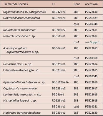


 Open access

