Introduction
In number of species, abundance and ecological diversity, fish of the genus Trematomus dominate the fauna of the High Antarctic shelf (DeWitt Reference DeWitt and Bushnell1971, Kock Reference Kock1992, Hureau Reference Hureau1994). Trematomus is composed of 11 species encompassing benthic, epibenthic and semipelagic lifestyles (Andriashev Reference Andriashev and Holdgate1970, Eastman Reference Eastman1993). Their radiation filled a wide range of habitats on the shelf and was probably the result of pelagization, an evolutionary trend whereby lineages originating from benthic ancestors have repeatedly evolved to live or at least feed in the water column (Klingenberg & Ekau Reference Klingenberg and Ekau1996). In ecomorphological analyses, Trematomus eulepidotus, T. lepidorhinus and T. loennbergii cluster as epibenthic species (Ekau Reference Ekau1988, Reference Ekau1991, Klingenberg & Ekau Reference Klingenberg and Ekau1996), living on or in close proximity to the bottom as documented by underwater photography and video (Ekau & Gutt Reference Ekau and Gutt1991, Gutt & Ekau Reference Gutt and Ekau1996). These species share similar body morphology, characterized by a streamlined appearance, moderate activity levels, absence of substrate contact adaptations and lower percentage weights in seawater than most other nototheniids (Eastman Reference Eastman1993, Wöhrmann Reference Wöhrmann, di Prisco, Pisano and Clarke1998). The most recent phylogenetic analysis of Trematomus, including 10 of 11 species, indicates that the genus is monophyletic and that T. lepidorhinus and T. loennbergii are sister species (Sanchez et al. Reference Sanchez, Dettaï, Bonillo, Ozouf-Costaz, Detrich and Lecointre2007). However T. eulepidotus is external to these species by several nodes and thus the epibenthic lifestyle was not attained by common ancestry, and the ecomorphological index of Ekau (Reference Ekau1988, Reference Ekau1991) does not reflect phylogeny but rather similar life styles that were independently acquired (Sanchez et al. Reference Sanchez, Dettaï, Bonillo, Ozouf-Costaz, Detrich and Lecointre2007).
Although the epibenthic species share a suite of morphological characters, they differ in diet, depth distribution and relative abundance. Dietary data indicate that T. eulepidotus is a zooplanktivore, preying primarily on euphausiids, hyperiids, copepods and pteropods, but occasionally on other crustaceans, polychaetes, chaetognaths and fish (Permitin & Tarverdieva Reference Permitin and Tarverdieva1978, Targett Reference Targett1981, Schwarzbach Reference Schwarzbach1988, Roshchin Reference Roshchin1991, Pakhomov Reference Pakhomov1997, Brenner et al. Reference Brenner, Buck, Cordes, Dietrich, Jacob, Mintenbeck, Schröder, Brey, Knust and Arntz2001). Conversely, T. loennbergii feeds mostly on benthic species, usually associated with the substrate, including polychaetes, isopods, decapods, amphipods, bivalves and fishes (Eastman Reference Eastman1985, Schwarzbach Reference Schwarzbach1988, La Mesa et al. Reference La Mesa, Vacchi, Castelli and Diviacco1997).
The epibenthic species of Trematomus have a circum-Antarctic distribution and occur sympatrically in the large shelf areas of the Weddell and Ross seas (DeWitt et al. Reference DeWitt, Heemstra, Gon, Gon and Heemstra1990). They exhibit reasonably disjunct depth distributions, with T. eulepidotus most common in shallow water, T. loennbergii in deep water, and T. lepidorhinus ranging from shallow to moderately deep water (Lannoo & Eastman Reference Lannoo and Eastman2000). Specific depth ranges for these three species in the Ross Sea are 130–344 m, 663–1191 m and 130–663 m, respectively (Eastman & Hubold Reference Eastman and Hubold1999, Lannoo & Eastman Reference Lannoo and Eastman2000). One species is usually more abundant than the others, perhaps because of these depth preferences (Eastman Reference Eastman1993). In the Weddell and Ross seas, for example, T. eulepidotus is the dominant epibenthic fish in shallower depths, whereas in deeper waters it is replaced by T. loennbergii (Ekau Reference Ekau1990, Hubold Reference Hubold1992, Eastman & Hubold Reference Eastman and Hubold1999, Vacchi et al. Reference Vacchi, Greco, La Mesa, Faranda, Guglielmo and Ianora1999a, Donnelly et al. Reference Donnelly, Torres, Sutton and Simoniello2004). Although the three species share the shelf habitat, they have different feeding strategies and depth preferences that overcome or partially mitigate interspecific competition.
Different reproductive strategies, consisting principally of a temporal mismatch in the spawning period (La Mesa et al. Reference La Mesa, Caputo and Eastman2006), also reduces interspecific competition among sympatric species of Trematomus in the Ross Sea. For example, the benthic species T. bernacchii and T. hansoni spawn, respectively, between October–December and January–February, whereas the semipelagic T. newnesi spawns between March–April (Dearborn Reference Dearborn1965, Shust Reference Shust, Skarlato, Alekseev and Liubimova1987, Vacchi et al. Reference Vacchi, Williams and La Mesa1996, La Mesa et al. Reference La Mesa, Caputo and Eastman2006). Most information on the reproductive biology of epibenthic species of Trematomus is from specimens collected in the Weddell Sea, and analyses have been based exclusively on macroscopic examination of gonads (Ekau Reference Ekau1991, Duhamel et al. Reference Duhamel, Kock, Balguerias and Hureau1993). These species have not been studied in the Ross Sea. Therefore, employing both macroscopic and histologic approaches, we: 1) investigate features of the reproductive biology of T. eulepidotus and T. loennbergii from the south-western Ross Sea, and 2) compare our findings with the literature to examine the hypothesis that these species employ a common reproductive strategy to avoid interspecific competition.
Materials and methods
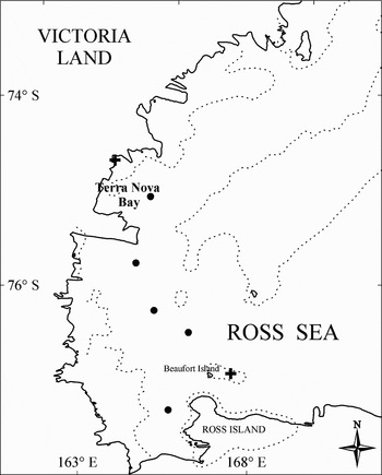
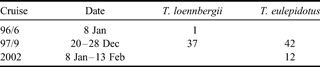
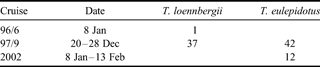
Samples of T. loennbergii were collected during cruises 96-6 and 97-9 of the RV Nathaniel B. Palmer in the south-western Ross Sea. Fish were caught at five stations between 663 and 1191 m depth (Fig. 1). Most specimens of T. eulepidotus were collected during cruise 97-9 near Beaufort Island at about 250 m depth (Fig. 1). All fish were caught by an otter trawl towed at 2–3 knots for 30–60 minutes (Eastman & Hubold Reference Eastman and Hubold1999). A few additional samples of T. eulepidotus were obtained off Terra Nova Bay between 8 January and 13 February 2002, during the XVII Italian Antarctic Expedition. Fish were caught at several stations located near the Italian station (74°41′42′′S, 164°07′23′′E), by trammel and gill nets set down to 100 m depth. Overall data on available fish samples are summarized in Table I.
Diagram of the south-western Ross Sea, showing sampling locations for Trematomus eulepidotus (crosses) and T. loennbergii (dots). Dotted line represents the 500 m isobath.

Number of fish samples available for the study.
In the laboratory, each specimen was measured for total length (TL, mm) and weighed (TW, g). After dissection, sex and stage of maturity were assessed on the basis of macroscopic appearance of gonads, according to the five point scale of maturity for nototheniid fish (Everson Reference Everson1977, Kock & Kellermann Reference Kock and Kellermann1991). After this procedure, the gonads were removed and weighed on an analytical balance with an accuracy of 0.01 g (Wg, g) and then stored in 70% alcohol.
As a measure of reproductive effort and stage of gonad maturity, the gonadosomatic index was calculated as the percentage of gonad to total body weight (GSI = 100 × Wg/TW) (Crim & Glebe Reference Crim, Glebe, Schreck and Moyle1990). To determine fish fecundity, all mature females (i.e. in stage 4 of macroscopic scale) were selected and their gonads processed as follows. A preliminary microscopic analysis indicated that the ovaries of these specimens exhibited a group-synchronous development, as two main cohorts of oocytes were easily distinguished by size (Wallace & Selman Reference Wallace and Selman1981, West Reference West1990). As a consequence, the gravimetric method was used to estimate fecundity (Murua et al. Reference Murua, Kraus, Saborido-Rey, Witthames, Thorsen and Junquera2003). According to this method, three subsamples of known weight (generally about 1 g each with an accuracy of 0.001 g) were sampled from three different parts of the ovary and the number of ripe oocytes counted under a stereomicroscope at low magnification. Hence, potential or absolute fecundity (F) was estimated applying the following relationship (Kock Reference Kock1989, Murua et al. Reference Murua, Kraus, Saborido-Rey, Witthames, Thorsen and Junquera2003):

where Wg is gonad weight, oi is the number of ripe oocytes in the ith subsample, wi is the weight of ith subsample and n is the number of subsamples. A chi-square test revealed no significant differences (P > 0.05) for numbers of ripe oocytes per gram (oi/wi) among different parts of the ovary.
The relative fecundity (Fr) was calculated as number of oocytes per gram of total body weight (TW). The relationship between fish size and absolute/relative fecundity was investigated applying linear regression analysis (Kartas & Quignard Reference Kartas and Quignard1984). Finally, in each mature female, the mean size of ripe oocytes was assessed by measuring the maximum diameter of 10 oocytes taken randomly from the subsamples used to estimate fecundity.
Based on the stage of macroscopic maturity of gonads, the proportion of individuals in stages 3–5 for each centimetre size class was recorded. The length at first spawning (Lm50), representing the length at which 50% of the population was mature (namely at stage 3–5, Kock Reference Kock1989), was determined by fitting a logistic equation to the proportion of fish in each size class (Ni & Sandeman Reference Ni and Sandeman1984):

where p is the estimated proportion in a size class, TL is the total length (mm) and α and β are coefficients. The values of coefficients were obtained by linearizing the equation through log-transformation of both terms:

Hence, the length at first spawning (Lm50) was estimated as the negative ratio of coefficients, -α/β, by substituting p = 0.5 in the linear equation. Unfortunately, the few immature specimens available in our sample did not allow estimation of the length at sexual maturity (Kock Reference Kock1989).
As far as the histological analysis is concerned, whole or subsamples of gonads were removed from each specimen and fixed in Bouin's solution for 12 hours. The samples were subsequently dehydrated in ethanol and embedded in paraffin. A series of cross-sections 10 µm thick were obtained from each sample, mounted on slides and stained with Mayer's haematoxylin-eosin following standard procedure (Beccari & Mazzi Reference Beccari and Mazzi1972). Sections were examined with a Nikon Eclipse 800 optical microscope at magnifications of 40–400x. Measurements of oocytes were made using the Nikon software package Lucia 4.51.
Ovarian follicles were classified according to six development stages: I = chromatin nucleolar, II = perinucleolar, III = cortical alveoli formation, IV = vitellogenic, V = mature, VI = postovulatory follicle (Wallace & Selman Reference Wallace and Selman1990, West Reference West1990).
The relative frequency of oocytes at different histological maturity stage was obtained pooling fish in the same stage of macroscopic maturity. In addition, the size frequency distribution of oocytes was determined for each of the histological maturity stages described.
As females exhibit group synchronous ovaries (see above), each specimen was staged on the basis of the most advanced stage of development observed in the ovary sections. For each stage of development, cellular and nuclear diameters (µm) were measured on 20 oocytes (if not otherwise indicated) and nucleoplasmic indices (NP) were calculated as follows:

where Vn is the nuclear volume and Vc is the cellular volume.
In males, the spermatogenic activity was assessed on the basis of the different types of gametocytes (from spermatogonia to spermatozoa) in the seminiferous lobules of each testis, and also the presence of spermatogonial mitoses. Maturity of testes was evaluated according to a five-point scale (Billard Reference Billard1986): I = immature stage (presence of spermatogonia and spermatogonial mitoses), II = early development stage (first meiotic division), III = advanced development stage (second meiotic division), IV = mature stage (presence of spermatozoa cysts), V = post-reproductive stage (presence of collapsed lobules and residual spermatozoa). Note that macroscopic and histologic stages of development are numbered with Roman and Arabic numerals, respectively.
Results
Trematomus eulepidotus
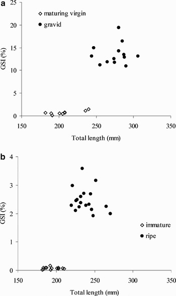
Overall, 23 females ranging between 182–306 mm TL and 47–327 g TW and 31 males ranging between 182–271 mm TL and 40–208 g TW were analysed. Applying the chi-square test for goodness of fit, the sex ratio was not significantly different from unity (χ2 = 1.18, df = 1, P > 0.1). On the basis of the macroscopic appearance of gonads, 10 females between 182–240 mm TL were considered to be maturing virgins (stage 2), with a GSI range between 0.2–1.4%. All other females (13 specimens) were gravid (stage 4), attaining a GSI between 11.0–19.4%. As for males, 13 specimens were relatively small (182–210 mm TL) and immature (stage 1), with a GSI range of 0.03–0.1%. All other males (18 specimens) were ripe (stage 4), exhibiting a GSI range between 1.9–3.6%. In plots relating GSI to fish size, both sexes showed an obvious gap between different stages of gonad maturity (Fig. 2a & b).
Plot of gonadosomatic index (GSI) in relation to fish size and stage of gonad maturity for Trematomus eulepidotus a. females, and b. males.

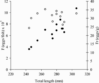
The length at first spawning (Lm50) of T. eulepidotus estimated by fitting the logistic function to the length-at-maturity data was, respectively, 210 mm TL in males and 240 mm TL in females. The absolute fecundity (F), estimated from 13 females ranging from 244 mm TL to 306 mm TL, varied between 3550–10736 eggs per individual (mean ± SE, 6637 ± 535) (Fig. 3). A positive relationship was found between F and fish size, as fecundity increased significantly with increasing length of females (F = -19331.5 + 94.4 TL, n = 13, r 2 = 0.76). Relative fecundity (Fr) in T. eulepidotus ranged between 24.3 and 35.3 eggs/g (mean ± SE, 30.8 ± 0.9) (Fig. 3), with little relationship with fish size.
Pattern of distribution of potential fecundity (F, •) and relative fecundity (Fr, ○) in relation to fish size for T. eulepidotus.

Size of mature oocytes, as measured in gravid females (stage 4) ranged widely between 1.9 mm and 2.7 mm (mean size 2.0–2.5 mm). However, on the basis of size distribution, mature oocytes were easily distinguished from all other previtellogenic oocytes dispersed within the ovarian stroma (see below), since there were few and they were of considerably smaller size (0.2–0.6 mm).
Trematomus loennbergii
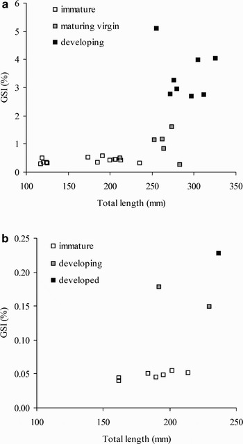
A total of 37 specimens were studied, with 27 females ranging between 117–326 mm TL and 7–278 g TW and 10 males ranging between 162–237 mm TL and 23–88 g TW. The sex ratio differed significantly from unity (χ2 = 7.81, df = 1, P < 0.005). On the basis of the macroscopic gonad maturity scale, 14 females between 117–235 mm TL were immature (stage 1), with a GSI range of 0.29–0.56%, whereas five females ranging between 253–283 mm TL were maturing virgins, attaining a GSI of 0.84–1.59%. All other specimens (eight), measuring between 255–326 mm TL, were in the developing stage (stage 3), exhibiting an increased value of the GSI (2.70–5.10%). Most males (seven) were small, ranging between 162–214 mm TL, and immature (stage 1), with a GSI of 0.04–0.05%. Of the other males investigated, two fish between 192–230 mm TL in resting stage (stage 2, GSI of 0.15–0.18%), whereas only one fish was developed (stage 3), with a GSI of 0.23%. The GSI-fish size relationship for males and females, as summarized in Fig. 4a & b, shows an increasing pattern of GSI across the stages of gonad maturity. The length at first spawning (Lm50) in females, estimated by fitting the logistic function to the length-at-maturity data, was c. 26 cm TL. The lack of males in advanced stage of maturity (only one in stage 3) did not allow evaluation of Lm50. Similarly, the absence of mature females did not enable us to determine the total and relative fecundity for this species as well. Indeed, when an ovary is sampled too early in the season (i.e. not in mature females), it may not be sufficiently developed to allow the identification of all oocyted destined to be spawned, providing a biased estimate of fecundity (Murua et al. Reference Murua, Kraus, Saborido-Rey, Witthames, Thorsen and Junquera2003).
Plot of gonadosomatic index (GSI) in relation to fish size and stage of gonad maturity for Trematomus loennbergii a. females, and b. males.

Histological analysis Trematomus eulepidotus
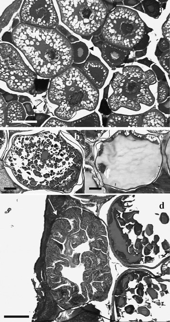
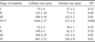
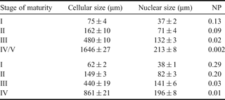
Histology revealed that females encompassed two development stages. Gonads of small fish (180–240 mm TL) were made up of oocytes in three stages of development, chromatin nucleolar (stage I), perinucleolar (stage II) and cortical alveoli formation (stage III) (Fig. 5a). With maturation, cellular size increased progressively, while the nucleoplasmic ratio decreased (Table II). All other females had relatively large ovaries, filled with oocytes in late vitellogenesis (stage IV–V) and few oocytes in the previtellogenic and endogenous vitellogenic stages (II and III respectively). The cytoplasm of large yolked oocytes was completely filled by yolk granules (Fig. 5b), which in some cases were fused giving these oocytes their characteristic transparency (Fig. 5c). Occasionally, yolked oocytes underwent resorption through atretic processes (Fig. 5d). No postovulatory follicles were recorded at this stage. The size-frequency distribution of oocytes in different stages of histological maturity was markedly discontinuous (Fig. 6), confirming the presence of group-synchronous ovaries in this species.
Photomicrographs of histological sections of Trematomus eulepidotus ovarian tissue. a. Maturing virgin female, with ovaries filled mainly with oocytes at cortical alveoli formation stage. Presence in the ovarian stroma of several previtellogenic oocytes, respectively, at chromatin nucleolar (arrow) and perinucleolar (arrowhead) stages. b. Gravid female, oocyte in late vitellogenesis, with cytoplasm completely filled by yolk granules. c. Late vitellogenic oocyte, in which yolk granules are fused together giving the characteristic transparency. d. Atretic oocyte (arrow), with chorion convoluted and invaded by granulosa cells. Scale bars 250 µm in all photos.

Relative frequency and size frequency distribution of oocytes at different histological stages of maturity in Trematomus eulepidotus. I = chromatin nucleolar, II = perinucleolar, III = cortical alveoli formation, IV/V = late vitellogenic, at = atretic oocytes.

Morphometry of oocytes at different stages of oogenesis (mean ± standard error) and nucleo-plasmic index (NP) in Trematomus eulepidotus (above) and T. loennbergii (below) from the Ross Sea. See text for description of each stage of maturity.
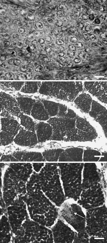
Males were in three different stages of maturity. All specimens smaller than 210 mm TL were immature (stage I), showing testes uniformly occupied by spermatogonial cysts (Fig. 7a), with evidence of spermatogonial mitoses. All other fish showed active spermatogenesis, although they were at different stages of development. At the earlier stage, testicular lobules consisted of cysts filled with spermatocytes I and II (stage III) (Fig. 7b), whereas in more advanced stages testes had cysts of spermatozoa (Fig. 7c).
Photomicrographs of histological sections of Trematomus eulepidotus testicular tissue. a. Immature male, characterized by testes made up by spermatogonial cysts with evidence of mitotic activity. Scale bar 10 µm. b. Early ripe male, showing testicular lobules with cysts composed of spermatocytes I and II. Scale bar 25 µm. c. Ripe male, with a cyst of spermatozoa beginning to fill the testicular lumen (arrow). Scale bar 25 µm.

Trematomus loennbergii
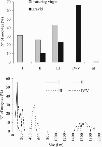
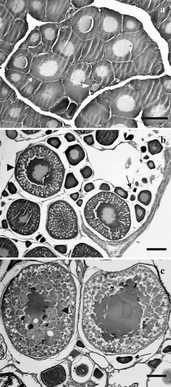
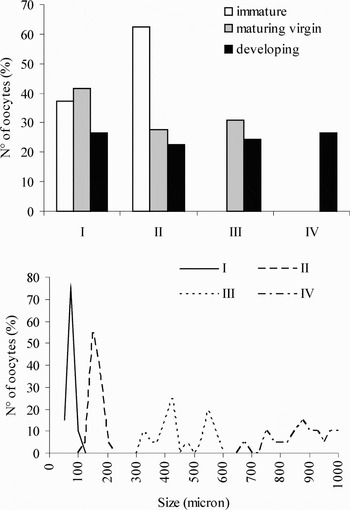
Small females ranging from 117–235 mm TL were immature, with ovarian follicles composed only of previtellogenic oocytes (chromatin nucleolar and perinucleolar, stages I and II) (Fig. 8a). In some larger specimens, batches of oocytes had undergone endogeneous vitellogenesis (cortical alveoli or stage III), although ovaries still contained previtellogenic oocytes (Fig. 8b). All other females had ovaries with large oocytes in early exogeneous vitellogenesis (stage IV), consisting of cytoplasm filled by yolk granules and a central nucleus (Fig. 8c). At this stage of development, several previtellogenic and endogeneous vitellogenic oocytes were still present (Fig. 8c), probably forming the reserve stock for the next spawning season. This is also supported by the wide gap in cellular sizes among oocytes in different stages of development (Table II), as is also the case in T. eulepidotus. Finally, the relative frequency and the size frequency distribution of oocytes is provided in Fig. 9.
Photomicrographs of histological sections of Trematomus loennbergii ovarian tissue. a. Immature female, with ovarian follicles consisting of only previtellogenic oocytes, respectively, at chromatin nucleolar (arrowhead) and perinucleolar (arrow) stages. Scale bar 100 µm. b. Maturing virgin female, with several previtellogenic oocytes (arrow) and few oocytes in endogenous vitellogenesis or cortical alveoli stage (arrowhead). Scale bar 200 µm. c. Developing female, showing two oocytes in active exogenous vitellogenesis (arrowhead), characterized by yolk granules filling the whole cytoplasm and a pale central nucleus, as well as previtellogenic oocytes (arrow). Scale bar 200 µm.

Relative frequency and size frequency distribution of oocytes at different histological stages of maturity in Trematomus loennbergii. I = chromatin nucleolar, II = perinucleolar, III = cortical alveoli formation, IV = vitellogenic

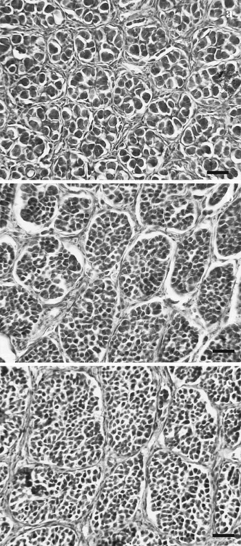
Most male T. loennbergii were immature, exhibiting testicular lobules made up of cysts of spermatogonia (Fig. 10a). All other specimens had testes at an early development stage (spermatocytes I or stage II) (Fig. 10b), except for a single fish with testicular lobules filled by germinal cells in the second meiotic division (spermatocytes II), or in the late development stage (stage III) (Fig. 10c).
Photomicrographs of histological sections of Trematomus loennbergii testicular tissue. a. Immature male, with testes filled exclusively by cysts of spermatogonia. b. Resting male, showing testicular lobules with cysts composed of spermatocytes I. c. Developed male, showing testicular lobules with spermatocytes II. Scale bars 25 µm in all photos.

Discussion
As is the case for most nototheniids (Everson Reference Everson and Laws1984, North & White Reference North, White, Kullander and Fernholm1987, Kock & Kellermann Reference Kock and Kellermann1991, Duhamel et al. Reference Duhamel, Kock, Balguerias and Hureau1993), gametogenesis in both epibenthic species investigated in the present study is a slow process, starting before the current spawning season and continuing until the next spawning season one year later. Indeed, maturing or gravid females of both Trematomus species are characterized by having group synchronous ovaries (sensu Wallace & Selman Reference Wallace and Selman1981), in which the more advanced batch of vitellogenic (T. loennbergii) or mature (T. eulepidotus) oocytes that will be ovulated in the current spawning season are widely separated in size from the smaller previtellogenic and endogenous vitellogenic oocytes, which form the reserve stock for the next spawning season. As a result, spawning occurs once a year from sexual maturity onward (or iteroparity), as is generally reported in Antarctic fish (Vanella et al. Reference Vanella, Calvo, Morriconi and Aureliano2005). On the basis of this pattern of oocyte maturation, females of both species probably release their mature eggs in a single spawning event or over a short period of time, and can be considered total spawners (Holden & Raitt Reference Holden and Raitt1974). This is also supported by the lack of postovulatory follicles in mature females with mature oocytes. When present, they indicate the species is a fractional spawner (Van der Molen & Matallanas Reference Van der Molen and Matallanas2004). As a consequence, the relatively prolonged spawning season observed in both species (see below) is mostly due to a probable size-related difference in spawning time within local population.
Considering our results on length at first spawning and literature data (DeWitt et al. Reference DeWitt, Heemstra, Gon, Gon and Heemstra1990), T. eulepidotus and T. loennbergii probably spawn for the first time relatively late in their life, i.e. at about 70–75% of their maximum length. A similar percentage was also reported for the other epibenthic species of Trematomus, T. lepidorhinus (Kock & Kellermann Reference Kock and Kellermann1991). Such a delayed sexual maturity is a common feature in Antarctic fishes. The reproductive effort, in terms of GSI, is generally very high, reaching 20–30% in females of T. eulepidotus (see Table III). Thus, delayed maturity is probably the result of either the considerable energy spent during spawning or the advantage of postponing sexual maturity until achieving a size sufficiently large to feed on the most abundant food resources available in shelf waters.
Reproductive characteristics of Trematomus eulepidotus in the High-Antarctic Zone.

The reproductive characteristics of T. eulepidotus are relatively well known, although most data are based only on macroscopic analysis of gonads. Most information on the reproductive biology of this species is based on specimens collected from the Weddell Sea (Lisovenko Reference Lisovenko, Skarlato, Alekseev and Liubimova1987, Ekau Reference Ekau1988, Reference Ekau1989, Reference Ekau1991, Duhamel et al. Reference Duhamel, Kock, Balguerias and Hureau1993) and from Davis and Mawson seas (Roshchin Reference Roshchin1991, Shandikov & Faleeva Reference Shandikov and Faleeva1992), where T. eulepidotus is the most common species amongst nototheniids collected both by benthic and midwater trawls. Additional data on absolute and relative fecundity of this species is from an Elephant Island population (Kock Reference Kock1989). Summarizing this data (Table III), it appears that T. eulepidotus spawns in the summer over an extended period lasting from December–March, and produces 2500–20 000 eggs per season depending on fish size. Interestingly, a batch of about 2000 eggs of T. eulepidotus were collected in the Weddell Sea on the bottom at 200 m (Ekau Reference Ekau1989), confirming the assumption that the eggs of Trematomus are demersal.
Unlike T. eulepidotus, knowledge of the reproductive biology of the other epibenthic Trematomus species is relatively scarce and restricted to the Weddell Sea (Ekau Reference Ekau1991, Duhamel et al. Reference Duhamel, Kock, Balguerias and Hureau1993). In particular, T. loennbergii spawns about 6000–13 000 eggs per season, whereas T. lepidorhinus produces 2200–20 000 eggs per season.
In general the epibenthic Trematomus species exhibit an overall similarity in some reproductive features, including delayed maturity, prolonged gametogenesis, group synchronous oocyte development, relatively low fecundity and a single spawning event per season. However, the reproductive strategy, in terms of spawning period, differs markedly among the species, producing a mismatch in the time of appearance of larvae in the environment. The present study and literature data indicate that T. eulepidotus is a summer spawner, maturing in summer between December and March (Table III), whereas T. loennbergii and T. lepidorhinus spawn, respectively, in autumn (Ekau Reference Ekau1991) or in winter (Kock & Kellermann Reference Kock and Kellermann1991, Duhamel et al. Reference Duhamel, Kock, Balguerias and Hureau1993). Accordingly, the timing of hatching of these species, which share the same habitat as adults, shows a chronological sequence. Hatching of T. eulepidotus take place in May–June in the Weddell Sea (Ekau Reference Ekau1989, Loeb et al. Reference Loeb, Kellermann, Koubbi, North and White1993), when yolk sac larvae attain 10–15 mm SL. On the other hand, hatching of T. loennbergii could be hypothesized to occur in spring (September–October), as some postlarvae ranging from 20–23 mm SL have been caught in the Ross Sea in late December (Vacchi et al. Reference Vacchi, La Mesa and Greco1999b). Finally, T. lepidorhinus hatches in November–December, both in the Ross Sea (Vacchi et al. Reference Vacchi, La Mesa and Greco1999b) and off the Antarctic Peninsula (Kellermann Reference Kellermann1990).
In conclusion, during the evolutionary process of pelagization, the epibenthic species of the genus Trematomus developed reproductive and other strategies which serve to mitigate interspecific competition. Ecologically, they differ both in feeding habits and depth preferences, factors probably linked to each other. Although they share a common process of gonadal maturation, they differ in the timing of spawning and larval hatching, consistent with avoiding food competition among the early life history stages.
Acknowledgements
This study was financially supported by the PNRA (Italian National Antarctic Research Program). JTE was supported by National Science Foundation grant ANT 04-36190. We are very grateful to two anonymous referees for their helpful comments.