Case report
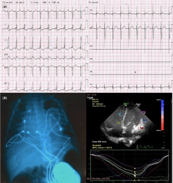
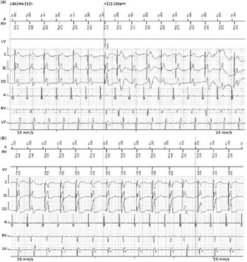
We present the case of a preterm girl (34 weeks of gestation, birthweight 1650 g) from a twin pregnancy obtained through in vitro fertilisation. At birth, the child had a heart rate of 90 bpm and was hypoxic with an oxygen saturation of 80%. The electrocardiogram revealed a Wenckebach atrio-ventricular block and the echocardiogram identified tetralogy of Fallot with hypoplastic pulmonary annulus but without anterograde pulmonary gradient shortly after birth. In the subsequent weeks, the pulmonary stenosis became important, with a maximum anterograde gradient of 108 mmHg. Four weeks after birth, the conduction disorder progressed to high-degree atrio-ventricular block with a ventricular rate of 50 bpm and, at a weight of 2380 g, a single chamber VVI pacemaker (Medtronic Sensia) was implanted through left thoracotomy, with a bipolar epicardial lead (Medtronic CapSure Epi) sutured on the apical-lateral wall of the left ventricle and the patient was discharged 1 week later, with a left ventricular ejection fraction of 50%. At the age of 4 months, the patient was admitted for severe hypoxic spells while on betablocker and a modified Blalock–Taussig shunt was performed using a 4-mm Goretex graft. One week later, the patient developed severe low-output syndrome and invasive mechanical ventilation together with inotropic treatment was necessary. On echocardiography, the shunt was permeable but the left ventricular systolic function was severely decreased with an ejection fraction of 25%, there was intraventricular (septal-to-posterior wall motion delay of 150 ms) and interventricular dyssynchrony (difference between pre-ejection times of the two ventricles 107 ms), severe mitral regurgitation (atrio-ventricular dyssynchrony) and left ventricular volume overload due to increased pulmonary return. The patient was started on adrenaline and milrinone infusion with persistent low output in the following 2 weeks, with a maximum creatin kinase-myocardial band of 287 U/l, lactate dehydrogenase 7033 U/l and aspartat aminotransferase 3809 U/l. In this context, given the impossibility to downscale the inotropic treatment, severe left ventricular dysfunction and intraventricular dyssynchrony, we opted for complete surgical correction of the congenital anomaly and upgrade to a triple-chamber pacemaker during the same procedure. The patient underwent surgery at the age of 5 months and 4000 g weight. The pulmonary annulus was enlarged with a transannular patch, the Blalock–Taussig shunt was interrupted and the ventricular septal defect was closed with a patch. Two additional bipolar epicardial leads were implanted, one on the right atrium and the other one on the right ventricle at the border between right ventricular outflow and inflow, and all three leads were connected to a triple chamber pacemaker (Biotronik Enitra 8 HF-T), placed subcutaneously above the right rectus abdominal muscle (Fig 1b). The pacemaker was programmed as DDD 110 bpm, with a sensed atrio-ventricular delay of 70 ms and a paced atrio-ventricular delay of 90 ms, interventricular (VV) delay 0 ms, optimised based on ECG and echocardiography (Fig 1a and c). Twenty-four hours after surgery, the patient had sustained ventricular tachycardia, resistant to overdrive pacing for which first lidocaine boluses, followed by mexiletine, and amiodarone were needed, with suppression of arrhythmia episodes after 3 days. All inotropic treatment (adrenalin, noradrenalin, milrinone) was stopped after 1 week, and the patient was haemodynamically stable, with a left ventricular ejection fraction of 45%, mild mitral regurgitation and a normal transmitral flow. During the following days, two important device-related events occurred. In the context of an infection with Klebsiella, the patient had sinus tachycardia interpreted as pacemaker-mediated tachycardia by the device, which correctly started the pacemaker-mediated tachycardia pacing protocol (Fig 2a), therefore we inactivated pacemaker-mediated tachycardia suppression function. Second, we observed that at a heart rate above 160 bpm, the pacemaker switched to right ventricular only pacing, losing the benefit of resynchronisation (Fig 2b). Unfortunately, this issue could not be solved through device programming, therefore, besides antibiotic treatment, we opted for propranolol and then ivabradine was associated in order to obtain sinus rates below 160 bpm. The evolution was favourable and the patient was evaluated 1 month, 6 months and 1 year after discharge, in good clinical status, with normal growth according to age and an improved left ventricular ejection fraction of 70% and mild mitral regurgitation.
( a ) Electrocardiogram after cardiac resynchronization therapy. ( b ) Thoracic x-ray after device implantation. ( c ) Apical four-chamber view with speckle tracking after resynchronization with optimal timing of contraction.

Device recorded intracardiac electrograms during (a) pacemaker mediated tachycardia supression protocol and (b) loss of left ventricular pacing at rates higher than 160 bpm

Discussion
Cardiac resynchronisation therapy is now a widely used therapy in adult patients with decreased left ventricular ejection fraction and bundle branch block, recommended for heart failure therapy after 3 months of optimal medical treatment. The use of cardiac resynchronisation therapy in paediatric patients is a lot less standardised as there is a great anatomical heterogeneity. Three larger studies on cardiac resynchronisation therapy in paediatric patients Reference Janoušek, Gebauer and Abdul-Khaliq1–Reference Cecchin, Frangini and Brown3 have shown that ventricular dyssynchrony due to single site pacing was the most frequent indication for cardiac resynchronisation therapy (44–77%), but in these cohorts the number of children with tetralogy of Fallot was low (<10%). At the same time, a decrease in LVEF was observed in less than 10% of children paced from the left ventricular apex or lateral wall. Reference Janoušek, van Geldorp and Krupičková4 Nevertheless, our patient developed biventricular dysfunction most probably both due to volume overload in uncorrected Fallot and left ventricular dyssynchrony due to pacing. In this context, as inotropic treatment could not be stopped, CRT at the time of total correction was considered the optimal solution. The greater challenge derived from cardiac resynchronisation therapy programming, as the software of triple chamber pacemakers is not adapted to paediatric heart rates. Therefore, even if sinus tachycardia at a rate of 160 bpm is not unusual in an infant, the pacemaker cannot pace using the left ventricular lead at that rate, so interventricular synchrony is lost. In this case, even if there was intermittently preserved atrio-ventricular conduction, Reference Bogdan and Glikson5 this was associated with bradycardia, so we aimed for permanent cardiac resynchronisation therapy pacing and we did not try to preserve intrinsic rhythm. In this context, balancing between physiologic tachycardia and the need to optimise cardiac resynchronisation therapy can be hard, and using ivbradine could be explored as an option. Further, cardiac resynchronisation therapy programming in children is a challenge not only due to physiology particularities but also due to technological limitations.
Acknowledgements
None.
Financial support
This case report received no specific grant from any funding agency, commercial or not-for-profit sectors.
Conflicts of interest
None.