Introduction
The genus Pseudocyphellaria (Lobariaceae, lichenized Ascomycota) is among the best known lichen genera in the Southern Hemisphere (Galloway Reference Galloway1986, Reference Galloway1992). However, during the past decades molecular evidence suggests that the genus, as traditionally understood, is polyphyletic (Miądlikowska & Lutzoni Reference Miądlikowska and Lutzoni2000, Reference Miądlikowska and Lutzoni2004; Thomas et al. Reference Thomas, Ryan, Farnden and Galloway2002; Stenroos et al. Reference Stenroos, Stocker-Wörgötter, Yoshimura, Myllys, Thell and Hyvönen2003; Wiklund & Wedin Reference Wiklund and Wedin2003). More recently, Moncada et al. (Reference Moncada, Lücking and Betancourt-Macuase2013, Reference Moncada, Reidy and Lücking2014) focused on the phylogeny of Lobariaceae, and confirmed the polyphyletic origin of Pseudocyphellaria. Furthermore, discrepancies among some species within the genus remain unresolved; many of them are related to the retention of incorrect names or uncertain taxonomic interpretations.
Some of these problems might be solved by studying more material, allowing for a better knowledge of intraspecific variation. One example of this concerns the southern South American endemic species, Pseudocyphellaria pilosella Malme and P. piloselloides (Räsänen) H. Magn., characterized by a foliose thallus, cyanobacterial photobiont, yellow medulla and pseudocyphellae, and tomentose upper surface. These two species were originally conceived as similar by Räsänen (Reference Räsänen1932), differing only in their reproductive mode (fertile vs. sorediate). Later, Galloway (Reference Galloway1986) cited both as counterparts. However, their diagnostic features and overlapping geographical distribution led us to reconsider whether these are well-defined species. Accordingly, we examined the morphological, anatomical, chemical, and molecular characters in the complex.
Materials and Methods
Specimens studied
Specimens of Pseudocyphellaria pilosella and P. piloselloides were collected by the authors in 2014 during field studies in Nothofagus forest in north-western Patagonia, Argentina. All the lichen material was deposited at BCRU.
In order to increase the sampling area, additional specimens from Llao-Llao (Río Negro Province, Argentina), Parque Nacional Puyehue and Parque Nacional Queulat (X and XI Regions, Chile) provided by S. Calvelo (hb. SC) and T. Wheeler together with P. Nelson (hb. Wheeler) respectively, were also included; within these were collections which exhibit intermediate forms, with both soredia and apothecia.
Digital images of the type specimens provided by the S and H herbaria were compared with the material collected from Argentina and Chile.
Morphological and anatomical observations were performed following standardized methods used in lichenology. Secondary chemistry of the material was studied with high performance thin-layer chromatography (HPTLC) using methods described by Arup et al. (Reference Arup, Ekman, Lindblom and Mattsson1993). Photomicrographs were taken with a digital camera USB 2.0.
Molecular studies
Total genomic DNA of P. pilosella and P. piloselloides was extracted from a total of 12 collections using the commercially available Ultraclean Microbial DNA isolation kit (Mo Bio Laboratories, Carlsbad, USA) according to the manufacturer’s protocols, and resuspended in 50 µl of ultrapure water. The internal transcribed spacer regions (ITS, including 5.8S) of nuclear ribosomal DNA were sequenced and used in phylogenetic analysis. The ITS regions were amplified with the following pair of fungal-specific primers: ITS1-F (Gardes & Bruns Reference Gardes and Bruns1993) and ITS4 (White et al. Reference White, Bruns, Lee and Taylor1990). The PCR reactions were performed in a 25 µl PCR mixture containing 5× GoGreen Buffer (5 µl), 10 mM of each PCR nucleotide mix (1·6 µl), 10 µM of upstream (1 µl) and 10 µM downstream primer (1 µl), 5 u/µl GoTaq DNA polymerase (Promega, USA) (0·125 µl), 2 ng of template DNA and nuclease-free water to final volume (25 µl). PCR amplification was carried out in a Veriti 96-well thermal cycler (Applied Biosystems Inc., USA) using the following profile: one cycle of 2 min at 94 °C; 35 cycles of 1 min at 94 °C, 55 s at 53 °C, 3 min at 72 °C; one cycle of a final extension for 10 min at 72 °C. DNA fragments were detected by staining with SYBR® Safe DNA gel stain (Invitrogen, Life Technologies, USA). Amplicons generated by PCR were sequenced by the Macrogen Inc. (Korea) sequencing service.
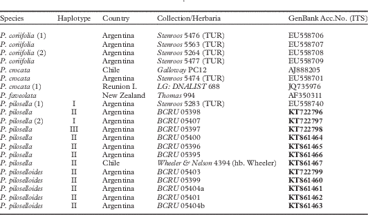
Sequences of three Pseudocyphellaria species were used as outgroups: two of them, P. coriifolia (Müll. Arg.) Malme and P. crocata (L.) Vain., are closely related to P. pilosella, and the third one, P. faveolata (Delise) Malme, is more distantly related, according to Moncada et al. (Reference Moncada, Reidy and Lücking2014) and the MycoBank website (http://www.mycobank.org/BioloMICSSequences.aspx?expandparm=f&file=all). Sequences of these species were downloaded from GenBank. The matrix consisted of four individuals of P. coriifolia, three of P. crocata, one of P. faveolata, eight of P. pilosella, and five of P. piloselloides (Table 1). Sequences were aligned and trimmed manually in Aliview (Larsson Reference Larsson2014). Genetic distances were calculated as uncorrected P-values in R (R Development Core Team 2011) with the seqinR package (Charif & Lobry Reference Charif and Lobry2007). Intermediate forms were not amplified due to low quality DNA.
List of the species, specimens, collection data and accession numbers for the marker ITS of Pseudocyphellaria used in the estimation of genetic distances and phylogenetic analyses. Newly obtained sequences for this study are in bold. Numbers in parentheses indicate specimens used in the calculation of genetic distances
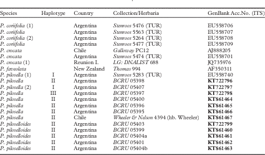
The phylogenetic reconstruction was carried out with maximum likelihood criterion using RAxML (Stamatakis Reference Stamatakis2014) with a Gamma model of rate heterogeneity. Equal sequences were collapsed and only one representative was left for the analysis to avoid redundancy. Branch support was attained through non-parametric bootstrap with 1000 replicates.
Results and Discussion
Molecular analyses
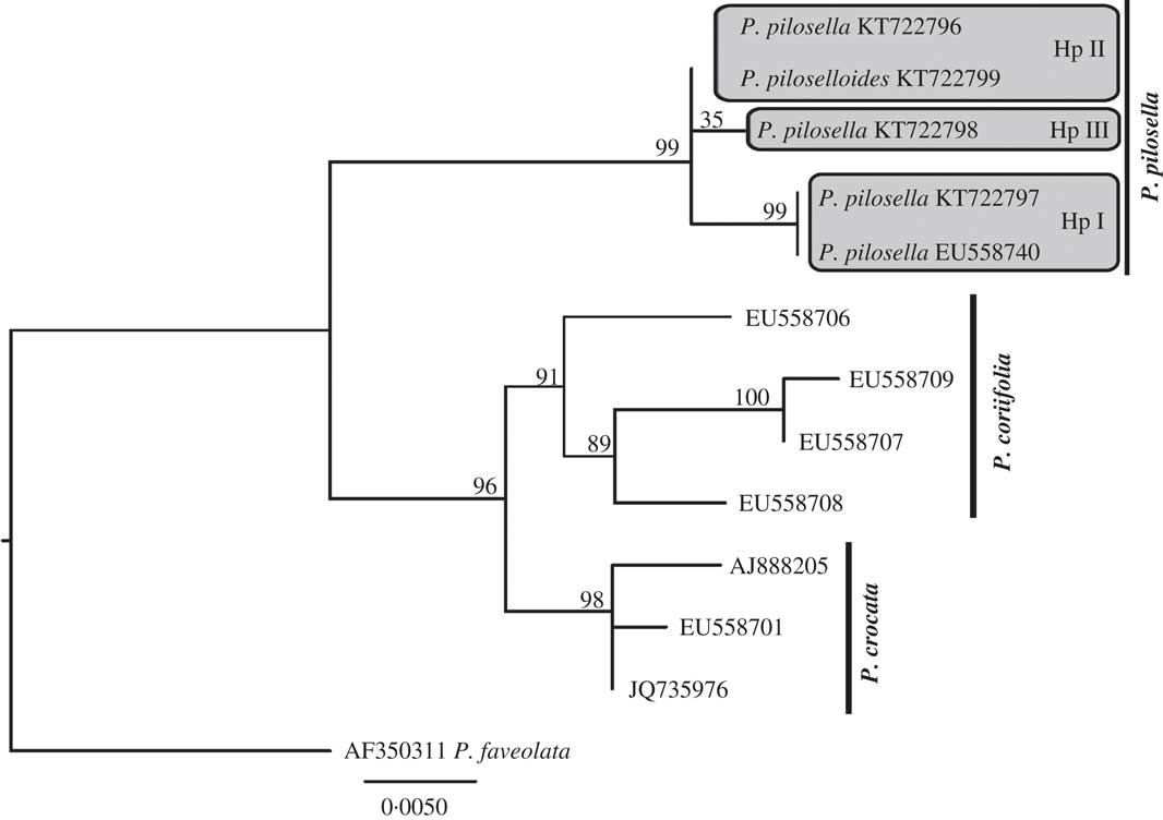
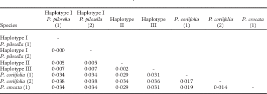
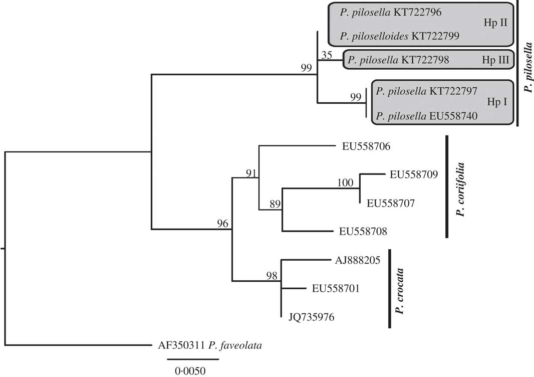
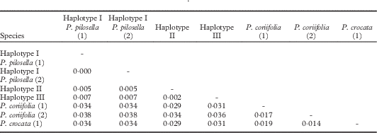
The total length of the ITS alignment consisted of 420 characters. In the maximum likelihood tree, Pseudocyphellaria coriifolia and P. crocata are sister taxa (with 96% bootstrap support) and both are monophyletic (with 91% and 98% bootstrap support respectively). All individuals of P. pilosella and P. piloselloides form a clade with 99% bootstrap support (Fig. 1). The 13 sequences of “P. pilosella-piloselloides” analyzed were grouped in three haplotypes (Table 2 and Fig. 1). Haplotype I was found in two individuals, both identified as P. pilosella; Haplotype II, represented in Fig. 1 by only two sequences, was found in 10 individuals, five identified as P. pilosella and five as P. piloselloides; Haplotype III includes only one individual, identified as P. pilosella (Table 1). The pairwise genetic distance between the three haplotypes ranges from 0·002–0·007 (Table 2), where the number of site differences between each pair of sequences in the alignment ranges from 1–4. When the sequences of these individuals of P. pilosella and P. piloselloides were compared with sequences from individuals of P. coriifolia and P. crocata the site differences between each pair of sequences in the alignment increased to a minimum of eight sites. The pairwise genetic distance between species used in this study ranges from 0·029–0·038. The average genetic distance within P. coriifolia was higher than that between P. pilosella and P. piloselloides (Table 2). Consequently, the phylogenetic results, together with low genetic distances of ITS sequences found between P. pilosella and P. piloselloides, confirm that they should be regarded as a single species.
Phylogenetic relationships of Pseudocyphellaria pilosella and P. piloselloides. Maximum likelihood tree based on ITS data with bootstrap branch support above branches. Haplotypes I, II, III (Hp I, Hp II, Hp III) are indicated in grey boxes.
Genetic distances of ITS sequences calculated as uncorrected P-values for haplotypes of Pseudocyphellaria pilosella and P. piloselloides and a comparison with outgroup species. See Table 1 for numbers in parentheses
Taxonomy
Pseudocyphellaria pilosella Malme
Bih. K. svenska VetenskAkad. Handl., ser. 3, 25 (6): 30 (1899); type: Argentina, Puerto Blest, ad lacum Nahuel Huapi, 1897, P. Dusén 180 (S!— holotype).
Pseudocyphellaria piloselloides (Räsänen) H. Magn., Acta Horti gothoburg 14: 7 (1940).—Cyanisticta piloselloides Räsänen, Ann. Bot. Soc. Zool.-Bot. Fenn. “Vanamo” 2 (1): 39 (1932); type: Chile, Fuegia, Fjordo Finlandia, Berberis ilicifolia, 28 February 1929, H. Roivainen (H!—lectotype).
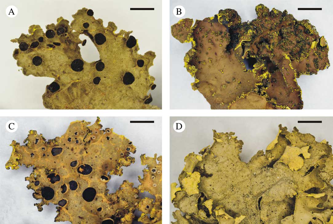
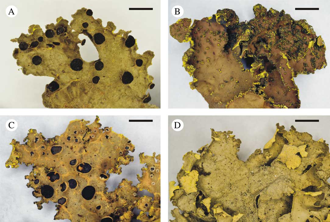
Thallus variability in Pseudocyphellaria pilosella. A, Argentina, Provincia Neuquén, Puerto Blest, M. I. Messuti et al. (BCRU 05407) (apotheciate thallus); B, Chile, Parque Nacional Puyehue, along trail to Volcán Puyehue, T. Wheeler & P. Nelson 5820 (hb. Wheeler) (sorediate thallus); C, Chile, Parque Nacional Queulat, T. Wheeler & P. Nelson 7082 (hb. Wheeler) (apotheciate and sorediate thallus); D, Argentina, Provincia Río Negro, Puerto Blest, M. I. Messuti et al. (BCRU 05400) (sterile and non-sorediate thallus). Scales: A–D=5 mm.
For a detailed description of the morphology and anatomy see Galloway (Reference Galloway1986: 144 and 147). A short emended description with significant diagnostic features of P. pilosella is proposed below.
Thallus foliose, submonophyllous to lobulate, margins entire, irregular or notched. Upper surface white-pubescent tomentose and shallowly faveolate. Medulla yellow. Lower surface white-tomentose. Pseudocyphellae yellow, scattered, round to irregular, conspicuous. Soralia absent or present, mainly marginal, rarely laminal, glomerulate to coralloid. Photobiont cyanobacterial (Nostoc).
Apothecia absent or present, subpedicellate, rounded; disc dark red-brown, epruinose. Pycnidia present only in non-sorediate specimens.
Secondary chemistry. Calycin (+), pulvinic acid (+), norstictic acid (±), gyrophoric acid (+) and two unidentified triterpenoids (+).
Distribution and habitat. Pseudocyphellaria pilosella s. lat. occurs in the southern region of Argentina (Neuquén, Río Negro and Tierra del Fuego Provinces) and in Chile (VIII, IX, X, XI, XII Regions). This corticolous species is an epiphytic component of the Valdivian rainforest and Magellanic forest, occurring on trunks and twigs of trees and shrubs. Our field observations in Parque Nacional Nahuel Huapi, which exhibits a sharp precipitation gradient along a short distance, suggest that fertile individuals are found only in more humid, shaded habitats, while sorediate individuals are associated with drier, open areas.
Taxonomical notes. Pseudocyphellaria pilosella and P. piloselloides, as traditionally interpreted, were an easily recognizable species pair. Both share a combination of characters, namely a yellow medulla, a tomentose upper surface, and a cyanobacterial photobiont, which distinguishes them from most species of Pseudocyphellaria s. lat. in southern South America. The two species were separated from each other by the presence or absence of apothecia and the presence or absence of soredia, and by a more laciniate thallus in P. pilosella in contrast to a submonophyllous one in P. piloselloides. Accordingly, the two species were considered as fertile-sorediate counterparts (Galloway Reference Galloway1986). Regarding the lobe shape, when the type material of both species (P. Dusén 180, S; H. Roivainen s/n, H) was observed it became evident that the thallus of P. pilosella is not laciniate. Furthermore, revision of the several specimens collected from Argentina and Chile reveals that the shape of the lobes of both species is extremely variable, from submonophyllous to laciniate. Moreover, we found individuals (S. Calvelo 1360, T. Wheeler & P. Nelson 5801, 7082) with intermediate forms bearing apothecia and soredia in the same thallus. Therefore, the cited differences between the species, mentioned by Galloway (Reference Galloway1986, Reference Galloway1992), were not reliable.
In addition, it has been suggested that species pairs with a sympatric distribution and intermediate forms should not be treated as separate species (Mattson & Lumbsch Reference Mattson and Lumbsch1989). Differences in reproductive mode (sorediate vs. fertile forms) can be associated with environmental conditions and, thus, part of the phenotypic plasticity of the taxon; therefore difference in reproductive mode alone appears not to be a sufficient reason to recognize different species (Crespo & Pérez-Ortega Reference Crespo and Pérez-Ortega2009). All these assertions are supported by molecular evidence, as shown in Table 2 and Fig. 1, and consequently P. piloselloides is reduced here to synonymy of P. pilosella.
Additional specimens examined. Chile: X Región: Parque Nacional Puyehue, along trail to Volcán Puyehue, 40·620°S, 72·145°W, on bark of Nothofagus dombeyi, 2008, T. Wheeler & P. Nelson 5801, 5820 (hb. Wheeler); ibid., 40·744°S, 72·296°W, on bark of Ribes sp., 2008, T. Wheeler & P. Nelson 5055 (hb. Wheeler); Parque Nacional Alerce Andino, camp at the western head of Laguna Sargazo, 41°30·631'S, 72°35·022'W, on Berberis spp., 2006, T. Wheeler & P. Nelson 1369 (hb. Wheeler). XI Región: Parque Nacional Queulat, 44·469°S, 72·548°W, on Weinmannia trichosperma, 2008, T. Wheeler & P. Nelson 6729 (hb. Wheeler); 44·471°S, 72·560°W, on bark of Nothofagus dombeyi, 2008, T. Wheeler & P. Nelson 7082 (hb. Wheeler); Reserva Nacional Río Simpson, trail from Sector Correntoso heading into R. N. Río Simpson, 45·4865°S, 72·251°W, on Fuchsia magellanica, 2008, T. Wheeler & P. Nelson 6161 (hb. Wheeler). — Argentina: Provincia Neuquén: Puerto Blest, 41°01'16·5''S, 71°49'26·4''W, on bark of Saxegothaea conspicua, 2014, M. I. Messuti et al. (BCRU 05394). Provincia Río Negro: Puerto Blest, trail to Puerto Alegre, 2004, S. Calvelo #23 (hb. SC); ibid., coast of Río Frías, on bark of Saxegothaea conspicua, S. Calvelo s. n. (hb. SC); S.C. de Bariloche, Llao-Llao, Villa Tacul, on bark of Lomatia hirsuta, 1993, S. Calvelo 1360 (hb. SC); ibid., Puente Romano, 41°02'40'3''S, 71°49'26·4''W, on bark of Lomatia hirsuta, 2014, A. Passo (BCRU 05402); ibid., on bark of Lomatia hirsuta, 2014, M. I. Messuti et al. (BCRU 05405, 05406, 5408).
This study was financially supported by UNComahue (Secretaría de Investigación, No B0177), CONICET (PIP 11220120100564) and ANPCyT (PICT 1036E2). We wish to thank Leena Myllys, curator of H, for providing images of type specimens, a bibliography and comments. The critical reviews of one anonymous reviewer and Arve Elvebakk have improved the paper considerably. We are grateful to Susana Calvelo, Peter Nelson and Tim Wheeler who kindly sent us additional P. pilosella and P. piloselloides specimens for examination. Thanks to María Fernanda Artola del Pozo for her assistance with the herbarium material.