Introduction
Endocrine-disrupting chemicals (EDCs) result in precocious puberty, altered sexual behaviour, reduced fertility and increased reproductive tract abnormalities and cancers in humans (Pelch et al., Reference Pelch, Beeman, Niebruegge, Winkeler and Nagel2011). Per- and polyfluoroalkyl substances (PFAS), an EDC component, are a large family of man-made fluorinated chemicals and ubiquitous environmental toxins with the potential to disrupt hormone function (Zhang et al., Reference Zhang, Tan, Pan, Xiong, Tian, Wu and Chen2018; Zota et al., Reference Zota, Geller, Romano, Coleman-Phox, Adler, Parry, Wang, Park, Elmi and Laraia2018).
PFAS contamination occurs mainly through contaminated food and water (Zhang et al., Reference Zhang, Tan, Pan, Xiong, Tian, Wu and Chen2018), and because of high environmental persistence, and precursor conversion, removal from contaminated water has been unsuccessful (Saliu et al., Reference Saliu, Michaud, Munoz, Dinh, Feder, Arrouy, Sappin-Didier, Watteau, Houot and Liu2025). In addition to contaminated food and water, occupational accidents can also be a source of contamination. It has been reported that PFOA, as a carcinogenic chemical, was detected in the blood of firefighters and the possible cause is firefighting equipment, dust at the fire site, etc (Mazumder et al., Reference Mazumder, Hossain, Jahura, Girase, Hall, Lu and Ormond2023). Although regulatory agencies are attempting to limit the production of perfluorooctane sulfonic acid (PFOS) and perfluorooctanoic acid (PFOA) – two primary PFAS compounds – it is necessary to be aware of the threat PFAS pose to human health (Ateia et al., Reference Ateia, Arifuzzaman, Pellizzeri, Attia, Tharayil, Anker and Karanfil2019). Despite the strict guidelines of developed countries, the production and release of PFOA continues worldwide due to the lack of strict monitoring of the production of these compounds in developing and underdeveloped countries. The tightening strictness of regulatory standards in developed countries reflects increasing concerns about the toxicity of this substance (Wee and Aris, Reference Wee and Aris2023).
Based on the American Red Cross database, Olsen et al. (Reference Olsen, Mair, Church, Ellefson, Reagen, Boyd, Herron, Medhdizadehkashi, Nobiletti, Rios and Butenhoff2008) reported higher statistically mean plasma concentrations of PFOS and PFOA in men than in women. Men retain higher levels of PFOA and PFOS in their system (Tarapore and Ouyang, Reference Tarapore and Ouyang2021).
PFAS have been identified as contaminants of concern with reproductive toxicity (Jensen and Leffers, Reference Jensen and Leffers2008). About 15% of couples have infertility, 30–50% of which are male-related (Del Giudice et al., Reference Del Giudice, Kasman, Ferro, Sciarra, De Berardinis, Belladelli, Salonia and Eisenberg2020). In recent years, EDCs have decreased fertility in men by affecting endocrine function and inducing epigenetic changes (Rocco et al., Reference Rocco, Durairajanayagam and Mottola2024). Several key studies have investigated the association between PFOA exposure and male reproductive health. A study by Joensen et al., (Reference Joensen, Bossi, Leffers, Jensen, Skakkebæk and Jørgensen2009) showed an inverse association between blood levels of PFOA in men and normal sperm counts (Joensen et al., Reference Joensen, Bossi, Leffers, Jensen, Skakkebæk and Jørgensen2009). According to a meta-analysis, PFOAs may be associated with reduced sperm motility (Wang et al., Reference Wang, Wei, Wu, Liu, Wang, Peng, Liu, Xu, Ap and Zhang2023). Zhang et al. showed that oral gavage of P. fulvus at doses of 5 to 25 mg/kg/day for 28 days can lead to damage to the seminiferous tubules and decreased testicular testosterone and progesterone levels in male rats (Zhang et al., Reference Zhang, Lu, Luo, Yan, Guo and Dai2014).
Some studies reported that PFOA resulted in an increase in ROS (Liu et al., Reference Liu, Yang, Wu, Zou, Pan, Zou, Liu, Xia, Wang and Zhang2015; Lu et al., Reference Lu, Pan, Sheng, Zhao and Dai2016; Han and Park, Reference Han and Park2023). Considering the adverse effects of excessive ROS production on gamete quality and fertility potential, antioxidant therapy has recently been proposed. Alpha-lipoic acid (ALA) is an antioxidant that has received much attention for its ability to reduce excess ROS and peroxide levels and prevent cell membrane damage. Today, many studies have been conducted on the four capabilities of ALA, including the ability to chelate metals, eliminate ROS, regenerate internal antioxidants and repair oxidative damage. ALA also plays an important role as a coenzyme in the Krebs cycle and mitochondrial energy production, especially in motile cells such as sperm (Najafi et al., Reference Najafi, Cheki, Amini, Javadi, Shabeeb and Musa2019). Given the extensive research in this field, it seems that this antioxidant has received much attention in the infertility field today (Di Tucci et al., Reference Di Tucci, Galati, Mattei, Bonanni, Capri, D.’Amelio, Muzii and Benedetti Panici2021).
Given the limited studies on the effect of PFOA on fertilization outcomes, this study investigated the effect of oral consumption of PFOA on sperm quality and in vitro fertilization (IVF) and the therapeutic role of ALA in improving its possible effects.
Materials and methods
Animal treatment
The study was approved by the Ethics Committee of the Iran University of Medical Sciences (No: IR.IUMS.AEC.1402.079). The animal experiment was in accordance with the guidelines of the national Animal Ethics Committees (AECs). Twenty adults male NMRI mice weighing 24–20 g were used. Free access to food and water and controlled housing conditions (12–12h dark/ light and 22°C ± 2°C) were provided for the animals. The male mice were randomly divided into four groups (n = 5 per group). The control group received no drug. The sham group received only DMSO (as a PFOA solvent). The model group received PFOA (10 mg/kg (Ajdary et al., Reference Ajdary, Minaeian, Mehdizadehkashi, Chaichian, Mehdizadeh, Derakhshan, Karimzadeh and Govahi2025; Liu et al., Reference Liu, Yang, Wu, Zou, Pan, Zou, Liu, Xia, Wang and Zhang2015)), and the treated group received ALA (100 mg/kg) and PFOA (10 mg/kg) with an interval of 6–8 h. The dose of 100 mg/kg was chosen because of its maximum anti-inflammatory effect (Kang et al., Reference Kang, Kim, Jung, Lee, Lee, Lee, Jang, Sung, Park and Kim2009). PFOA was dissolved in DMSO to prepare a solution with a concentration of 2 mg/ml. ALA was also dissolved in distilled water to obtain a 20 mg/mL solution. The solutions were then administered in a fixed volume of 5 ml/kg. A 20 g mouse received 0.1 ml of the above solutions, which was equivalent to the desired doses. To ensure accurate dosing, mice were weighed daily, and the gavage volume was calculated based on their weight. The mice were treated daily for 35 consecutive days by gavage.
Evaluation of serum testosterone levels, testicular weight and histopathology
After the treatment period, blood was collected from the mice and serum was separated by centrifugation at 3000 rpm for 15 min. Then, serum testosterone concentrations were measured using ELISA according to the kit manufacturer’s protocol (DRG Instruments GmbH, Germany).
The testicles were removed from each mouse, and their weight was measured using a digital scale with an accuracy of 0.0001 g. For histological examination, the left testicles were fixed in 10% formalin for 72 h. After preparing 5 μm microscopic sections (microtome; Didsabz, DS8402, Iran), the slides were deparaffinized and dehydrated, then stained with haematoxylin–eosin (H&E). Then, the image was prepared using a light microscope (400×). Histopathological evaluation was performed based on Johnsen’s scoring system (Thanh et al., Reference Thanh, Van, Cong, Le Minh and Vu2020).
Sperm preparation
To sperm preparation, after separating and fragmenting with a pair of syringes, each epididymis tail was placed in a preheated Petri dish containing 1 ml of human fallopian tube fluid (HTF) containing 4 mg/ml BSA, and for the sperm to exit into the culture medium, they were incubated for 10 min in an incubator at 37°C. Part of this sample was used for sperm analysis and the DNA fragmentation index (DFI) assay. Then the remaining sperm suspension, free of excess tissue, was removed and centrifuged (3 min at 2000 rpm). The supernatant was discarded, and 1 ml of HTF medium with 4 mg/ml BSA was added to pellet under 5% CO2 at 37°C, to achieve a suspension with a concentration of ∼ 16 × 106 sperm/ml. Then it was placed in an incubator for 60 min to allow the sperm to undergo the capacitation stage and the motile sperm to move upwards. Capacitated and motile sperm were used for IVF (Govahi et al., Reference Govahi, Eghbali, Ajdary, Amjadi, Nazari, Kazorgah and Mehdizadeh2024).
Sperm analysis
Sperm concentration and motility assay
Sperm characteristics, including sperm motility (progressive and non-progressive motility), sperm movement pattern (including: VAP, VCl, VSL) and sperm concentration, were measured using a computer-assisted sperm analysis (CASA) device, including a phase contrast microscope (NikonTM, Eclipse E-200, Japan) connected to a camera (BaslerTM, A312 FC, Germany) along with Version 5.1 SCA software (Microptic Co., Spain) with mouse sperm settings. Before analysis, sperm dilution (via 10 μl physiological solution) and pipetting into a Makler Counting Chamber were performed. By the CASA system, a minimum of seven fields of the Makler Counting Chamber were counted, and the total motility and concentration were reported (Adamkovicova et al., Reference Adamkovicova, Toman, Martiniakova, Omelka, Babosova, Krajcovicova, Grosskopf and Massanyi2016)
Eosin–nigrosin staining for viability assessment
Eosin-nigrosin staining was used to determine sperm viability. Dead sperm become permeable to eosin due to membrane damage. 20 μl of sperm were dissolved with the same volume of eosin solution on a slide. After 20–30 s, 20 μl of nigrosin was added to it. Then, the viability percentage was calculated using a light microscope (magnification: 400×) (Kamali et al., Reference Kamali, Shahrooz, Najafi and Razi2019).
SpermBlue staining for morphology assessment
The evaluation was performed according to the manufacturer’s protocol. After mixing 10 μl of the sperm with 10 μl of the SpermBlue stain (Microptic S.L., Spain), incubation was performed at 37°C for 10 min. The prepared smear was dried, and then the abnormalities in the head, midsection and tail were examined with a light microscope (magnification: 1000×).
Sperm DNA fragmentation evaluated by Sperm Chromatin Structure Assay (SCSA)
To measure sperm DNA integrity, sperm samples were diluted to approximately 1–2 × 10⁶/ml. They were then treated with a buffer at pH 1.2 to open the DNA strands at the break site. Next, the sperm were stained with acridine orange (AO). The binding of AO to double-stranded DNA results in the emission of green fluorescent dye, whereas the binding of AO to single-stranded DNA results in the emission of red fluorescent dye. The ratio of red fluorescence to total fluorescence was then reported as DFI. Then, DFI was compared between groups (Evenson, Reference Evenson2022).
Investigation of the IVF rate
To stimulate ovulation, 6–8-week-old female mice were injected intraperitoneally with 7.5 units of Folligon pregnant mare serum gonadotropin (PMSG, MSD, Intervet, Australia) and 46–48 h later with 10 IU of human chorionic gonadotropin (hCG, Daro Farseh, Tehran, Iran). Cumulus–oocyte complexes (COCs) were collected 13–14 h after HCG injection. The separation time of the COCs coincided with the end of the male mouse preparation period and sperm isolation. COCs were placed in drops (50 μl) of HTF containing 4 mg/ml BSA. The COCs, which seemed to contain MII oocytes based on the expansion of the surrounding cumulus cells, were transferred to culture medium. Then, capacitated sperm (∼6.2 µL for the control group. For other groups, calculations were made based on the sperm concentration in the respective group) were added to each drop to achieve a final concentration of 2 × 10⁶ sperm/mL in each droplet. The volume of added sperm was monitored 15–20 min after its addition. After 4–6 h, fertilized oocytes were removed from the sperm-containing drops and examined for the presence of male and female pronuclei (2PN). Then, the fertilized oocytes were washed and placed in a cleavage medium (Sigma Chemical Company). Finally, the number of two-cell embryos (2Cell) was counted the next morning. The embryos were also monitored until the third day, and the fertilization rate was calculated (Movahed et al., Reference Movahed, Shabani, Hosseini, Shahidi and Salehi2020).
Statistical analysis
Following the normality test of Shapiro–Wilk, data were analysed through analysis of variance (ANOVA) by post hoc Tukey’s test in SPSS software (version 16). Data were reported as mean ± standard deviation (SD), and p ≤ 0.05 was considered significant.
Results
Evaluation of serum testosterone levels, testicular weight and histopathology
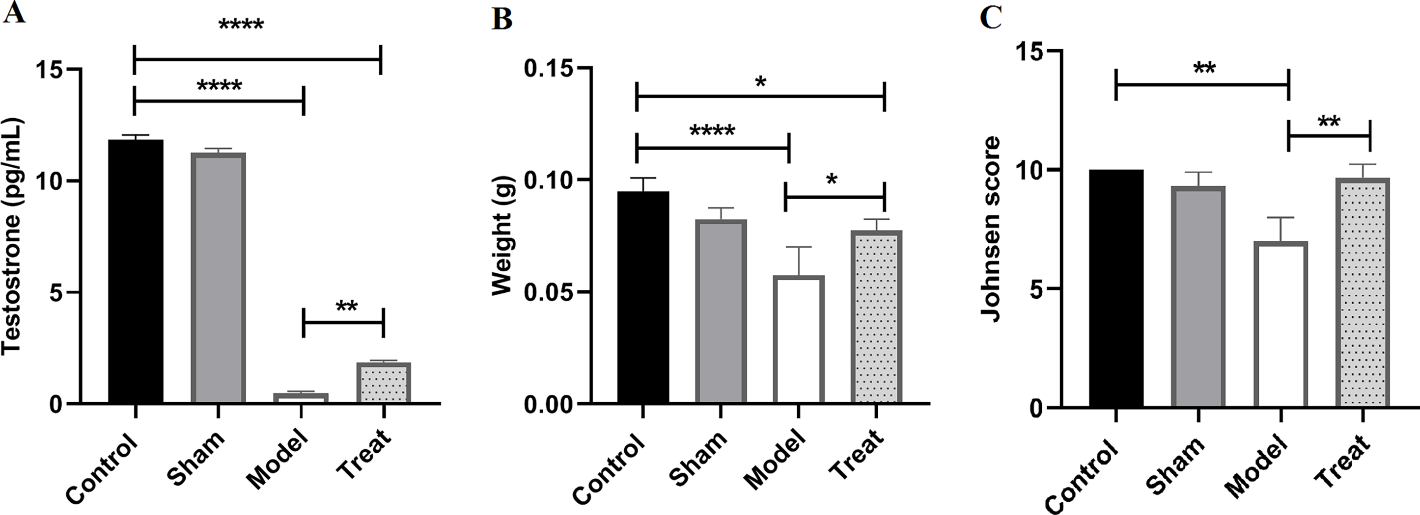
Under the influence of PFOA, the testosterone level and testicular weight in the model group significantly decreased compared to the control group (p < 0.0001). However, in the treated group, this decrease was largely compensated by ALA and the hormone level and testicular weight in this group increased statistically significantly compared to the model group (p < 0.01, p < 0.05). But its level was still significantly lower compared to the control group (p < 0.0001, p < 0.05), indicating that ALA partially but not completely repaired the damage (Figure 1A and B).
A: the testosterone level in the model group significantly decreased compared to the control group (****p < 0.0001), and in the treated group it increased statistically significantly compared to the model group (**p < 0.01). B: The testicular weight in the model group significantly decreased compared to the control group (****p < 0.0001), and in the treated group it increased significantly compared to the model group (*p < 0.05). Testosterone level and testicular weight in the treat group were still significantly lower compared to the control group. C: Johnsen scoring. In the model group, this score significantly decreased compared to the control group. In the treat group, this score significantly increased compared to the model group (**p < 0.01) and was not significantly different from the control group.

Figure 1. Long description
Panel A: A bar graph displays testosterone levels in picograms per milliliter (pg/mL) across four groups: Control, Sham, Model, and Treat. The y-axis represents testosterone levels, and the x-axis lists the groups. The Control and Sham groups have the highest testosterone levels, significantly higher than the Model group. The Treat group shows a statistically significant increase in testosterone levels compared to the Model group but remains lower than the Control group. Panel B: A bar graph shows testicular weight in grams (g) for the same four groups. The y-axis represents weight, and the x-axis lists the groups. The Control group has the highest weight, significantly higher than the Model group. The Treat group shows a significant increase in weight compared to the Model group but is still lower than the Control group. Panel C: A bar graph presents Johnsen scores for the four groups. The y-axis represents the Johnsen score, and the x-axis lists the groups. The Control and Treat groups have higher scores compared to the Model group, with the Treat group showing a significant increase but not differing significantly from the Control group.
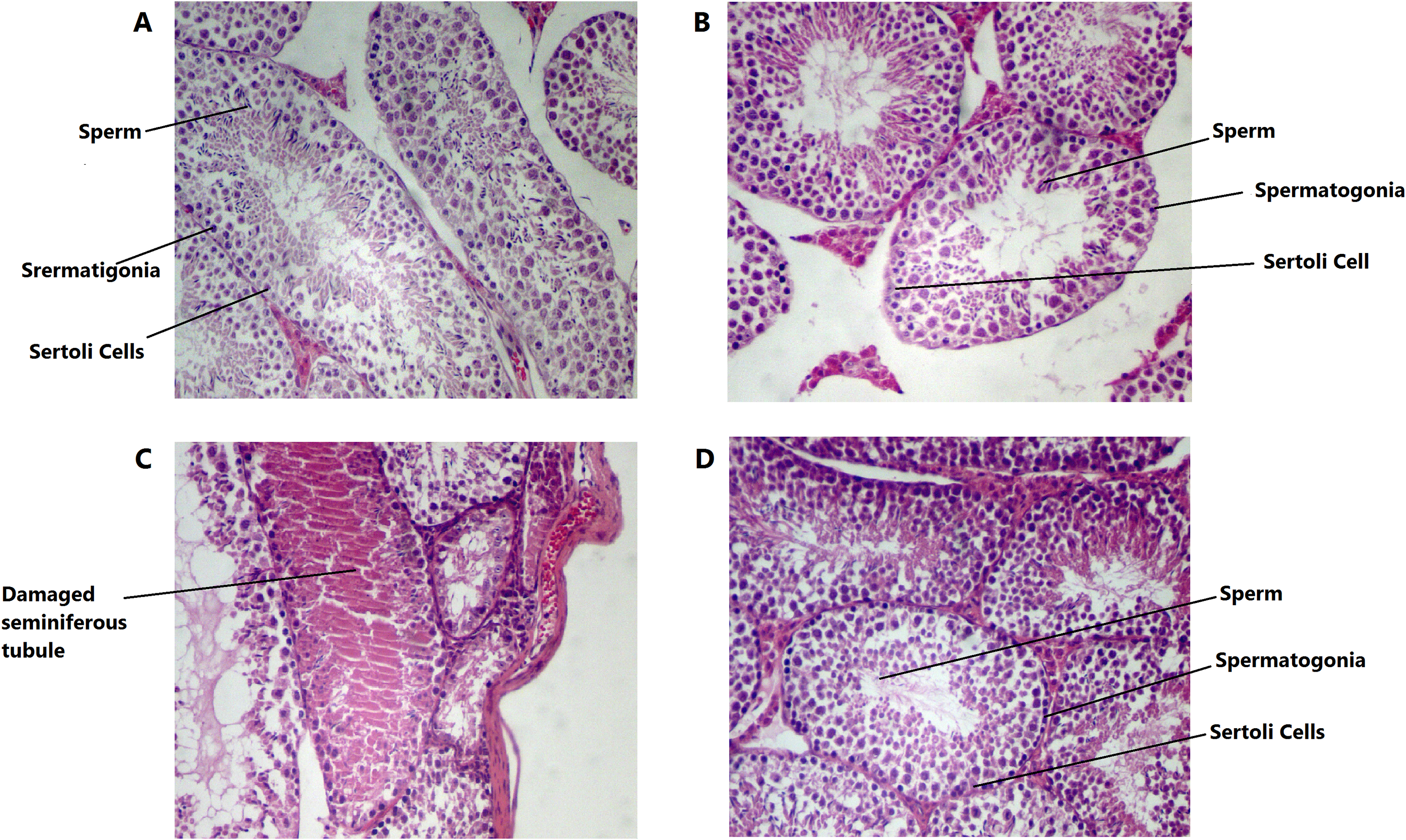
Histological classification of seminiferous tubular cross sections was performed according to the Johnsen scoring system. This grading is from grade 1 to grade 10. Grade 1 is no cells present in the tubular section, and grade 10 is complete spermatogenesis with many spermatozoa. The results of this study showed that in the model group, this score decreased to a range of 7, which was significantly different from the control group (p < 0.01). There are no spermatozoa in this grade, but there are spermatids. Some of the seminiferous tubules have also been damaged. In the treatment group, this damage was compensated and the score returned to a range close to 10, which was statistically significantly different from the model group (p < 0.01) but not from the control group (Figure 1C). Histological images of the control, sham, model and treat groups are shown in Figure 2.
H&E staining of seminiferous tubules in the experimental groups (A: Control; B: Sham; C: Model; D: Treat).

Sperm parameters analysis
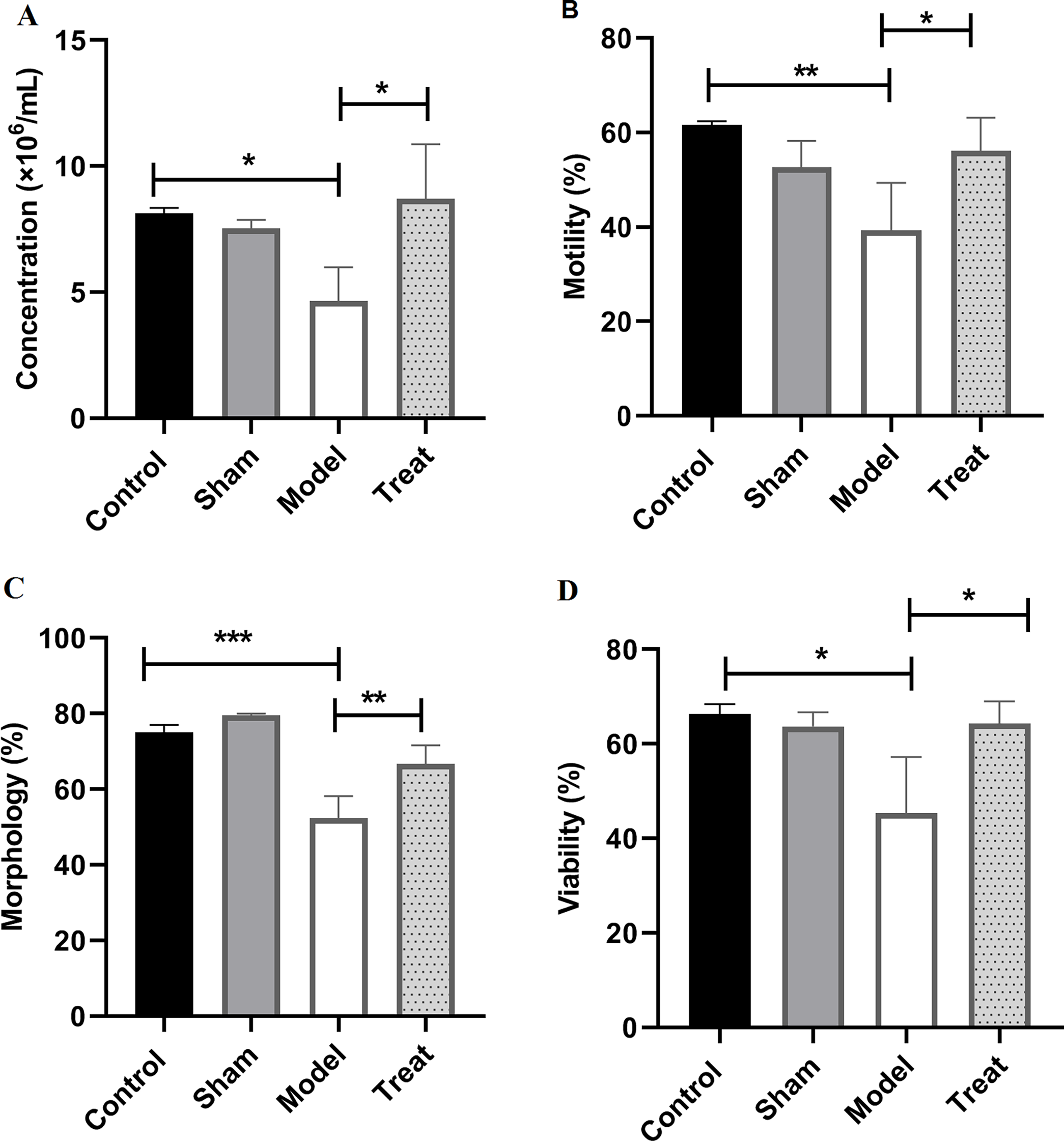
In this study, sperm parameters, including sperm concentration, morphology, total motility and viability, were examined. The results showed that all parameters in the model group (treated with PFOA) decreased significantly compared to the control group. However, in the treated group, this decrease was largely compensated by ALA, so that its amount was significantly increased compared to the model group, but not statistically significantly different from the control group. P < 0.05 was considered significant (Figure 3).
Sperm analysis results showed parameters of concentration, morphology, total motility and viability in the model group significantly decreased compared to the control group. In the treated group, this rate increased significantly compared to the model group and was not significantly different from the control group (p > 0.05). (*p < 0.05/**p < 0.01/***p < 0.001).

SCSA results
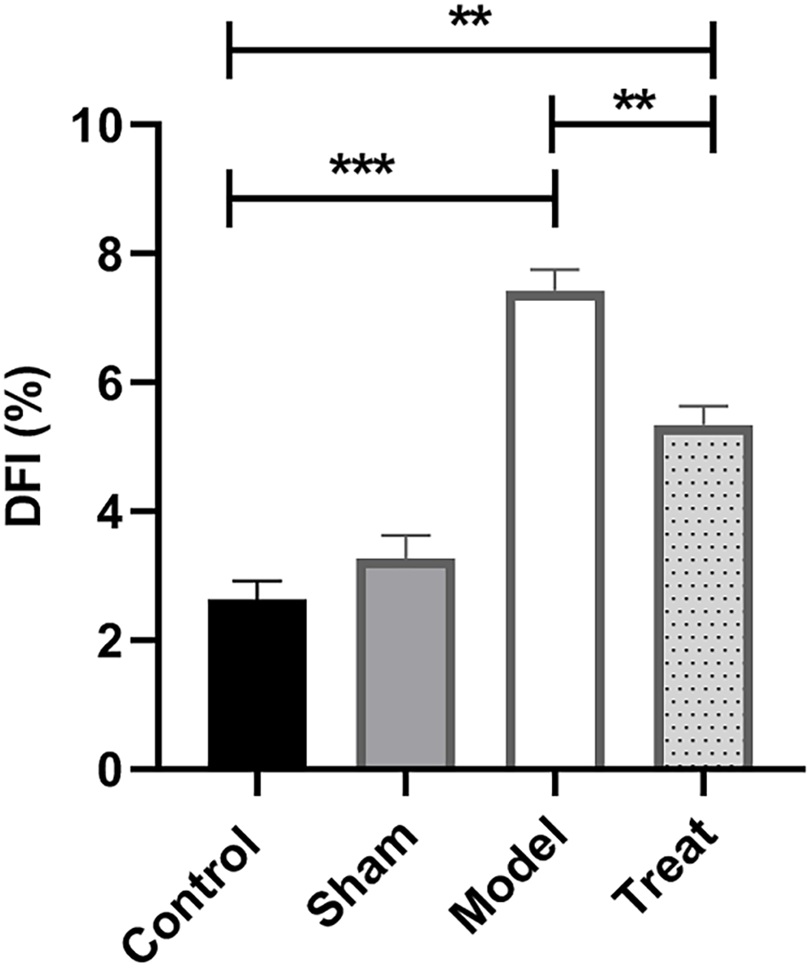
As shown in Figure 4, DFI in the model group (treated with PFOA) was significantly higher than in the control group (p < 0.001). However, in the treated group, this increase was largely compensated by ALA and DFI in this group decreased statistically significantly compared to the model group (p < 0.01). But its level was still significantly higher compared to the control group (p < 0.01), indicating that ALA partially but not completely repaired the damage (Figure 4).
SCSA results showed the DFI in the model group significantly increased compared to the control group (***p < 0.001). In the treated group, it decreased significantly compared to the model group (**p < 0.01), but its level was still significantly higher compared to the control group (**p < 0.01).

IVF results
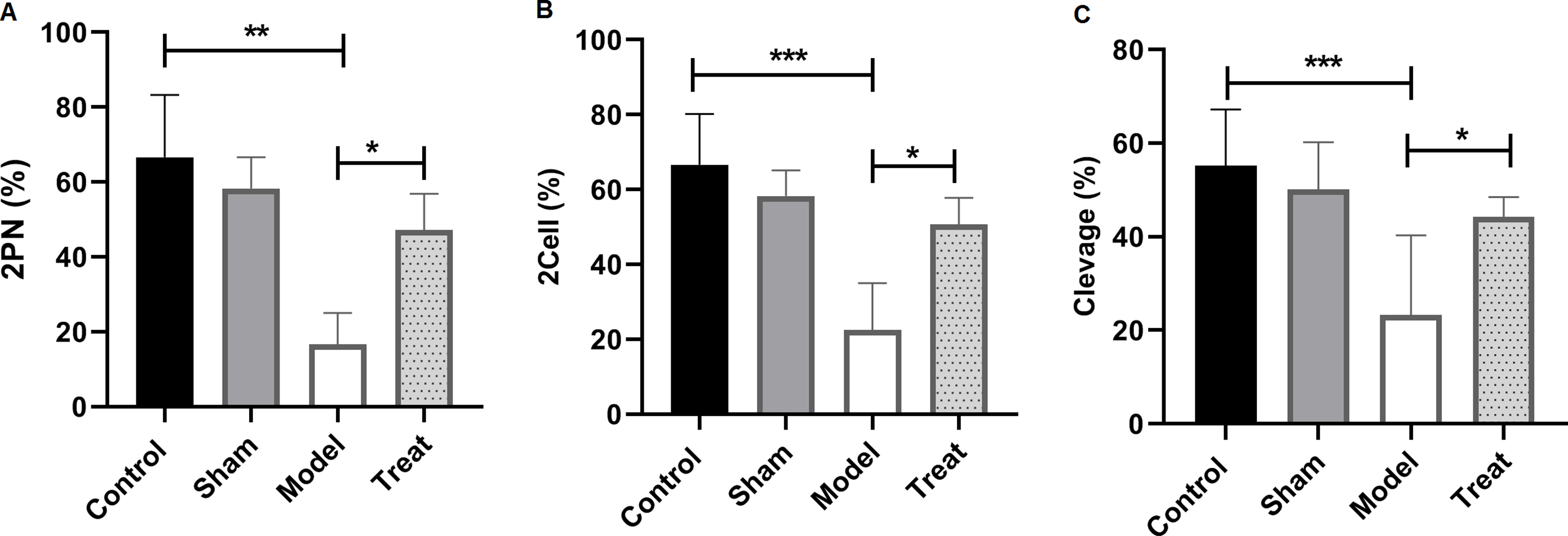
In this study, in vitro fertilization was investigated. and the fertilization rate was measured in stages: the 2PN, 2-cell and 8-cell (cleavage). The results showed that at all these stages, fertilization rate in the model group (treated by PFOA) significantly decreased compared to the control group (p < 0.001/p < 0.01). However, in the treated group, this decrease was largely compensated by ALA, so that its amount was significantly increased compared to the model group (p < 0.05) and not statistically significantly different from the control group (p > 0.05) (Figure 5).
The fertilization rate at 2PN, 2-cell and cleavage stages in the model group significantly decreased compared to the control group (***p < 0.001/**p < 0.01). In the treated group, this rate increased significantly compared to the model group (*p < 0.05) and was not significantly different from the control group (p > 0.05).

Discussion
As a bioaccumulative environmental pollutant, PFOA can have hazardous effects on the health of the human reproductive system (Zhang et al., Reference Zhang, Han, Qiu, Zhao, Gao, Chu and Dai2023). The tightening of standards and the change in the recommended safety level from 70 ng/L to 0.004 ng/L by the United States Environmental Protection Agency (USEPA) reflects increasing concern about the toxicity of this substance (Wee and Aris, Reference Wee and Aris2023; Saliu et al., Reference Saliu, Michaud, Munoz, Dinh, Feder, Arrouy, Sappin-Didier, Watteau, Houot and Liu2025). This study investigated the adverse effects of PFOA on testicular function and IVF outcomes and the role of ALA in reducing these effects. Exposure of male mice to PFOA significantly reduced sperm concentration and quality, testicular weight, testosterone levels, IVF rates at the 2PN, two-cell and cleavage stages and increased sperm DFI. While simultaneous administration of ALA significantly reduced these adverse effects.
Studies show that male fertility is declining, and synthetic chemicals are a major candidate for causing it. Despite the ban on PFOA production in developed countries, regulatory gaps in developing and underdeveloped countries and its continued production continue to expose the human population to baseline levels of this substance (Wee and Aris, Reference Wee and Aris2023). According to the Globally Harmonized System of Classification and Labelling of Chemicals, PFOA accumulates in all tissues and damages organs over time (Wang et al., Reference Wang, Zhang, Wu, Wei, Xu, Wang and Wang2021). The presence of PFOA in testis, serum and semen and its adverse effects on sperm quality have also been reported (Sun et al., Reference Sun, Wen, Wang, Deng, Zhang, Fu, Yuan and Zhang2023).
A decrease in sperm count is an indicator of the reproductive toxicity of a substance (Marty et al., Reference Marty, Erraguntla, North, Barranco, Kirman, Cagen, Rushton, Shen, Koehler and Budinsky2021). In this study, sperm concentration was reduced in the PFOA group compared to the control group. In a study on male mice, gavage with PFOA at doses of 2.5, 5 and 10 mg/kg for 14 days resulted in decreased sperm count and damage to the seminiferous tubules (Liu et al., Reference Liu, Yang, Wu, Zou, Pan, Zou, Liu, Xia, Wang and Zhang2015). Also, maternal exposure to PFOA has had negative effects on the sperm count of male offspring. Motility and morphology are important factors in the success of IVF. In line with our study, some studies have shown that PFOA leads to defects in sperm motility and morphology and decreases in the percentage of progressive and normal sperm. In a 28-day gavage study, PFOA at the 5 mg/kg dose resulted in sperm damage and a negative effect on concentration, motility and morphology (Zhang et al., Reference Zhang, Lu, Luo, Yan, Guo and Dai2014), and it was consistent with the results of our study at the 10 mg/kg dose. Also, sperm incubated with this substance have shown decreased motility and reduced ability to penetrate artificial cervical mucus (Sun et al., Reference Sun, Wen, Wang, Deng, Zhang, Fu, Yuan and Zhang2023). It seems that this substance alters the progressive sperm percentage by disrupting the sperm membrane (Šabović et al., Reference Šabović, Cosci, De Toni, Ferramosca, Stornaiuolo, Di Nisio, Dall’Acqua, Garolla and Foresta2020).
In this study, a negative effect of PFOA on testicular tissue, testosterone levels, testicular weight and sperm DFI was observed. The effect of a mixture of EDCs (including PFAS) on male fertility was examined. No association was found between maternal EDC blood levels and fetal exposure to these substances and sperm morphology, testosterone levels and DFI, but, in line with our study, testicular weight decreased (Hull et al., Reference Hull, Hougaard, Toft, Petersen, Flachs, Lindh, Ramlau-Hansen, Wise, Wilcox and Liew2026). Also, in an investigation of the chronic exposure effects of PFOA in zebrafish, the results were consistent with our study, and harmful effects of this substance on plasma testosterone levels, testicular tissue and sperm concentration and motility were observed (Wang et al., Reference Wang, Huo, Khan, Ma and Mai2024). In the study by Zhang et al. (Reference Zhang, Han, Qiu, Zhao, Gao, Chu and Dai2023), PFOA also caused testicular tissue damage and decreased testosterone levels. However, testicular weight, unlike body weight and epididymis, did not decrease significantly. Therefore, the testicular weight coefficient increased, which is a sign of PFOA toxicity (Zhang et al., Reference Zhang, Han, Qiu, Zhao, Gao, Chu and Dai2023).
In a study conducted by Zhang et al. (Reference Zhang, Lu, Luo, Yan, Guo and Dai2014), the effect of doses of 0, 0.31, 1.25, 5 and 20 mg/kg of PFOA on the reproductive system of male mice showed that doses of 5 and 20 mg/kg PFOA, in addition to damage to the seminiferous tubules, also reduced testosterone levels in a dose-dependent manner. In our study, a dose of 10 mg/kg was investigated to fill the dose–response gap in the above study and, in accordance with the trend observed there, testicular tissue damage and a decrease in testosterone levels were also observed at this dose. Also, the negative effect on testicular weight was observed only at the 20 mg/kg dose, and the decrease in testicular weight at 10 mg/kg in our study further reinforced the dose-dependent nature of PFOA on testicular weight. They showed that PFOA reduces testosterone levels by downregulating CPY11A1 expression (Zhang et al., Reference Zhang, Lu, Luo, Yan, Guo and Dai2014).
Our results showed a decrease in fertilization rates in the PFOA-treated sperm group. It seems that PFOA has a negative impact on IVF rates directly by increasing DFI and reducing sperm quality and indirectly by affecting testosterone levels and testicular tissue. The influence of sperm quality on fertilization rate is undeniable. Liao et al.’s study reported that sperm concentration and motility were strongly correlated with the 2PN and cleavage rates (Liao et al., Reference Liao, Huang, Zhang, Chen, Chen, Li and Ai2020). Since testosterone is one of the hormones that initiates and maintains spermatogenesis and a significant positive correlation has been observed between testosterone and sperm motility, testosterone levels are associated with human semen parameters and a decrease in its levels may negatively affect the IVF rate (Meeker et al., Reference Meeker, Godfrey-Bailey and Hauser2007). There are discrepancies in the results of studies, but several studies have shown that steroidogenic enzymes, serum testosterone and testicular gonadotropin receptor expression level decreased in response to PFOA, leading to hormonal imbalance in the spermatogenesis pathway. Also, this substance contributes to the destruction of the blood–testicular barrier and harm to sperm through damage to the Sertoli cell (Tarapore and Ouyang, Reference Tarapore and Ouyang2021). Another mechanism of PFOA is the activation of molecular pathways leading to oxidative stress (Wang et al., Reference Wang, Huo, Khan, Ma and Mai2024). Guo et al. (Reference Guo, Zhao, Zhao, Huang, Ding, Huang, Meng, Li, Wei and Zhang2021) reported that PFOA leads to DNA damage, impaired mitochondrial function and increased oxidative stress (Guo et al., Reference Guo, Zhao, Zhao, Huang, Ding, Huang, Meng, Li, Wei and Zhang2021), and oxidative stress in sperm is associated with reduced fertilization rates and embryo development in IVF (Ribas-Maynou et al., Reference Ribas-Maynou, Yeste and Salas-Huetos2020).
Abnormal elevation of ROS activates the lipid peroxidation cascade, disrupts apoptosis factors and alters sperm membrane and DNA integrity (Abbasi et al., Reference Abbasi, Molavi, Tavalaee, Abbasi and Nasr-Esfahani2020), subsequently decreasing motility and viability and increasing DNA fragmentation, impairing sperm quality and male fertility (Shaygannia et al., Reference Shaygannia, Ghandehari-Alavijeh, Tavalaee and Nasr-Esfahani2020). Antioxidants effectively improve sperm quality by reducing oxidative stress (Abbasi et al., Reference Abbasi, Molavi, Tavalaee, Abbasi and Nasr-Esfahani2020). ALA, with its antioxidant properties, can be a treatment option. In this study, ALA was able to reduce the harmful effects of PFOA on sperm quality and function, testicular tissue, testosterone levels and testicular weight and improve fertilization rates. Zhang et al. (Reference Zhang, Han, Qiu, Zhao, Gao, Chu and Dai2023) also investigated the therapeutic effect of lipoic acid on PFOA-treated rat testis. They used two doses of lipoic acid in their study and measured testosterone levels. Their results showed that PFOA significantly reduced testosterone levels, and the effect of lipoic acid on testosterone at a low dose (0.05 g/kg) was not significantly different from the PFOA group, but at a higher dose (0.10 g/kg), testosterone levels increased significantly compared to the PFOA group. Lipoic acid also increased the reduced sperm count in rats treated with PFOA and healed damaged testicular tissue (Zhang et al., Reference Zhang, Han, Qiu, Zhao, Gao, Chu and Dai2023). ALA can help damaged sperm with its ability to suppress oxygen-free radicals and reduce oxidized antioxidants (Abbasi et al., Reference Abbasi, Molavi, Tavalaee, Abbasi and Nasr-Esfahani2020). In a study of the effects of this substance in men with varicocele, sperm DNA fragmentation and sperm parameters improved (Abbasi et al., Reference Abbasi, Molavi, Tavalaee, Abbasi and Nasr-Esfahani2020). A multicenter study also showed that this substance improves sperm quality in sub-fertile men (Canepa et al., Reference Canepa, Dal Lago, De Leo, Gallo, Rizzo, Licata, Anserini, Rago and Scaruffi2018). Zhang’s study (therapeutic effect of lipoic acid on rat testis treated with PFOA in two doses of 0.05 and 0.1 g/kg) also examined key indicators of oxidative stress and mitochondrial damage, including malondialdehyde (MDA), glutathione peroxidase (GSH), superoxide dismutase (SOD), succinate dehydrogenase (SDH) and lactate dehydrogenase (LDH). Their results showed that the aforementioned parameters changed under the influence of PFOA in favour of increased oxidative stress and mitochondrial damage (increased MDA and decreased GSH, SOD, SDH and LDH). However, a high dose of lipoic acid compensated for these damages and reduced oxidative stress (Zhang et al., Reference Zhang, Han, Qiu, Zhao, Gao, Chu and Dai2023). In a study of the protective effect of this substance on frozen-thawed sperm, it was also shown that ALA leads to an improvement in sperm capacity by reducing ROS (Shaygannia et al., Reference Shaygannia, Ghandehari-Alavijeh, Tavalaee and Nasr-Esfahani2020). In our study, the damage caused by PFOA on sperm parameters and DFI levels was also improved by using ALA. In addition, the fertilization, 2-cell and cleavage rates were increased and improved in the group treated with ALA and PFOA compared to the group treated with PFOA. In fact, ALA was able to reduce the harmful effects of PFOA and increase the fertilization rate by increasing sperm quality, improving hormonal conditions and testicular tissue. Considering the improvement in Johnson score from 7 in the model group to 10 in the treatment group, ALA can be effective in improving mild to moderate structural damage in the testis. However, the effectiveness of ALA in conditions of complete stem cell depletion (which was not investigated in the study) requires further investigation. We did not assess liver or kidney structure/function in this study. Previous studies have documented PFOA-induced hepatotoxicity and nephrotoxicity in animal models, but whether these systemic effects contribute to impaired spermatogenesis remains unknown. Future studies should investigate this possibility. Also, in this study, ROS levels or antioxidant activity were not measured. Therefore, future investigation in this field seems necessary to understand the mechanism of action of ALA. Also, our findings in mice should not be directly generalized to humans and careful human studies are needed.
Our findings showed that ALA can improve the adverse impact of PFOA on sperm and fertilization results. Therefore, the use of ALA to improve IVF outcomes deserves further attention. Given the persistent presence of PFOA in the environment and its associative (not causal) link with reduced sperm quality, further research and larger studies in this area seem necessary.
Data availability statement
Data is provided within the manuscript.
Acknowledgements
The authors would like to thank Iran University of Medical Sciences (IUMS), Tehran, Iran, for their cooperation throughout the period of study.
Author contributions
Azam Govahi: Investigation-Equal, Methodology-Equal, Writing – original draft-Equal, Writing – review & editing-Equal; Hadis Gholipour: Data curation-Equal, Methodology-Equal, Writing – original draft-Equal, Writing – review & editing-Equal; Marziyeh Ajdary: Data curation-Equal, Writing – review & editing-Equal; Roya Hassani: Data curation-Equal, Writing – review & editing-Equal; Maryam Mirani: Formal analysis-Equal; Mehdi Mehdizadeh: Formal analysis-Equal; Rana Mehdizadeh: Formal analysis-Equal; Mahnaz Ashrafi: Conceptualization-Equal, Project administration-Equal, Supervision-Equal, Writing – review & editing-Equal.
Competing interests
The authors declare that they have no competing interests.
Funding information
This work was supported by the Iran University of Medical Sciences (Grant no: 26541).
Ethical Standards
This study was approved by the ethics committee of Iran University of Medical Science (IR.IUMS.AEC.1402.079).
Consent for publication
All authors consent to the publication of this study.