I. INTRODUCTION
Titanium and its alloys are often used as biomedical implants owing to their excellent mechanical properties, such as their relatively low modulus, excellent fatigue strength, and formability.Reference Liu, Chu and Ding1,Reference Topolski, Bochniak, Lagoda, Ostachowski and Garbacz2 Another property is its biocompatibility, considering that titanium is biologically inert and corrosion resistant due to the thin oxide layer of titanium oxide (TiO2), which forms automatically on its surface. For these reasons, titanium is commonly used in biomedicine, for example, in hip and knee prostheses or dental implants.Reference Oldani, Dominguez and Fokter3,Reference Zhu, Bao, Chen and Zhao4 However, biofilms often form on the surface of these materials.
Biofilms are highly organized biological systems composed of different types of micro-organisms, mainly bacteria, which first adhere individually on a surface, before expanding and maturating, thus producing a matrix of extracellular polymeric substances that makes the colonies highly resistant to antibiotics. Indeed, some micro-organisms, like Pseudomonas aeruginosa and E. coli (Gram-negative bacteria), or Candida albicans and Cryptococcus albidus (fungi), can stay inactive on the surface of the material for a long period until irreversibly forming a biofilm. As a consequence, the surface becomes a potential source of contamination by pathogenic micro-organisms, releasing toxins and leading to inflammatory and infectious processes.Reference Feng, Wu, Chen, Cuiz, Kim and Kim5,Reference Wan, Raman, Hea and Huang6
Once implants or prostheses are infected, the patient undergoes a revision surgery, given, in most cases, the inefficiency of antibiotics. This procedure is time consuming, stressful, and extremely expensive.Reference Kurtz, Lau, Watsom, Schmier and Parvizi7 An untreated infected implant can lead to its failure, which has dramatic consequences for the patient.Reference Inman, Gallegos, Brause, Redecha and Christian8 Implant removal can also increase morbidity and even cause mortality.Reference Zmistowski, Karam, Durinka, Casper and Parvizi9 Complications by periprosthetic joint infections are estimated to be on 2.5% of cases for primary hip and knee arthroplasties and up to 20% for revision arthroplasties.Reference Lentino10 Therefore, revision surgery may not be the best strategy to avoid infections. Rather, implants with antimicrobial properties, acting directly in vivo, should be considered.
Antimicrobial agents play an essential role in the control of infectious diseases. They present both immediate and long-term advantages.Reference Brady, Lisay, Yurkovetskiy and Sawan11,Reference Ferrara, Courson and Paulson12 Some elements, such as silver and copper, may play an interesting antimicrobial effect, mainly explained by the slow release of cations that are toxic to bacteria.Reference Liu, Sonshine, Shervani and Hurt13–Reference Wang, Jin, Qasim, Gao, Peng, Li, Feng and Chu16 The mechanisms of action of silver against bacteria are not completely understood at the moment, but it seems that silver nanoparticles and their ions generate reactive oxygen species, which lead to cell wall perforation, protein oxidation, and DNA damage, ultimately leading to the bacteria death.Reference Wang, Jin, Qasim, Gao, Peng, Li, Feng and Chu16,Reference Ballottin, Fulaz, Cabrini, Tsukamoto, Durán, Alves and Tasic17 For this reason, silver and copper have been intensively experimented for preventing wound,Reference Brady, Lisay, Yurkovetskiy and Sawan11,Reference Boateng and Matthews18–Reference Saint, Elmore, Sullivan, Emerson and Koepsell20 biomedical devices,Reference Liu, Sonshine, Shervani and Hurt13–Reference Perelshtein, Applerot, Perkas, Wehrschuetz-Sigl, Hasmann, Guebitz and Gedanken21 and textilesReference Knetsch and Koole22 contamination. However, silver is the most popular metal among other heavy metals, as mentioned in several studies,Reference Liao, Ou, Cheng, Lin and Peng23–Reference Sioshansi and Tobin26 presenting no toxicity for human cells, at low concentrations.
Here, an antibacterial agent such as silver was introduced into the original titanium implant by using surface treatment, to combine the mechanical and biocompatible properties of titanium and the antibacterial properties of silver. This study concentrates on low-energy ion implantation, forming a gradient of implanted particles a few nanometers below the surface.Reference Hirvonen27 In relation to conventional ion implantation, this method presents some advantages, such as high degree of ionization, low polarization energies (<5.0 keV) in the ion acceleration stage, reduction in process time (less than 1 h), and increase of the surface area of the material to be modified (about 200 cm2). These conditions may vary according to the availability of the reactor. The versatility of this method allows the process to be carried out on an industrial scale without restrictions.Reference Pezzagna, Meijer and Goorsky28,Reference Poon and Burd29
However, besides inhibiting bacterial adhesion, the material is also favorable to osseointegration once surface modification does not present cytotoxic effects. The potential impact of silver nanoparticles on cytotoxicity in individuals at the cellular level has motivated several recent studies that seek to elucidate their effects on human cells.Reference Wataha, Lockwood and Schedle30–Reference Echeverrigaray, Echeverrigaray, Delamare, Wanke, Figueroa, Baumvol and Aguzzoli32 Within this context, some of the aspects studied include cell uptake and intracellular distribution, cytotoxicity, genotoxicity, and immune responses. Furthermore, the main factors that may influence such in vitro and in vivo effects are related to dose, exposure time, size, shape, surface chemistry, and cell type.Reference Wataha, Lockwood and Schedle30–Reference Echeverrigaray, Echeverrigaray, Delamare, Wanke, Figueroa, Baumvol and Aguzzoli32
Low-energy ion implantation is an innovative process combining the simple equipment of physical vapor deposition and the conventional ion plating process, which consists in bombarding the substrate with accelerated ions of the species to implant. This process is called ion plating diversified (IPD). The main advantages of the IPD process are the possibility of ion concentration control, lower work temperature, short-time process, good reproducibility, versatility of ion species, and low-energy implantation.33
In view of all these facts, the aim of this research was to study the correlation between the implantation parameters and the bactericidal action in titanium samples by modifying the surface by applying the IPD process.
II. EXPERIMENTAL
A. Substrates
Unalloyed commercially pure titanium (CP Ti) grade 1 samples were cut in squares with initial dimensions (20 × 20 × 0.05 mm). The chemical composition of CP Ti grade 1 is in accordance with the specifications set by ASTM F67Reference Duraccio, Mussano and Faga34 [N: 0.03, C: 0.10, H: 0.0125, Fe: 0.20, O: 0.18, balance Ti (wt%)]. Cleaning was conducted by immersion in acetone PA in an ultrasound for 30 min and then dried in a hot air stream and submitted to the implantation process. At the end of the surface treatment, the samples were stored in vacuum desiccators.
B. Low-energy ion implantation
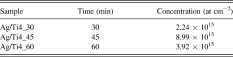
The ion implantation was made by the IPD process and it was accomplished in a reactive low voltage ion plating-type device.33 The vacuum in the reactor during implantations was kept at 5.0 × 10⁻⁵ Pa, and the base pressure was 1.0 × 10⁻⁵ Pa. The silver pellets evaporated in the process with a purity of 99.9% were provided by Kurt J. Lesker Company (Jefferson Hills, Pennsylvania). The reactor was vented down with commercial nitrogen gas. Operating conditions IPD are described in Table I. The regular silver doses were obtained under these process conditions. The name of the samples was chosen according to the implantation energy and ion implantation time used in the experiments. The influence of the implantation time is related to the concentration of silver ions and, for this reason, the sample with a 60 min implantation time was chosen to perform the main biological assays. In addition, this condition was the one that presented the best bactericidal efficiency.
Conditions of ion plating at low energy (BIAS = 4.0 keV and emission current = 25 mA) and silver concentration on CP Ti after ion implantation at different times.

C. Characterization of the modified surface
1. Monte Carlo simulations
The concentration of silver ion in-depth profiles was simulated using Monte Carlo method, by using software Stopping and Range of Ion in Matter (SRIM 2013, Morrisville, North Carolina).Reference Ziegler, Ziegler and Biersack35 The simulation was carried out to compare with high energy implantation (100 keV).
2. Rutherford backscattering spectrometry
The composition of silver implanted in CP Ti samples was obtained by Rutherford backscattering spectrometry (RBS). The equipment used was 3 MV Tandem accelerator. Mono-energetic ion helium at 2 MeV of energy was used to collide with samples. The detection angle was 165° and the beam was normal incidence angle with the sample.
3. Glow discharge optical emission spectroscopy
The in-depth chemical profiles were obtained by glow discharge-optical emission spectroscopy (GD-OES). A GD Profiler-2 from Horiba Jobin-Yvon (Longjumeau, France) was used, with a radiofrequency source operating at 13.56 MHz. The technique was applied in the qualitative mode. The power used was 15 W and the argon gas pressure was 650 Pa.
4. Wettability
The contact angle was measured by the sessile drop method, in which a drop of deionized water was deposited on top of the titanium samples with a microsyringe. The drop was observed with a low magnification lens. The contact angle was determined by using Surftens 3.0 image analysis software (OEG, Frankfurt, Germany). The measurement was taken in three different regions of the sample. In each region, five points were measured and the error was calculated with the standard deviation with these points.
5. Surface profilometry
Surface roughness of the CP Ti and Ag/Ti4_60 samples were measured using the profilometric module of the UMT Tribolab from Bruker Company (Billerica, Massachusetts). The measurement was taken in two different regions of the sample.
6. X-ray photoelectron spectroscopy
A VG CLAM4 hemispherical analyzer (VG Systems Ltd., East Grinstead, United Kingdom) with a Mg Kα X-ray source (hν = 1253.6 eV) was used to determine the bonding and chemical composition of silver implanted in the CP Ti samples.
D. Bactericidal effect evaluation
1. Agar diffusion test
The bactericidal efficiency of the Ag/Ti4_45 sample was tested against E. coli (ATCC-25922), which are fecal coliform bacteria that have been extensively used to indicate the presence of human pathogens in water. First, the bacterial culture was diluted in a 0.1% (m/v) peptone salt solution to reach the suspension turbidity at λ = 565 nm of the value of 0.5 on the McFarland scale, which corresponds to the bacteria concentration of approximately 1.0 × 10⁹ colony forming units per milliliter (CFU/mL). Next, this solution was spread uniformly over the solidified nutrient agar gel (M1461, Himedia, West Chester, Pennsylvania) contained in a Petri dish. At the end of this procedure, the sample was placed over the nutrient agar and incubated at 37 °C for 48 h to evaluate the size of the zone of inhibition formed around, under, and over the Ag/Ti4_45 sample.
2. Treatment of the industrial wastewater
An amount of 100 mL of industrial wastewater with an initial E. coli count of 5.0 × 100 CFU/mL was transferred to five Erlenmeyer flasks, each one containing four pieces (1.0 × 1.0 cm) of the CP Ti sample with implanted silver. Subsequently, the Erlenmeyer flasks were incubated in an orbital shaker at 180 rpm with controlled temperature (25.0 ± 1.0 °C) for 5, 15, 30, 90, and 240 min, respectively. At the end of this procedure, the industrial wastewater of each Erlenmeyer flask was filtered and a new total coliform count was performed by the method 991.14 of the Association of Official Analytical Chemists (AOAC) (2012).
E. MG-63 cell lines on Ag/Ti4_60 and pure CP Ti samples
1. Exposition of Ag/Ti4_60 samples to cell medium
Ag/Ti4_60 samples were first exposed to the cell Dulbecco’s Modified Eagle’s medium (DMEM) according to standard procedures in ISO 10993-5-2009-2. The extraction solution was prepared by using Ag/Ti4_60 (3.0 cm2/mL) in the DMEM culture medium supplemented with 10% fetal bovine serum (FBS) and 1% penicillin/streptomycin (P/S). The culture medium was placed in contact with the plates for 24 h at 37 °C in humidified atmosphere with 5% CO2 to obtain the extraction solution.
2. Cell viability and proliferation
MG-63 (osteosarcom cell lines) cells were seeded at a density of 5.0 × 10⁴ cells/mL in 100 μL of the DMEM culture medium supplemented with 10% FBS and 1% P/S and incubated in cell greenhouse at 37 °C in humidified atmosphere with 5% CO2. After 24 h, the cells were treated with the extraction solution of AgTi4_60 for 1, 5, and 7 days, respectively. DMEM was used for negative control, in which cells were exposed to the medium without previous contact with extraction solution of Ag/Ti4_60. Negative control samples should not induce metabolic changes on cells. For positive control, 5% dimethyl sulfoxide (DMSO) was used. DMSO at high concentrations, such as the one used in this study, is known as deleterious to cell metabolism, inducing cell death among other molecular and biological transformations. The culture medium was removed and 1.0 mg/mL of 3-(4,5-dimethylthiazol-2-yl)-2,5-diphenyltetrazolium bromide (MTT) in the FSB-free medium and P/S was added to the wells and incubated for 2 h. MTT solution was withdrawn and dissolved in 100 μL of DMSO. Spectrophotometric measurements at 570 nm were performed on a microplate reader (Max190 spectra, Molecular Devices). The results were expressed as percent viability. The absorbance of the negative control represented 100% viability and the values of the treated cells were calculated as a percentage of the control.
Statistical significance was assessed using t-test and one-way analysis of variance (ANOVA) with the multiple mean comparison test (Tukey) to assess statistical differences in the case of normal distribution. Significance was accepted for P < 0.05 using Statistical Package for Social Sciences (SPSS, version 19.0, IBM, Armonk, New York) software for Windows.
3. Cell morphological analysis
MG-63 cells were plated, treated, and exposed to the extraction solution as previously mentioned in the MTT assay. The medium was removed and the cells were fixed with methanol for 5 min and Giemsa’s solution was added for 10 min on the same plate. Subsequently, the sample was visualized in optical microscopy (BX43, Olympus, Shanghai, China) with 200× magnification.
Likewise, MG-63 were seeded on Ag/Ti4_60 and pure CP Ti samples in 24-well plates at the same density as above, for 1 and 5 days, respectively. Cells were then fixed with 3% glutaraldehyde solution in PBS (v/v) for 15 min, with subsequent dehydration with 30, 50, 70, 90, and 100% (v/v) ethanol for 10 min. Afterwards, the samples were kept in a desiccator until the analysis was performed. The samples were gold-coated in sputtering equipment and examined with a Shimadzu SSX-550 electronic microscope (Kyoto, Japan) in the high vacuum mode with the maximum beam voltage set at 15 kV.
III. RESULTS AND DISCUSSION
A. Modified surface analysis
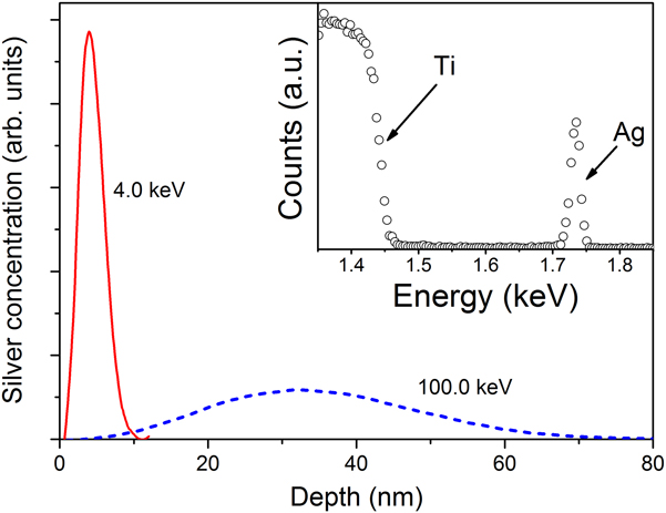
A comparison between simulated profiles of the same amount of silver ion implantation in titanium at 4.0 and 100.0 keV is shown in Fig. 1. For 4.0 keV energy, the highest dose of implanted silver ions is approximately 4 nm deep. On the other hand, with 100.0 keV, the implanted ions are distributed along 70 nm below the surface.
In-depth silver concentration profiles inside titanium through self-bias low-voltage, calculated by SRIM software package. The inset shows the RBS spectrum.
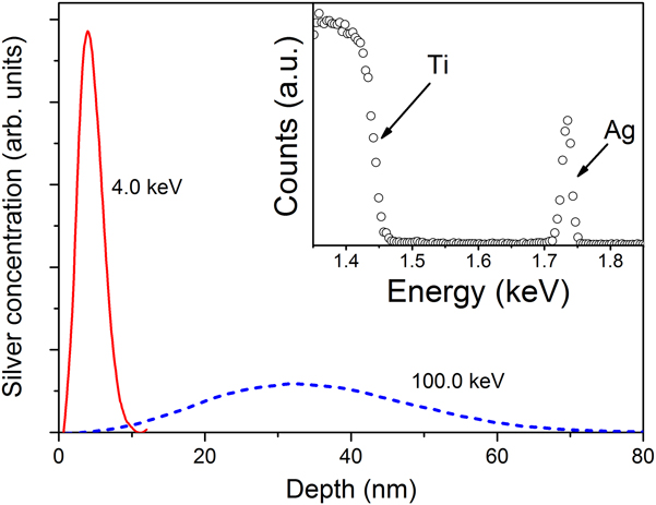
The results from the simulation were important to provide us an estimate to guide the definition of the self-bias voltage used at the low-energy ion implantation. In the inset of Fig. 1, the RBS spectrum shows the characteristic signals of titanium (substrate) and silver incorporated in the samples. The same tendency was showed in Monte Carlo simulation results. Table I shows the silver concentration on CP Ti after ion implantation. In addition, the increased process time increased the concentration of silver atoms.
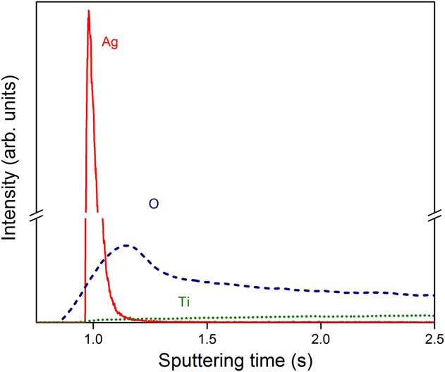
Figure 2 shows the chemical composition profile in-depth by the GD-OES technique. As can be observed, silver is on the surface of the sample, and on the bulk region, the titanium profile stands out. The profiles of silver and titanium overlap and the titanium profile begins before the silver profile, confirming that the silver is implanted on the surface of the titanium.
In-depth chemical composition profile by the GD-OES spectrum for the Ag/Ti4_60 sample.
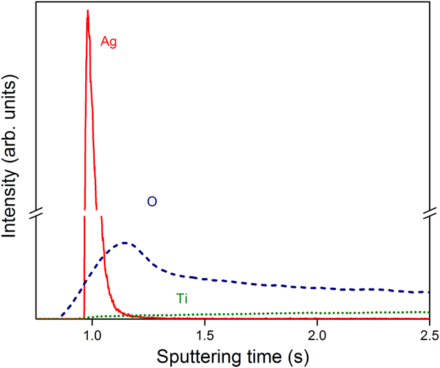
The titanium profile did not present an abrupt growth, which would indicate the presence of a film. However, it presented a gradual growth, which is a sign that the implantation has occurred. The oxygen detected is possibly in the form of native oxides of silver and titanium, which is due to the fact that silver has a low ionization energy and the titanium forms a passive layer of oxide when exposed to an oxidizing medium at room temperature.Reference Strnad, Petrovan and Russu36 Furthermore, the oxygen on the surface is an impurity that can influence the antibacterial properties. The results obtained with GD-OES are in agreement with the Monte Carlo simulation and RBS results.
The contact angle for Ag/Ti4_60 (102.7° ± 1.0) is lower than the pure substrate (107.6° ± 1.5), which indicates that the titanium surface becomes less hydrophobic after silver implantation.Reference Zeraik and Nitschke37 In other words, it is easier to form a thin layer of water on the surface, capable of hindering the initial anchoring of bacteria that could initiate the formation of a biofilm.Reference Zareidoost, Yousefpour, Ghaseme and Amanzadeh38 On the other hand, the reduction of hydrophobicity favors the adhesion of cells and, consequently, the osseointegration of the biomaterial.Reference Zeraik and Nitschke37
The roughness R α values of the CP Ti and Ag/Ti4_60 samples were 0.193 and 0.205 µm, respectively. Surface roughness has a significant influence on cell response. Previous studies have shown that chemical etching of a titanium implant surface significantly enhances osseointegration and increases the strength of osseointegration. The extent of the bone implant interface is positively correlated with an increasing roughness of the titanium surface.Reference Zareidoost, Yousefpour, Ghaseme and Amanzadeh38,Reference Calderon, Cavaleiro and Carvalho40 The implanted sample (Ag/Ti4_60) presents basically the same roughness than CP Ti, which indicates that the treated sample represents a condition for inhibiting the initial anchoring of bacteriaReference Zareidoost, Yousefpour, Ghaseme and Amanzadeh38 and favors the adhesion of cells (osseointegration).Reference Zeraik and Nitschke37
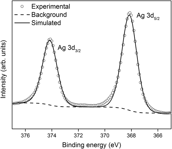
The X-ray photoelectron spectroscopy (XPS) analysis confirmed the chemical state of implanted silver in CP Ti (Ag/Ti4_60). The silver high-resolution spectrum was deconvoluted and shows two separated peaks (Fig. 3). The binding energy around 368.3 eV (Ag 3d 5/2) and 374.2 eV (Ag 3d 3/2) indicate that all the silver present in the sample is in metallic state.Reference Ni, Zhang, Chen, Zhan, Huo and Zuo41–Reference Rai, Yadav and Gade43
Deconvolution of the high resolution Ag 3d XPS spectrum of the Ag/Ti4_60 sample.

The ionization energy of the metallic silver is low since even contact with moisture or body fluid provides its ionization.Reference Chang, Wang and Daniel45 Thus, silver can easily dissociate and act in the effect against the proliferation of bacteria on titanium surfaces. This is an important result for the antimicrobial activity of the implanted samples.Reference Fontenoy and Kamel44,Reference Chang, Wang and Daniel45
Additionally, as shown in the RBS spectra, the samples showed no significant amount of contaminants, namely oxygen, which could damage the surface activity if bonded with the implanted silver.Reference Fontenoy and Kamel44
B. Bactericidal effect
1. Agar diffusion test
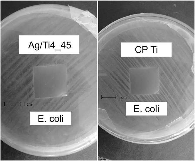
Figure 4 shows the image of the agar diffusion test for the Ag/Ti4_45 and CP Ti samples. The Ag/Ti4_45 sample exhibited an inhibitory activity against E. coli since no micro-organism growth was observed. If this inhibitory activity does not occur, then the sample would suffer a displacement on nutrient agar due to the growing of bacteria below the substrate. This preliminary result indicates that an intermediate amount of silver (lower than the maximum) can be efficient for bacterial reduction tests. This test is only qualitative, indicating the possibility for using the samples to further bactericidal experiments.
Agar diffusion test for CP Ti and Ag/Ti4_45 samples.

2. Treatment of the industrial wastewater
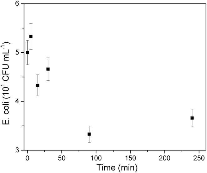
The Ag/Ti4_45 sample was used in the treatment of industrial wastewater. This sample was chosen because it had an indication of bactericidal efficiency in the agar diffusion tests as well as the incorporation of the average amount of silver according to the characterization results. Figure 5 shows the counting of E. coli as a function of time. A trend is observed: a first increase of bacterial concentrations during the first 15 min, followed by a decrease of bacterial concentrations. This trend shows that there is no action of silver during the first minutes, in which the bacteria proliferate because of the incubating conditions favorable to their expansion. However, the decrease in bacterial concentrations clearly proves the antibacterial efficacy.
Bacterial reduction as a function of immersion time in industrial wastewater.

The first increase in bacterial concentrations shows the delayed action of silver, which could be explained considering the time for silver nanoparticles to leave the substrate and diffuse in the environment to attack bacteria. The delay of silver and ion release could be responsible for this latent period of silver inactivity.
A regain in the bacterial concentration is observed after the bacterial-killing phase. This bacterial expansion succeeding to an antibacterial phase of action of silver could be due to the exhaustion of active silver ions, either because the density of silver in the treated surface was not sufficient to have a long lasting effect or because the silver ions have already eliminated a certain amount of bacteria and are outnumbered. There might be cycles of periodic antibacterial phases and latent phases, silver being unavailable when it is already active, killing bacteria.
3. Evaluation of the amount of Ag leached after effluent treatment
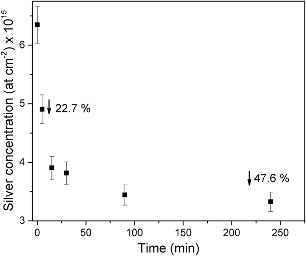
The Ag/Ti4_45 sample used in the treatment of the industrial effluent was analyzed by RBS to trace a profile of the amount of silver remaining as a function of time that is permanent in contact with the effluent. This profile is shown in Fig. 6, which shows that the concentration of silver in the samples is lower than the initial concentration, and the longer the immersion time, the lower the remaining amount of silver.
Quantification of silver in samples after treatment of industrial wastewater.

In the first 5 min, approximately 22.7% of the initial dose of implanted silver was leached and at the end of 240 min, the reduction due to leaching was 47.6%. It means that only 52.4% of the initial dose of silver remains incorporated into the titanium substrate.
These data indicate that even with more than half the initial silver dose in the samples, perhaps the depth of the silver particles remaining relative to the surface is such that the energy required to ionize them and cause them to detach from the substrate is greater than the energy available in the system. Another possible justification for the rates of removal to be not as efficient as what is reported in the literature would be the presence of oxygen in the samples, as verified by the GD-OES analysis. When the silver atoms are bound, forming AgO, for example, the energy required for ionization is much greater than when it is available in the Ag0 state.Reference Chang, Wang and Daniel45
C. Morphology and cytotoxicity tests
1. Morphology of cells
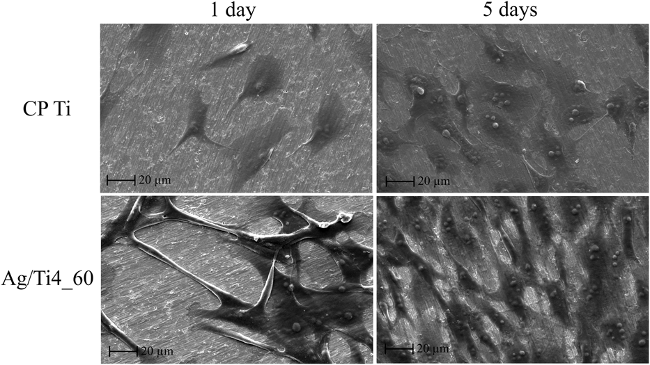
The Ag/Ti4_60 sample showed an increase in cell distribution compared to pure titanium (CP Ti), with at least the double of cell population, as can be seen visually and comparatively in Fig. 7, with a 500× magnification. In the micrographs, the clearest part with grooves corresponds to the titanium substrate and the cells are observed as prolonged and darker structures in the images. It is possible to observe that in 5 days, almost the entire titanium surface was covered by cells.
Comparison of the morphology of MG63 cells on CP Ti and Ag/Ti4_60 samples after 1 and 5 days of incubation, respectively.

The increase of cell adhesion in the treated sample may be explained due to the reduction of hydrophobicity of the samples caused by the silver implantation process.Reference Gadelmawla, Koura, Maksoud, Elewa and Soliman39,Reference Chang, Wang and Daniel45
2. Morphological alterations of MG-63 lines after extract exposition
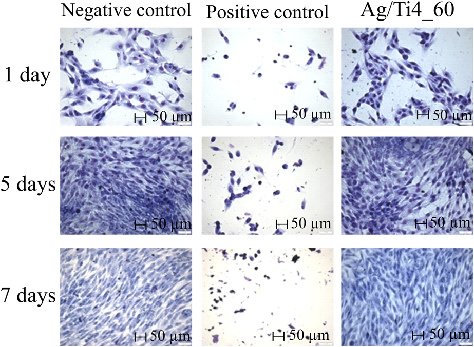
The morphology of MG-63 cells in contact with the Ag/Ti4_60 extract during the period of 1, 5, and 7 days were rounded or prolonged, enveloped in abundant, well defined and intact cytoplasm, characterizing a healthy cell culture (Fig. 8). On the other hand, MG-63 cells in contact with positive control (5% DMSO) during the same period of incubation presented a reduced number of cells, with severe altered morphology, suggesting a cytotoxic activity compared to the sample extract.
Cell morphology treated for 1, 5, and 7 days compared to negative control (DMEM culture medium supplemented with 10% FBS and 1% P/S) and positive control (DMEM culture medium supplemented with 10% FBS, 1% P/S, and 5% DMSO). The samples were visualized in optical microscopy with 200× magnification. *50 µm – Magnification scale bar.

3. Cell viability and proliferation
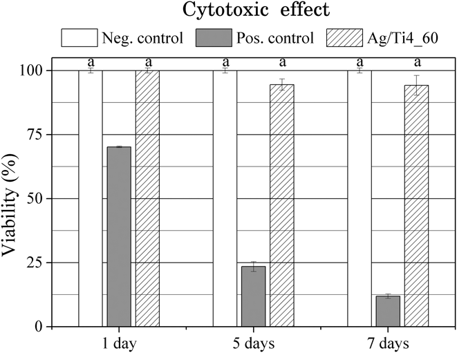
Figure 9 shows a comparison of the cytotoxic effect between the Ag/Ti4_60 sample with positive and negative controls. There was no reduction of cell viability during the period of 1, 5, and 7 days in the presence of the extract of the treated sample in comparison to the negative control, which represents the best cellular condition. In analogy to what was observed in the Giemsa staining test, it should be noted that the positive control with 5% DMSO showed a marked reduction in cell viability when compared to the negative control and the treatments made with the extract of the Ag/Ti4_60 sample.
Cytotoxic effect of Ag/Ti4_60 on the viability of MG-63 cells. Cells were treated for 1, 5, and 7 days. Positive control (cells treated with 5% DMSO). Results were obtained from three independent experiments. Each bar represents the average percentage of viability ± SD relative to negative control (culture medium), which was normalized to 100%. Letters correspond to statistically significant differences by using the ANOVA-Tukey test (P ≤ 0.05).

It is important to emphasize that the sample used in this evaluation is the one that presented the highest concentration of implanted silver. The Ag/Ti4_60 sample showed no evidence of toxicity to human cells. The process proposed in this study can be considered as biologically viable, once that in lower Ag/Ti4_45 silver concentrations, there were indications of bactericidal action. Therefore, these concentrations are in a safe range for the use of artifacts treated as prostheses or implants of the dental area.
IV. CONCLUSIONS
The results show that the low-energy silver ionic implantation method by IPD allows controlling the process parameters and, together with the simulations, results in different levels of surface modification aiming at the bactericidal action. Additionally, the process results in samples without contaminants, namely oxygen. Not only the concentration of silver implanted but also the topography of the surface is determinant in the action against micro-organisms.
The choice of parameters to obtain a bactericidal and nontoxic biomaterial for human cells should consider the ideal combination “energy + silver concentration”. These parameters promote changes on the surface topography, which influences the wettability and roughness and, as a consequence, modifies the response of this surface in the adhesion of both unwanted micro-organisms and human cells.
The low-energy ion implantation by IPD is a reproducible process that allows a wide control of the implantation parameters that can be modified as needed. This versatility allows the process to be applied on an industrial scale, where process conditions (energy, time, and maintenance) make it industrially viable.
ACKNOWLEDGMENTS
The authors are grateful to the Materials Graduate Program (PGMAT-UCS), the University of Caxias do Sul—UCS, and the Federal University of Rio Grande do Sul - UFRGS. We also thank LCMic-UCS. Part of this work was developed in collaboration with the Ion Implantation Laboratory (Institute of Physics, Federal University of Rio Grande do Sul, Porto Alegre, RS, Brazil). This research did not receive any specific grant from funding agencies in the public, commercial, or not-for-profit sectors.