INTRODUCTION
Strongyloidiasis is a human intestinal infection that affects between 30 and 100 million people worldwide. Recently, it was included in the list of neglected tropical diseases (Olsen et al. Reference Olsen, van Lieshout, Marti, Polderman, Polman, Steinmann, Stothard, Thybo, Verweij and Magnussen2009). Chronic infections in endemic areas can remain asymptomatic for decades through the autoinfective cycle of the filariform larvae L3 (Grove, Reference Grove1996; Siddiqui and Berk, Reference Siddiqui and Berk2001). Diagnosis of these chronic infections is sometimes cumbersome, requiring more sensitive diagnostic methods, particularly in low-level infections and immunocompromised patients (Marcos et al. Reference Marcos, Terashima, DuPont and Gotuzzo2008; Paula and Costa-Cruz, Reference Paula and Costa-Cruz2011).
A diagnosis of strongyloidiasis is suspected when suggestive clinical signs and symptoms are observed, particularly eosinophilia. Currently, a definitive diagnosis is usually achieved by fecal larval detection using parasitological methods, using concentration techniques or stool culture on agar plate medium (Liu and Weller, Reference Liu and Weller1993). These methods are laborious and time consuming, requiring expertise and repeated stool collection and examination over a number of consecutive days (Concha et al. Reference Concha, Harrington and Rogers2005). In contrast, several immunological tests, showing variable sensitivity and specificity, have been studied for diagnosis of Strongyloides stercoralis infection. However, the use of these methods has certain limitations due to either cross-reactive antibodies from other helminthic infections or serological positivity resulting from previous Strongyloides infections (Siddiqui and Berk, Reference Siddiqui and Berk2001; Mejia and Nutman, Reference Mejia and Nutman2012).
As DNA detection becomes increasingly utilized in the diagnosis of parasite infections, this method has the potential sensitivity to overcome the problems faced in diagnosing human strongyloidiasis (ten Hove et al. Reference ten Hove, van Esbroeck, Vervoort, van den Ende, van Lieshout and Verweij2009). Polymerase chain reaction (PCR)-based techniques have been developed and used to detect different intestinal parasites in fecal samples (ten Hove et al. Reference ten Hove, van Esbroeck, Vervoort, van den Ende, van Lieshout and Verweij2009). Recently, detection of parasite DNA in fecal samples using real-time PCR has proved to be a sensitive and specific method for the diagnosis of S. stercoralis infections (Verweij et al. Reference Verweij, Canales, Polman, Ziem, Brienen, Polderman and van Lieshout2009; Kramme et al. Reference Kramme, Nissen, Soblik, Erttmann, Tannich, Fleischer, Panning and Brattig2011). The evaluation and standardization of such techniques is important to overcome the limitations of the current diagnostic methods (Olsen et al. Reference Olsen, van Lieshout, Marti, Polderman, Polman, Steinmann, Stothard, Thybo, Verweij and Magnussen2009; Moghaddassani et al. Reference Moghaddassani, Mirhendi, Hosseini, Rokni, Mowlavi and Kia2011).
Nevertheless, there are few reports concerning conventional PCR use in the diagnosis of strongyloidiasis (Nilforoushan et al. Reference Nilforoushan, Mirhendi, Rezaie, Rezaian, Meamar and Kia2007; Moghaddassani et al. Reference Moghaddassani, Mirhendi, Hosseini, Rokni, Mowlavi and Kia2011; Repetto et al. Reference Repetto, Soto, Cazorla, Tayeldin, Cuello, Lasala, Tekiel and González Cappa2013). In this study, a conventional PCR test was applied to detect S. stercoralis DNA in stool samples and its performance was evaluated in samples from patients with strongyloidiasis.
MATERIALS AND METHODS
Samples collection
Stool samples from 103 patients of Hospital das Clínicas da Faculdade de Medicina da Universidade de São Paulo (HC-FMUSP) were collected and examined by Lutz (Reference Lutz1919), Rugai et al. (Reference Rugai, Mattos and Brisola1954) and agar plate culture (Koga et al. Reference Koga, Kasuya, Khamboonruang, Sukhavat, Ieda, Takatsuka, Kita and Ohtomo1991). After carrying out the parasitological assays, the stool samples were preserved in 70% ethanol at 4 °C. These samples included: (1) 33 stool samples positive for S. stercoralis by at least one parasitological method; (2) 30 stool samples positive for other parasite infections detected by routine methods [Schistosoma mansoni (n = 7), hookworm (n = 4), Ascaris lumbricoides (n = 3), Hymenolepis nana (n = 1), Enterobius vermicularis (n = 1), Giardia lamblia (n = 4); Endolimax nana (n = 4), Blastocystis sp. (n = 2); Entamoeba coli (n = 1) and 3 poly-infected samples (S. mansoni, A. lumbricoides, E. coli, Blastocystis sp. and E. nana/hookworm, S. mansoni, E. nana and Blastocystis sp./hookworm, E. coli and E. histolytica/Entamoeba dispar)]; (3) 40 stool samples, collected from healthy patients (based on clinical observation), which were copronegative and negative by parasitological methods, and patient history that showed no evidence of contact with S. stercoralis infection or previous treatment for strongyloidiasis.
This study protocol was approved by the local Research Ethics Committee of the Clinical Hospital of the University of São Paulo School of Medicine under protocol no. 0123/10.
DNA extraction
Approximately 500 mg of stool sample preserved in 70% ethanol were vigorously shaken, centrifuged and the pellet was washed twice with phosphate-buffered saline (0·01 mol L−1, pH 7·2). DNA was extracted from the pellet using the commercial QIAamp® DNA stool MiniKit (Qiagen, Hilden, Germany), with slight modifications, which included incubation at 56 °C, overnight, following the addition of lysis buffer containing proteinase K. DNA was eluted with 100 μL elution buffer and quantified using a NanoDrop ND-1000 UV-VIS spectrophotometer v.3.2.1 (NanoDrop Technologies, Wilmington, DE, USA).
Conventional PCR
Two sets of primers located on the 18S ribosomal RNA gene obtained at the GenBank were used for S. stercoralis DNA amplification. These primer sets were obtained from S. stercoralis (accession AF279916) and Strongyloides venezuelensis (accession AJ417026) sequences. The species-specific primers were: forward (5′-GAATTCCAAGTAAACGTAAGTCATTAGC-3′) and reverse (5′-TGCCTCTGGATATTGCTCAGTTC-3′) (Verweij et al. Reference Verweij, Canales, Polman, Ziem, Brienen, Polderman and van Lieshout2009). The genus specific primers were: forward (5′-AAAGATTAAGCCATGCATG-3′) and reverse (5′-GCCTGCTGCCTTCCTTGGA-3′) (Dorris et al. Reference Dorris, Viney and Blaxter2002).
PCR was performed in a Master cycler ep gradient S thermocycler (Eppendorf, Hamburg, Germany). The reaction was performed in a final volume of 25 μL containing 10 mm dNTPs, 20 pmol of each primer, 1·5 mm MgCl2, 50 mm KCl pH 8·4; 0·5 U of Platinum Taq DNA polymerase (Invitrogen, Life Technologies, Carlsbad, CA, USA) and 100 ng of extracted DNA. An initial denaturation step at 95 °C for 5 min, was followed by 40 cycles of 95 °C for 30 s (denaturation), 55 °C (annealing species specific primers) or 60 °C (annealing genus specific primers) for 30 s and 72 °C for 30 s (extension), followed by a final extension step at 72 °C for 5 min. The PCR products were loaded on 2% agarose gel containing ethidium bromide and submitted to electrophoresis in 1× TAE buffer. Ultraviolet light was used to visualize the stained DNA bands.
In the same reaction tube, the human β-globin gene was amplified as an internal amplification control, using the following primers (forward 5′ACACAACTGTGTTCACTAGC3′ and reverse 5′CAACTTCATCCACGTTCACC 3′).
The size of the PCR products for species-specific, genus-specific and human β-globin was 101, 392 and 110 bp, respectively.
Negative controls (PCR mix without DNA template) were included in each amplification run.
Controls
Filariform larvae of S. stercoralis were collected from positive agar plates by washing the surface with 70% ethanol and DNA extracted from them was used as positive control during molecular assays.
The DNA obtained from the filariform larvae of S. stercoralis were amplified with conventional PCR using the species-specific and genus-specific primers. The PCR products were submitted to sequencing. Sequences obtained were aligned by using BioEdit program (Biological Sequence Alignment Editor) (http://www.mbio.ncsu.edu/bioedit/page2.html) and compared with previously reported sequences from GenBank (http://www.ncbi.nlm.nih.gov/GenBank/tbl2asn2), using the BLAST (Basic Local Alignment Search Tool).
Data analysis
Statistical analysis was performed using the software Stata version 11 (Stata Corporation, Texas, USA). The concordance among the results of the parasitological methods and PCR assay was carried out analysis of Kappa coefficient (κ). Statistical significance was set at P < 0·05.
RESULTS
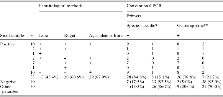
Regarding the parasitological methods, 29 of the 33 samples were positive using agar plate culture and only 4 were positive for the other methods (Table 1).
Comparison of the results obtained with parasitological and conventional PCR methods for the detection of Strongyloides stercoralis in stool samples

n (total samples in relation to parasitological methods); +(positive samples); −(negative samples); κ 0·66* and κ 0·59**.
PCR assay was first evaluated using DNA isolated from S. stercoralis larvae and performed using species-specific and genus-specific primer pairs. DNA fragments of the expected sizes (101 and 392 bp, respectively) were obtained (Fig. 1). The sequences of PCR products from DNA obtained from filariform larvae of S. stercoralis using species-specific primers showed high similarity while the sequences obtained using genus-specific primers showed less similarity in the database.
PCR products on 2·0% agarose gel. (A) Amplification of the 18S gene with species-specific primer pairs (101 bp). Lane M, DNA size markers (100 bp ladder; Fermentas); lane 1, negative control, lane 2, S. stercoralis larvae DNA; lanes 3–5, stool samples of patients infected with S. stercoralis; lanes 6–8, stool samples with other parasite infections; lanes 9–11, negative stool samples; (B) Amplification of the 18S gene with genus-specific primer pairs (392 bp). Lane M, DNA size markers (100 bp ladder; Invitrogen); lanes 1–5, stool samples of patients infected with S. stercoralis; lane 6, stool samples with other parasite infections; lane 7, negative stool samples; lane 8, negative control; lane 9, S. stercoralis larvae DNA.

PCR sensitivity was determined to detect S. stercoralis DNA obtained from experimentally spiked stools (10, 5, 2 and 1 larvae per 500 mg stool). PCR sensitivity was able to detect 1 larva per 500 mg stool with both primers.
The sequences of PCR products from DNA obtained from filariform larvae of S. stercoralis using species-specific primers showed high similarity while the sequences obtained with genus-specific primers showed less similarity in the database.
The results obtained using species- and genus-specific conventional PCR methods are shown in Table 1. Species- and genus-specific PCR were positive in 28 (87·9%) and 26 (78·8%) samples, respectively. The β-globin gene amplification (internal control, 110 bp) was observed in all samples evaluated.
Among the 40 stool samples negative by parasitological methods, DNA amplification was observed in 7 (17·5%) using species-specific primers and 2 (5·0%) using genus-specific primers (Table 1).
In 30 stool samples positive for other parasite infections, DNA amplification of S. stercoralis was detected in 4 (13·3%) using species-specific primers; however, amplification was observed in 9 samples (30·0%) with genus-specific primers, including samples infected with S. mansoni (n = 2), hookworm (n = 2), A. lumbricoides (n = 1), H. nana (n = 1), G. lamblia (n = 1) and poly-infected samples (n = 2).
DISCUSSION
Strongyloidiasis is one of several neglected diseases that show worldwide distribution, with endemic areas in developing countries, where chronic infections are life threatening (Marcilla et al. Reference Marcilla, Garg, Bernal, Ranganathan, Forment, Ortiz, Muñoz-Antolí, Dominguez, Pedrola, Martinez-Blanch, Sotillo, Trelis, Toledo and Esteban2012). In the majority of uncomplicated cases of strongyloidiasis, the intestinal worm load is very low and the output of larvae is minimal (Moghaddassani et al. Reference Moghaddassani, Mirhendi, Hosseini, Rokni, Mowlavi and Kia2011). Using conventional methods, the detection rate can be low and multiple samples have to be examined to achieve adequate sensitivity (Paula and Costa-Cruz, Reference Paula and Costa-Cruz2011). Thus the development of highly sensitive diagnostic tests to detect mild cases of strongyloidiasis is crucial to prevent potentially fatal infections.
PCR has been proposed for the detection of both protozoan and helminthic infections in fecal samples (Verweij et al. Reference Verweij, Canales, Polman, Ziem, Brienen, Polderman and van Lieshout2009; Basuni et al. Reference Basuni, Muhi, Othman, Verweij, Ahmad, Miswan, Rahumatullah, Aziz, Zainudin and Noordin2011; Taniuchi et al. Reference Taniuchi, Verweij, Noor, Sobuz, Lieshout, Petri, Haque and Houpt2011), and has been shown to be a highly sensitive and specific method compared with the routine approach of microscopy and antigen-based methods. In this study, sensitivity similar to that obtained by parasitological methods for the detection of S. stercoralis infection was achieved using conventional PCR.
The sensitivity of the agar plate culture method was 87·9%. Studies have demonstrated the high efficiency of agar plate culture in the diagnosis of strongyloidiasis, even when only a few worms are present (Koga et al. Reference Koga, Kasuya, Khamboonruang, Sukhavat, Ieda, Takatsuka, Kita and Ohtomo1991; Inês et al. Reference Inês Ede, Souza, Santos, Souza, Santos, Silva, Silva, Teixeira and Soares2011). In the present study, two patients were negative by agar plate culture and positive by the Rugai method. This is in agreement with observations reported by Inês et al. (Reference Inês Ede, Souza, Santos, Souza, Santos, Silva, Silva, Teixeira and Soares2011), suggesting the need for concurrent use of these methods to improve the sensitivity of parasitological methods. Our study combined the parasitological methods to determine the number of stool samples positive for S. stercoralis.
When determining the contribution of a more sensitive method in strongyloidiasis diagnosis, the choice of primer pairs is extremely important. The target DNA sequence for many diagnostic PCRs of intestinal parasites is the small subunit rRNA. This gene is present in all eukaryotes, occurs in high copy numbers and sequence data are available for the majority of intestinal parasites (Stensvold et al. Reference Stensvold, Lebbad and Verweij2011). Species-specific and genus-specific primers were evaluated using DNA from S. stercoralis larvae, which produce fragments of 101 and 392 bp. This region has been described in the literature as a diagnostic marker species-specific within the genus Strongyloides sp. (Hasegawa et al. Reference Hasegawa, Hayashida, Ikeda and Sato2009; Verweij et al. Reference Verweij, Canales, Polman, Ziem, Brienen, Polderman and van Lieshout2009; Janwan et al. Reference Janwan, Intapan, Thanchomnang, Lulitanond, Anamnart and Maleewong2011; Repetto et al. Reference Repetto, Soto, Cazorla, Tayeldin, Cuello, Lasala, Tekiel and González Cappa2013).
The DNA extraction procedure is fundamental to increasing the sensitivity of the S. stercoralis PCR assay in stool samples. To obtain the optimum performance for DNA extraction, it was necessary to increase the time of action of proteinase K, which facilitated the action of buffers during DNA extraction. The cuticles of nematodes are extremely resistant to environmental reagents, conferring a barrier to chemical, enzymatic and physical compromise within the host organism (Dawkins and Spencer, Reference Dawkins and Spencer1989). Repetto et al. (Reference Repetto, Soto, Cazorla, Tayeldin, Cuello, Lasala, Tekiel and González Cappa2013), for the same reason, showed optimum performance of the in-house method of DNA isolation making a variation in DNA extraction protocol, however, this variation was exhaustive. The extraction of DNA used in this study, with a few modifications in the instructions proposed by the manufacturer, showed better results than those presented by Repetto et al. (Reference Repetto, Soto, Cazorla, Tayeldin, Cuello, Lasala, Tekiel and González Cappa2013) using only the kit for extracting DNA in fecal samples, and higher sensitivity than studies proposed by Schär et al. (Reference Schär, Odermatt, Khieu, Panning, Duong, Muth, Marti and Kramme2013) and Verweij et al. (Reference Verweij, Canales, Polman, Ziem, Brienen, Polderman and van Lieshout2009).
Different factors have to be considered when comparing the parasitological methods with the PCR assay. Schär et al. (Reference Schär, Odermatt, Khieu, Panning, Duong, Muth, Marti and Kramme2013) reported that the sample input volume of parasitological methods and PCR assays differs by a factor of 10, with considerable increases in sensitivity for the parasitological methods. In contrast, in our study, the PCR assay using species-specific primers showed sensitivity values near to those demonstrated by the agar plate culture method, and a higher sensitivity when compared with Rugai and Lutz methods.
Sensitivity and specificity of the conventional PCR assay in the present study were similar to those reported in studies using real-time PCR (Verweij et al. Reference Verweij, Canales, Polman, Ziem, Brienen, Polderman and van Lieshout2009; Kramme et al. Reference Kramme, Nissen, Soblik, Erttmann, Tannich, Fleischer, Panning and Brattig2011; Schär et al. Reference Schär, Odermatt, Khieu, Panning, Duong, Muth, Marti and Kramme2013). When cost-effectiveness is measured the conventional PCR is more applicable than qPCR.
In recent literature, the limits detection were 4 larvae per 100 mg stool (Janwan et al. Reference Janwan, Intapan, Thanchomnang, Lulitanond, Anamnart and Maleewong2011) and 100 Strongyloides larvae per 200 mg stool (Kramme et al. Reference Kramme, Nissen, Soblik, Erttmann, Tannich, Fleischer, Panning and Brattig2011). The limits detection of the conventional PCR sensitivity was of 1 larva per 500 mg stool for both primers.
Another factor that should be considered in PCR assays using fecal samples is the presence of substances which can inhibit DNA amplification by PCR (Repetto et al. Reference Repetto, Soto, Cazorla, Tayeldin, Cuello, Lasala, Tekiel and González Cappa2013). The false-negative conventional PCR results were not caused by inhibition of the amplification by fecal components, because amplification of the internal control was observed in all samples. The DNA amplification in stool samples negative by parasitological methods may be due to failure in the sensitivity of these methods, since only one sample was obtained. It is reported that the analysis of a single stool sample has a sensitivity of only 15–30%; however, sensitivity increases to nearly 100% when seven consecutive daily stool specimens are examined (Uparanukraw et al. Reference Uparanukraw, Phongsri and Morakote1999). On the other hand, failure of the parasitological methods to detect these samples could be explained by the intermittent excretion of larvae due to chronic infections or by the presence of dead larvae. In another hand, PCR does not depend on parasite viability for detection of S. stercoralis (Verweij et al. Reference Verweij, Canales, Polman, Ziem, Brienen, Polderman and van Lieshout2009).
In this study, PCR sensitivity was associated with lower specificity, characterized by DNA amplification in stool samples positive for other parasitic infections. The presence of these bands could have occurred because the genus-specific primers amplified different regions of the 18S ribosomal RNA gene, instead of the expected nematode-specific region and could be due to DNA competition for PCR amplification in mixed samples (Marra et al. Reference Marra, Chiuso-Minicucci, Machado, Zorzella-Pezavento, França, Ishikawa, Amarante, Sartori and Amarante2010 ). Cases that DNA amplified in stool samples positive for other parasitic infection with both primers were helminths, except for a single sample positive for Blastocystis spp. The species-specific primer was able to amplify DNA of nematodes, all of them hookworms. This occurs commonly in stool samples obtained from areas endemic for these parasites, as in Brazil. DNA competition may occur even when the amplified products are approximately of the same length (Kreike and Lehner, Reference Kreike and Lehner1995), as observed by lower similarity in the database of sequences obtained with genus-specific primers.
The study showed that conventional PCR employing species-specific primer pairs provides a specific, sensitive molecular method for S. stercoralis diagnosis in human stool samples. Further studies are required to determine the effect of multiple stool sampling on the detection rate of S. stercoralis infections by PCR-based methods. In addition, conventional PCR for S. stercoralis can be applied in association with stool sample examinations in epidemiological studies to improve the diagnosis of strongyloidiasis, particularly in immunocompromised individuals who are at risk of fatal disease.
FINANCIAL SUPPORT
This research was supported by Fundação de Amparo a Pesquisa do Estado de São Paulo (FAPESP 2010/51110-2), Brazil.