




No CrossRef data available.
Article contents
Repair of aortic coarctation using temporary ascending to descending aortic bypass in children with poor collateral circulation
Published online by Cambridge University Press: 20 January 2005
Abstract
Aortic coarctation can now be repaired surgically with excellent results. Even though rare, injury to the spinal cord resulting in paraplegia remains a major concern. Preoperative evaluation showing the absence of collateral circulation is valuable in order to introduce protective actions. This report describes our experience using a temporary bypass from the ascending to the descending aorta bypass in children undergoing surgical correction of aortic coarctation in the setting of poorly developed collateral circulation.
Between 1990 and 2002, we undertook direct surgical repair in 56 patients with isolated aortic coarctation, 20 as neonates, 11 as infants, and 25 during childhood. From 1998 onwards, we introduced preoperative evaluation of the collateral circulation with magnetic resonance imaging. From that time, we placed a temporary bypass from the ascending to the descending aorta, using a polytetrafluoroethylene tube of 4 to 8 mm diameter, whenever distal pressures were shown to be 25 mmHg or less after test clamping, or when magnetic resonance imaging revealed absence of collateral circulation.
We found excellent correlations between the direct intra-operative measurements of distal pressure and the findings at magnetic resonance imaging. Following introduction of the temporary bypass, we observed no neurological complications, nor were there any complications related to bypass. Freedom from restenosis was 96%.
Preoperative magnetic resonance imaging, therefore, can accurately visualize poor collateral circulation in children with aortic coarctation. The use of a temporary bypass can possibly eliminate the risk of neurological sequels following direct repair of coarctation in children with poorly developed collateral circulation. The temporary bypass is both easy to apply and safe.
Information
- Type
- Original Article
- Information
- Copyright
- © 2004 Cambridge University Press
It has long been known that patients with untreated coarctation of the thoracic aorta have reduced life spans because of associated complications.1 Surgical repair of coarctation was first described in 19452 and, since then, has been the procedure of choice, with excellent results. Injury to the spinal cord, nonetheless, with paraplegia as result, is a major concern following surgical repair, albeit that in large series the risk has been reported to be low. In small neonates and infants, the incidence is said to be less than 0.5%.3, 4 In older children and adults, however, and for patients undergoing surgery for recurrent coarctation, the risk is higher, and has been reported at 2.6%.5 The mechanism for development of the injury is believed to be compromised flow of blood to the spinal cord, either due to prolonged aortic occlusion, the interruption of vital collateral blood vessels, or most often because of the absence of adequate collateral circulation.3, 6 Even though the extent of collateral blood vessels is usually proportional to the severity of the coarctation, exceptions do occur. The collateral flow is usually through the intercostal arteries,7 but these collateral vessels may not always be readily visible on standard angiographic examination. Recent studies have demonstrated that the collateral vessels are better detected using velocity mapping of magnetic resonance images.8 More frequent use of preoperative magnetic resonance angiography now gives an opportunity to evaluate the collateral circulation prior to surgery, thus giving the surgeon the possibility better to plan any necessary precautions to avoid ischaemia of the spinal cord during surgery. At surgery immediately following test aortic cross-clamping, direct measurements of the blood pressure distal to the clamp can also be used to identify a poorly developed collateral circulation, even though the critical pressure has not yet been established.9 In this respect, various groups have suggested construction of shunts,10–12 various bypasses,13–15 or bypass of the left heart using a centrifugal pump16 to prevent spinal ischaemia in patients with a distal systolic pressure of 50 mmHg or lower.16, 17
We have now tried to evaluate the relationship between preoperative mapping of collateral circulation using magnetic resonance imaging and distal pressure measurements obtained following aortic cross-clamping in children with aortic coarctation. We also describe our experience using a temporary bypass placed between the ascending and descending aorta in patients whom we knew to have poorly developed collateral circulation.
Materials
Patients
Between January1990 and June 2002, we undertook surgical correction of 67 children with aortic coarctation. Of these, 4 patients (6%) had complex intracardiac anomalies, and 7 patients (10%) presented with aortic coarctation combined with ventricular septal defect. We excluded these cases from the subsequent analysis. The remaining 56 children were the focus for this report, and of these, 23 had a patent arterial duct that was closed surgically at the time of the operation.
Of the children at the time of the operation, 20 (36%) were neonates younger than 1 month, 11 (20%) were infants aged from 1 month to 1 year, and 25 were children aged from 1 year to 15 years. The distribution according to age, gender, weight, urgency, as well as the pressure gradient measured across the site of coarctation, is summarized in Table 1.
Table 1. Distribution according to age, gender, urgency of the operation and pressure gradient measured over the coarctation at surgery (mmHg) in 56 patients undergoing direct surgical repair.

From January 1998, we have performed preoperative magnetic resonance imaging in these patients, evaluating in particular the collateral circulation. This cohort now includes 21 patients. We also performed direct intraoperative measurements of distal pressures in all patients during the period of study.
Also from January, 1998, we have inserted a temporary bypass from the ascending to the descending aorta during the period of aortic cross-clamping in all patients in whom we measured a peak systolic pressure distal to the coarctation that was 25 mmHg or lower than the pressures measured proximally. We also used this temporary bypass in all those patients in whom the preoperative resonance images showed poor collateral circulation, either using T1-weighted spin-echo, phase-contrast magnetic resonance angiography, or contrast enhanced 3-dimensional magnetic resonance imaging.
Surgery
In all the patients, we resected the coarctation and achieved a direct end-to-end anastomosis. With the patient positioned in the right lateral decubitus position, we made a left postero-lateral thoracotomy through the 4th intercostal space. With a rib spreader in place, the left lung was retracted anteriorly, a manoeuver which readily exposes the site of coarctation, the distal part of the aortic arch, and left subclavian artery, as well as the proximal part of the descending thoracic aorta. Thereafter, the mediastinal pleura is opened over the descending thoracic aorta. After heparinization, using 100 units/kg, we carry out test aortic clamping distal to the duct, measuring pressures distal to the clamp. The coarctation is then resected, the duct is divided and its pulmonary end is over-sewn, and an end-to-end anastomosis is completed using a running suture of 6-0 polydioxanonesulphate (Ethicon, Sommerville, New Jersey, USA) without tension. In 13 patients (23%), with no evidence of hypoplasia of the aortic arch, the proximal aorta was incised towards the base of the origin of the left subclavian artery, the so-called “Brom plasty”, prior to fashioning the end-to-end anastomosis. In patients with a moderate degree of hypoplasia of the aortic arch, we incise the inferior border of the aortic arch and perform an extended end-to-end anastomosis. The mean period of aortic cross clamping was 14 ± 5 min, with a range from 8 to 22 min.
Temporary ascending to descending aortic bypass
From January 1998 we inserted a temporary bypass from the ascending to the descending aorta in all patients with a poorly collateral circulation as indicated by the preoperative magnetic resonance imaging, and for those where we found a distal aortic pressure of 25 mmHg or less after test clamping of the aorta. Through an upper longitudinal pericardial incision, we prepared the ascending aorta and, after heparinization, we placed a small Cooley clamp between its proximal and median segments. Having made a 4 to 5 mm aortotomy, we anastomosed a polytetrafluoroethylene tube of 4 to 8 mm internal diameter end-to-side to the ascending aorta. The distal end of the tube is then sutured end-to-side to the descending aorta distal to the coarctation, again using lateral clamping (Fig. 1). The pressure measured distally after placement of the graft and subsequent aortic cross clamping was 59 ± 9 mmHg, ranging from 40 to 60 mmHg. Once the shunt is in place, the operation proceeded as described above. Once the repair is finished, and the aortic clamps removed, the temporary bypass is removed, and the arteriotomies closed under lateral clamping. The average additional operating time required for creation and dismantling the temporary bypass was 16 ± 3 min. The blood pressures measured in the right arm did not change either during the period of temporary shunting or during the repair of the ascending aorta.

Figure 1. Artist's drawing showing the temporary shunt placed to provide a bypass from the ascending to the descending aorta during direct repair of aortic coarctation in children. We used a polytetrafluoroethylene tube graft with an inner diameter from 4 to 8 mm for the temporary shunt.
Statistics
We used the Wilcoxon two sample test for comparison between measurements of distal pressure and presence or absence of collateral circulation. Actuarial survival and freedom from restenosis was calculated using the Kaplan-Meier method.
Results
There was no hospital death. In 35 patients (62%), the distal pressure measured intraoperatively was greater than 46 mmHg, while in 21 patients the distal pressures were 45 mmHg or less. In 13 patients, the distal aortic pressure after test occlusion of the aorta was 25 mmHg or lower and, of these, 12 were children aged between 2 and 15 years old, with the other patient being a neonate.
We encountered one episode of postoperative paraplegia, occurring in a child undergoing surgery in 1997. Retrospective analysis revealed that this child had a less tight coarctation, with a distal pressure after aortic cross clamping of only 22 mmHg, indicating a poorly developed collateral circulation. Since our introduction of routine preoperative magnetic resonance imaging, and the insertion of temporary shunts in selected cases, no further neurological deficits have been encountered after repair of the aortic coarctation. Other postoperative complications have also been rare.
During follow up, one child died 4 months after the repair of aortic coarctation, immediately following a reoperative procedure for closure of a previously undiagnosed ventricular septal defect. Actuarial rates of survival at 5 and 10 years, respectively, were 97%. Restenosis occurred in only one patient 6 years after the repair, giving an actuarial freedom from restenosis of 96%.
Analysis of subgroups
In 7 of the 21 patients undergoing surgery since the change of strategy in 1998, we found distal intraoperative pressures of 25 mmHg or less.
In this group of 21 patients, there was an excellent correlation between preoperative magnetic resonance imaging showing good or underdeveloped collateral circulation and intraoperative measurements of the distal pressures (Fig. 2: p = 0.003). We show examples of magnetic resonance angiography revealing well developed as opposed to poor collateral circulation in Figures 3 and 4.

Figure 2. Relationship between magnetic resonance imaging findings regarding well developed as opposed to underdeveloped collateral circulations and direct intra-operative measurements of distal pressure after test cross-clamping in 21 patients undergoing repair of coarctation.

Figure 3. Preoperative magnetic resonance imaging and resonance angiography in a child with a narrow coarctation of the aora (arrow), left panel, but with well-developed collateral circulation and a dilated internal thoracic artery (arrows), right panel.

Figure 4. Preoperative magnetic resonance imaging and resonance angiography in a child with a less severe coarctation of the aorta, left panel, with no collateral circulation visualized on resonance angiography, middle panel, even after spiral reconstruction, right panel.
In all of the 7 patients with distal pressures lower than 25 mmHg and poor collateral circulation as seen on the preoperative magnetic resonance images, we inserted a temporary bypass from the ascending to the descending aorta. These patients comprised 3 boys and 4 girls, with a mean age of 3.9 ± 2.6 years, ranging from 1 day to 8 years, with six of them being children and the other a neonate. In this latter patient, an urgent operation was required at the age of 1 day. The distal arterial pressures after cross clamping were 19.1 ± 2.2 mmHg, with a range from 17 to 22 mmHg. No shunt related mortality or morbidity was observed.
Discussion
Surgical correction of coarctation of the aorta in children has now become a standard procedure, with excellent results and few complications. In such children, resection and end-to-end anastomosis is by far the commonest procedure employed. Ischaemia of the spinal cord, with paraplegia as result, even though rare, has emerged as a special problem, and continues to be a major concern in patients undergoing surgical repair of their coarctation.3, 17 The exact cause of the damage to the spinal cord remains to be clarified, even though there is a consensus that this complication arises because of reduced flow of blood to the spinal cord. This is promoted by situations that may fail to stimulate the development of the usual amount of collateral circulation. Such situations include coarctation in infancy, coarctation proximal to the left subclavian artery, coarctation associated with stenosis of the origin of left subclavian artery, and possibly in patients with something less than severe narrowing of the coarcted area.17 Hughes and Reemtsma9 had already recommended in the 1970s that the distal aortic pressure should be measured at the time of aortic occlusion as an indicator of the adequacy of collateral circulation. The critical pressure indicating inadequate collateralisation, however, has still to be established. In our small series, a total of 13 patients had distal aortic pressures of 25 mmHg or lower. Of these 13 patients, 12 were children aged between 2 and 15 years, and only one was a neonate. These findings correspond with earlier reports.17 Recently Wada and associates18 have proposed the monitoring of somatosensory evoked potentials, together with measurements of pressures of cerebrospinal fluid during surgery, to detect poor collateral circulation. Since the absence of or poor development of collateral circulation is a major contributing factor for neurologic complications, it follows that a more precise preoperative evaluation would allow better planning of the optimal surgical strategy prior to the intervention. Such preoperative evaluation has previously been performed with angiography to obtain accurate information about the morphology of coarctation. It has now been demonstrated that cross-sectional echocardiography combined with Doppler interrogation,19 as well as magnetic resonance imaging, can accurately establish the anatomy and severity of coarctation, so angiography is rarely performed nowadays. Using magnetic resonance imaging, the condition of the collateral circulation can be identified by conventional T1-weighted spin echo images, or with magnetic resonance angiography.20 Contrast enhanced 3-dimensional magnetic resonance angiography of the thoracic vessels offers yet another way to visualize collaterals, a technique that is easily performed in children, even in infants.21 Quantification of collateral flow with magnetic resonance velocity mapping has recently been added as an excellent technique if abundant collaterals are not visualized with the other techniques.9, 22 In our patients, we now routinely use magnetic resonance imaging as a preoperative tool, and we have found an excellent relationship between magnetic resonance imaging findings and low distal pressure measurements distal to aortic occlusion after test clamping of the aorta.
As a consequence of this improved preoperative evaluation, which provides an accurate evaluation of the state of the collateral circulation, we now institute preventive measures in all patients with an underdeveloped collateral circulation, using a temporary bypass from the ascending to the descending aorta for this purpose. The bypass has been described earlier as a permanent solution for treatment of complex aortic coarctation, with some authors advocating an approach via a median sternotomy, and others using a left thoracotomy as did we when fashioning our temporary shunt.23, 24 Various other techniques have also been described for temporary bypassing the area of coarctation to avoid neurologic sequels.10, 16 We have found that our temporary bypass is easy to construct, requires no additional operative access, allows an unobstructed operative field, is not associated with any complications related to the bypass, and prolongs the operative time by no more than approximately 15 min. In contrast to partial bypass using a centrifugal pump, our approach does not require femoral cannulation, which could be cumbersome in neonates, nor does it require full heparinization, thus avoiding the inflammatory response associated with extracorporeal circulation. Since the introduction of our temporary shunt, we have not experienced any neurological incidents during or after surgical repair of coarctation in children. Our long-term follow-up corresponds with earlier reports, confirming the low incidence of re-stenosis after simple resection and end-to-end anastomosis.25, 26
We conclude, therefore, that preoperative magnetic resonance imaging can accurately visualize any underdeveloped collateral circulation in children with aortic coarctation. The use of a temporary bypass from the ascending to the descending aorta provides a safe means of performing direct repair of aortic coarctation in children with such an underdeveloped collateral circulation. It is easy technically to insert the shunt, which is without associated morbidity.
References
Crafoord C, Nylin G. Congenital coarctation of the aorta and its surgical treatment. J Thorac Cardiovasc Surg 1945; 14: 347–361.Google Scholar
Brewer LA III, Fosburg RG, Mulder GA, Verska JJ. Spinal cord complications following surgery for coarctation of the aorta: a study of 66 cases. J Thorac Cardiovasc Surg 1972; 64: 368–381.Google Scholar
Keen G. Spinal cord damage and operations for coarctation of the aorta: etiology, practice, and prospects. Thorax 1987; 42: 11–18.Google Scholar
Westaby S, Parnell B, Pridie RB. Coarctation of the aorta in adults. Clinical presentation and results of surgery. J Cardiovasc Surg (Torino) 1987; 28: 124–127.Google Scholar
Krieger KH, Spencer FC. Is paraplegia after repair of coarctation of the aorta due principally to distal hypotension during aortic cross clamping? Surgery 1985; 97: 2–7.Google Scholar
Barnes RW, Rittenhouse EA, Kongtahworn C, Doty DB, Rossi NP, Ehrenhaft JL. Reversed intercostal arterial flow in coarctation of the aorta. Ann Thorac Surg 1975; 19: 27–32.Google Scholar
Holmqvist C, Stahlberg F, Hanséus K, et al. Collateral flow in coarctation of the aorta with magnetic resonance velocity mapping: correlation to morphological imaging of collateral vessels. J Magn Reson Imaging 2002; 15: 39–46.Google Scholar
Hughes RK, Reemtsma K. Correction of coarctation of the aorta: manometric determination of safety during test occlusion. J Thorac Cardiovasc Surg 1971; 62: 31–33.Google Scholar
Gott VL. Heparinized shunts for thoracic vascular operations. Ann Thorac Surg 1972; 14: 219–220.Google Scholar
Alexander JC Jr. Maintenance of distal aortic perfusion by heparin-bonded shunt during repair of coarctation of the aorta with minimal collateral circulation. Ann Thorac Surg 1981; 32: 304–306.Google Scholar
Lam CR, Arciniegas E. Surgical management of coarctation of the aorta with minimal collateral circulation. Ann Surg 1973; 178: 693–697.Google Scholar
Kouchoukos NT, Lell WA, Karp RB, Samuelson PN. Hemodynamic effects of aortic clamping and decompression with a temporary shunt for resection of the descending thoracic aorta. Surgery 1979; 85: 25–30.Google Scholar
Robicsek F, Hess PJ, Vajtai P. Ascending-Distal Abdominal aorta bypass for treatment of hypoplastic aortic arch and atypical coarctation in the adult. Ann Thorac Surg 1984; 37: 261–263.Google Scholar
Cartier R, Orszulak TA, Pairolero PC, Schaff HV. Circulatory support during crossclamping of the descending thoracic aorta. Evidence of improved organ perfusion. J Thorac Cardiovasc Surg 1990; 99: 1038–1047.Google Scholar
Wong CH, Watson B, Smith J, Hamilton JR, Hasan A. The use of left heart bypass in adult and recurrent coarctation repair. Eur J Cardiothorac Surg 2001; 20: 1199–1201.Google Scholar
Servais LJ, Rivelli SK, Daxchy BA, Christophe CD, Dan B. Anterior spinal artery syndrome after aortic surgery in a child. Pediatric Neurology 2001; 24: 310–312.Google Scholar
Wada T, Yao H, Miyamoto T, Mukai S, Yamamura M. Prevention and detection of spinal cord injury during thoracic and thoracoabdominal aortic repairs. Ann Thorac Surg 2001; 72: 80–85.Google Scholar
Smallhorn JF, Huhta JC, Adams PA, Anderson RH, Wilkinson JL, McCartney FJ. Cross-sectional echocardiographic assessment of coarctation in sick neonates and infant. Br Heart J. 1983; 50: 349–361.CrossRefGoogle Scholar
Von Schulthess GJ, Higashino SM, Higgins SS, Didier D, Fisher MR, Higgins CB. Coarctation of the aorta: MR imaging. Radiology 1986; 158: 469–474.Google Scholar
Julsrud PR, Breen JF, Flemlee JP, Warnes CA, Connolly HM, Schaff HV. Coarctation of the aorta: collateral flow assessment with phase contrast MR angiography. Am J Roentgenol 1997; 169: 1735–1742.Google Scholar
Steffens JC, Bourne MW, Sakuma H, O'Sullivan M, Higgings CB. Quantification of collateral blood flow in coarctation of the aorta by velocity encoded cine magnetic resonance imaging. Circulation 1994; 90: 937–943.Google Scholar
Grinda JM, Macé L, Dervanian P, Folliguet TA, Neveux J-Y.Bypass of complex forms of isthmic aortic coarctation in adults. Ann Thorac Surg 1995; 60: 1299–1302.Google Scholar
Karolczak MA, McKay R, Arnold R. Right-sided intrathoracic bypass graft for complex recurrent coarctation of the aorta. Eur J Cardio-thorac Surg 1989; 3: 278–281.Google Scholar
Corno AF, Botta U, Hurni M, et al. Surgery for aortic coarctation: a 30 years experience. Eur J Cardio-thoracic Surg 2001; 20: 1202–1206.Google Scholar
Lerberg DB, Hardesty RL, Siewers RD, Zuberbuhler JR, Bahnson HT. Coarctation of the aorta in infants and children: 25 years experience. Ann Thorac Surg 1982; 33: 159–170.Google Scholar
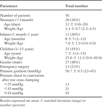
Table 1.

Artist's drawing showing the temporary shunt placed to provide a bypass from the ascending to the descending aorta during direct repair of aortic coarctation in children. We used a polytetrafluoroethylene tube graft with an inner diameter from 4 to 8 mm for the temporary shunt.

Relationship between magnetic resonance imaging findings regarding well developed as opposed to underdeveloped collateral circulations and direct intra-operative measurements of distal pressure after test cross-clamping in 21 patients undergoing repair of coarctation.

Preoperative magnetic resonance imaging and resonance angiography in a child with a narrow coarctation of the aora (arrow), left panel, but with well-developed collateral circulation and a dilated internal thoracic artery (arrows), right panel.

Preoperative magnetic resonance imaging and resonance angiography in a child with a less severe coarctation of the aorta, left panel, with no collateral circulation visualized on resonance angiography, middle panel, even after spiral reconstruction, right panel.
 You have
Access
- 13
- Cited by