Introduction
Inflammatory bowel disease (IBD) is a chronic relapsing inflammation afflicting any part of the entire bowel wall, which presents as either ulcerative colitis (UC) or Crohn's disease. IBD is the result of a deregulated, aberrant and even inappropriate overactivation of the immune response in the intestinal wall due to defects in barrier function between the intestinal epithelium and mucosal immune system (Klotz & Schwab, Reference Klotz and Schwab2005; Jung et al., Reference Jung, Kim, Kim, Kong, Choi, Yang and Kim2006). IBD affects about 12% of the population worldwide (Halvorson et al., Reference Halvorson, Schlett and Riddle2006). Unfortunately, there is no complete cure for IBD and the major therapeutic goals are to decrease the relapse episodes and to enhance the quality of life of patients (Gionchetti et al., Reference Gionchetti, Amadini, Rizzello, Venturi and Campieri2002).
Ulcerative colitis invariably affects the rectum and may extend proximally in a confluent pattern to involve the entire colon (Friend, Reference Friend2005). Moreover, long-standing extensive UC is associated with the development of UC-related colorectal carcinoma (Ekbom et al., Reference Ekbom, Helmick, Zack and Adami1990). Intracolonic instillation of acetic acid (4–10%) can induce acute colitis in rats, mice and guinea-pigs. The inflammation is characterized by histopathological changes that are very similar to those of UC. This model of induced colitis shows fast development, as well as long-lasting stable effects within 8–12 weeks (Fabia et al., Reference Fabia, Willen, Ar'Rajab, Andersson, Ahren and Beng-mark1992; Qin et al., Reference Qin, Wu, Tong, Sung, Xu and Bian2011). Recently, novel markers have been used for better assessment of colonic injury. One of the promising markers is pentraxin 3 (PTX3) – a key protein in modulating tissue inflammation (Agrawal et al., Reference Agrawal, Singh, Bottazzi, Garlanda, Mantovani and Kishore2009).
Helminths are known to skew the immune response towards T-helper cell 2 (Th2), which is characterized by Th2-related cytokines that typically include interleukin (IL)-4, IL-5 and IL-13 (Finkelman et al., Reference Finkelman, Shea-Donohue, Morris, Gildea, Strait, Madden, Schopf and Urban2004). Moreover, helminths can interact with the host's adaptive immune response by down-regulating T- and B-cell responses via the induction of regulatory T cells (Treg) or the anti-inflammatory cytokines IL-10 and transforming growth factor (TGF)-β that can regulate both Th1 and Th2 responses (Maizels & Yazdanbakhsh, Reference Maizels and Yazdanbakhsh2003). This immunomodulation is hypothesized to be beneficial for both the human host and the parasite; it could protect helminths from being eradicated and, at the same time, protect the host from excessive pro-inflammatory responses that may lead to organ damage (Harnett & Harnett, Reference Harnett and Harnett2006).
The gut is the major site for the induction of Treg. Recent findings suggest that some gut commensals, including lactobacilli, bifidobacteria and helminths, play a major role in the induction of Treg in gut lymphoid follicles. CD4+, CD25+, Foxp3+ Treg can suppress and modulate the excessive immune response. Therefore, this represents an essential homeostatic mechanism by which the host can tolerate the massive burden of innocuous antigens within the gut without responding through inflammation (Maloy et al., Reference Maloy, Salaun, Cahill, Dougan, Saunders and Powrie2003; Guarner, Reference Guarner2009).
In recent years, experimental and clinical evidence has accumulated to demonstrate that infection with parasitic worms could protect hosts from IBD. For example, the intestinal helminths Trichinella spiralis (T. spiralis), Trichuris muris and Hymenolepis diminuta protected rodents from induced colitis (Fox et al., Reference Fox, Beck, Dangler, Whary, Wang, Shi and Nagler-Anderson2000; Khan et al., Reference Khan, Blennerhasset, Varghese, Chowdhury, Omsted, Deng and Collins2002). Likewise, infecting IBD patients with Trichuris suis resulted in clinical amelioration of both Crohn's disease and UC (Summers et al., Reference Summers, Elliott, Urban, Thompson and Weinstock2005). The mechanisms of the therapeutic effects of helminths are still not clearly understood. Also, there are many issues that must be elucidated, such as the effective dosage, potential toxicity and inoculation times.
Using an experimental mouse model, the aims of this study were to determine if the underlying mechanism of the host immune regulation inherent to T. spiralis infection involves Treg recruitment, as well as to gain insight about time-related interactions between intestinal nematode infection and colitis in order to ameliorate experimentally induced colitis.
Materials and methods
Animals and experimental design
Male Swiss albino mice weighing 20–25 g were used. They were subdivided into four groups: group I, 20 mice infected with T. spiralis; group II, 20 mice infected with T. spiralis then subjected to induction of colitis 21 days post-infection; group III, 20 mice subjected to induction of acetic acid colitis; and group IV, 20 mice subjected to induction of colitis followed by T. spiralis infection on the third day post-colitis. Another group of 10 normal mice served as a negative control group. A total of five mice from groups II, III and IV were killed by the end of the second and fourth weeks post-colitis and five mice from group I were killed by the end of the second and fourth weeks post-infection. The colon was removed for histopathological, immunohistochemical staining for localization of Foxp3-expressing regulatory cells and assay of PTX3 levels in colonic tissues.
Mice were infected with T. spiralis larvae at a dose of 200 larvae orally, according to Dunn & Wright (Reference Dunn and Wright1985). For induction of colitis, mice were deprived of food the night before induction of colitis then 0.2 ml of 5% acetic acid diluted with saline was instilled into the colon of the mice using a 1 mm diameter enemator, 3.5 cm proximal to the anus. After 20 s of surface contact, the acidic solution was withdrawn and the lumen flushed with 1 ml saline (Luo et al., Reference Luo, Zhong, Cao and Cui2011).
Histopathology and immunohistochemistry
The entire colon was isolated, rinsed with phosphate-buffered saline (PBS) and assessed macroscopically for erythema, bleeding, adhesions or ulcers (Mahgoub et al., Reference Mahgoub, El-Medany, Hager, Mustafa and El-Sabah2003). Parts of the colon from all the studied specimens were fixed in 10% buffered formalin and subsequently processed in alcohols and embedded in paraffin, then submitted to haematoxylin and eosin staining for histopathological examination. The severity of the intestinal lesions was graded using a scale previously described by Al-Chaer et al. (Reference Al-Chaer, Kawasaki and Pasricha2000): 1+ (mild), infiltration of a limited number of neutrophils in the lamina propria with minimal or no interstitial oedema; 2+ (moderate), infiltration of moderate numbers of neutrophils in the lamina propria and moderate interstitial oedema; and 3+ (severe), diffuse infiltration of moderate to large numbers of neutrophils in the lamina propria with severe interstitial oedema.
Immunohistochemistry was carried out for the demonstration of Foxp3-expressing cells using mouse anti-Foxp3 monoclonal antibody (catalogue no. A00744, GenScript, Piscataway, New Jersey, USA; dilution 1:300). After peroxidase blockage and microwave antigen retrieval (using citrate buffer at pH 6.0), 3–5 μm tissue sections were incubated with the primary antibody overnight at 4°C. For the negative control, the primary antibody was replaced with PBS. Rabbit anti-mouse horseradish peroxidase-conjugated secondary antibody was added, followed by incubation for 40 min at room temperature. The colour was developed using diaminobenzidine as a chromogen. Slides were extensively washed with PBS after each step. Finally, they were counter-stained with Mayer's haematoxylin. Cells showing a distinct cytoplasmic/nuclear staining of Foxp3 were considered positive.
The stained sections were assessed for immunoreactivity. The sections were scanned using the 4 × objective lens to allocate the areas of highest intensity of inflammation and the highest intensity of staining. The percentage of positive lymphocytes was determined in ten high-power fields (/10 HPFs) and the mean percentage for each case was recorded. The cases were then scored as follows: 0, no immunoreactivity for Foxp3; 1, less than 25% of lymphocytes stained at the hot areas of inflammation; 2, 25–50% of lymphocytes stained; and 3, more than 50% of lymphocytes stained. The background staining in the necrotic areas and areas of suppuration was neglected during scoring. The mean score of each group was expressed as a mean value ± standard deviation.
Assessment of PTX3 by enzyme-linked immunosorbent assay
The colonic tissues were lysed with Tris-ethylenediaminetetraacetic acid (EDTA) (10 mm Tris-HCl and 1 mm EDTA, pH 7.4), then the tissue was homogenized and centrifuged at 11,000 g for 10 min at 4°C (Castagliulo et al., Reference Castagliulo, Karalis, Valenick, Pasha, Nikulasson, Wlk and Pothoulakis2001). The supernatant was collected, filtered and used for the quantitative determination of PTX3 concentrations by a commercially available enzyme-linked immunosorbent assay (ELISA) kit (Quantkine® Human Pentraxin 3/TSG-14 Immunoassay, catalogue no. DPTX30, R&D Systems Inc., Minneapolis, Minnesota, USA) according to the manufacturer's instructions (Bottazzi et al., Reference Bottazzi, Bastone, Doni, Garlanda, Valentino, Deban, Maina, Cotena, Moalli, Vago, Salustri, Romani and Mantovani2006). This kit is known to react with mouse PTX3 which has about 82% amino acid sequence homology with human PTX (Goujon et al., Reference Goujon, McWilliam, Li, Valentin, Squizzato, Paern and Lopez2010; Sievers et al., Reference Sievers, Wilm, Dineen, Gibson, Karplus, Li, Lopez, McWilliam, Remmert, Söding, Thompson and Higgins2011). Plates were read at 405 nm in an automatic ELISA reader. According to the manufacturer, the mean detection limit for PTX3 is 0.025 ng/ml.
Data analysis
Quantitative data regarding mean ± standard deviation were analysed by analysis of variance using the software package Graphpad Instat version 3 (http://www.graphpad.com/scientific-software/instat/). P values lower than 0.05 were considered statistically significant.
Results
Gross and histopathological examination of the colon
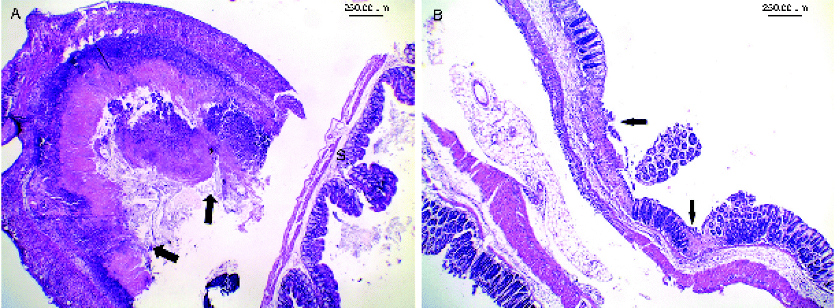
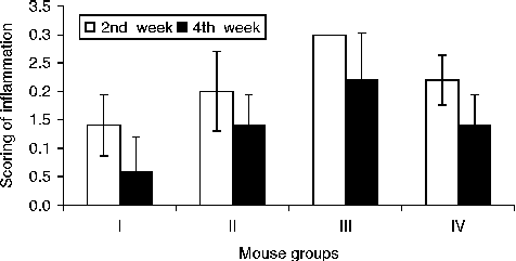
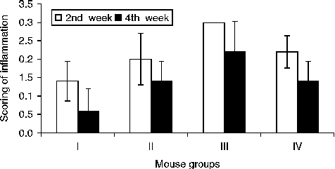
The animals with acetic acid-induced colitis (group III) showed the severest form of inflammation, where the colon (especially the rectum and lower one-third of the colon) exhibited marked gross inflammation in the form of hyperaemia, adhesions and mucosal ulcerations. Microscopically, the colon showed severe inflammatory changes with extensive ulceration, dense neutrophilic infiltrate (fig. 1A) and crypt abscesses, as well as marked goblet cell depletion. The mean score of this group was 3 ( ± 0) at the second week post-colitis (i.e. the maximal score), then declined to 2.2 ( ± 0.84) at the fourth week post-colitis.
(colour online) Histopathological inflammatory changes in the colon ( × 40) of mouse groups II (T. spiralis + colitis) and III (colitis only) after 2 weeks to show (A) severe inflammation with mucosal ulceration and suppuration (arrows) in group III compared with the unaffected part of the small intestine (S), and (B) moderate inflammation with mucosal ulcers (arrows) in group II.

Induced colitis was best ameliorated in group II (Trichinella + colitis) with moderate gross and microscopic inflammation as well as focal ulceration (fig. 1B). The inflammatory changes became milder with time, the mean score for inflammation declining from 2 ( ± 0.7) at the second week post-colitis to 1.4 ( ± 0.55) at the fourth week post-colitis. The severity of inflammation also declined in group IV (colitis + Trichinella). However, amelioration was less compared with that in group II, especially at 2 weeks post-colitis. The mean score of inflammation decreased from 2.2 ( ± 0.44) at the second week post-colitis to 1.4 ( ± 0.54) at the fourth week post-colitis.
On the other hand, group I (Trichinella only) displayed the least inflammation intensity. The colon showed no, or minimal, gross inflammation. Mild to moderate inflammatory infiltrate of the mucosa, formed mainly of eosinophils with eosinophilic cryptitis (intra-epithelial eosinophils), was detected with mild goblet cell depletion. The mean score for the intensity of the inflammation was 1.4 ( ± 0.55) at the second week post-infection and declined to 0.6 ( ± 0.55) at the fourth week post-infection.
Analysis of the mean histopathological scoring of inflammation (fig. 2) showed a statistically significant difference between all groups at both the second and fourth weeks (P < 0.05). In view of these data, it was evident that T. spiralis infection ameliorated the severe inflammation of the colon induced by acetic acid instillation. The amelioration was more evident when T. spiralis infection preceded the induction of colitis.
Mean scores of histopathological changes in mouse groups I (T. spiralis only), II (T. spiralis + colitis), III (colitis only) and IV (colitis +T. spiralis) after weeks 2 (white bars) and 4 (black bars); significant differences in scores (P < 0.05) are shown between groups II and IV, and between both groups and group III.
Immunohistochemistry
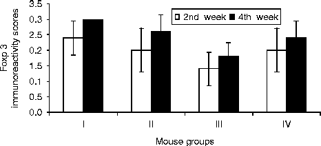
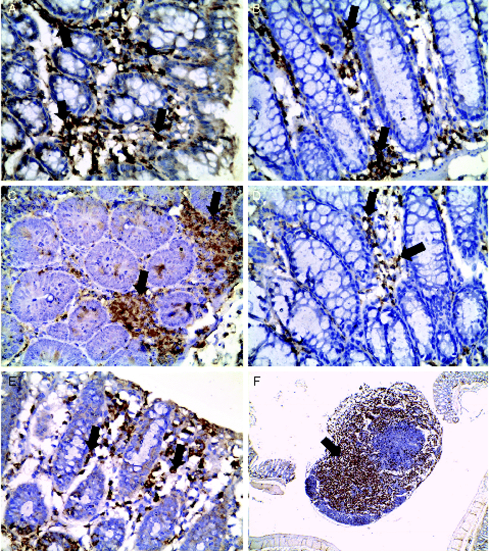
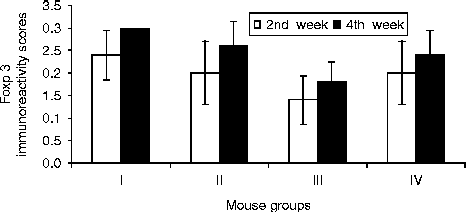
The Foxp3 stain (mostly cytoplasmic) was expressed in the lymphocytes at the areas of inflammation. The highest positivity was detected within the hot zones of inflammation (the rectum and the lower one-third of the colon). Some of the macrophages of the submucosa in the hot areas showed staining for the marker. The immunohistochemical scoring of the four groups is represented in fig. 3, where a statistically significant difference between the four groups was found only at the second killing episode (fourth week, P < 0.05). It was generally observed that the immunostaining for Foxp3 increased with the lapse of time, i.e. higher score at fourth week than at the second week. Also, at the first episode of animal killing, the positivity spanned the whole thickness of the mucosa (fig. 4B), whereas at the second episode of animal killing, the positive cells were more concentrated at the basal part of the mucosa, forming aggregates of Foxp3-positive lymphocytes (fig. 4C).
Mean immunoreactivity scores of Foxp3 in mouse groups I (T. spiralis only), II (T. spiralis + colitis), III (colitis only) and IV (colitis +T. spiralis) after weeks 2 (white bars) and 4 (black bars) to show significant increases in scores (P < 0.05) in groups II and IV compared to group III at 4 weeks.
(colour online) Immunoreactivity of Foxp3 in the colon of mice (A–E; × 400) and the mesenteric lymph nodes (F; × 40) of: (A) group I (T. spiralis only) to show marked superficial staining with the preservation of mucin (arrows) after week 4; (B) group II (T. spiralis + colitis) with moderate diffuse positivity of the mucosa (arrows) after week 2; (C) group II (T. spiralis + colitis) showing basal aggregates of positive lymphocytes (arrows) after week 4; (D) group III (colitis only) with mild mucosal positivity (arrows) after week 4; (E) group IV (colitis +T. spiralis) with moderate mucosal positivity (arrows) after week 4; and (F) group II showing intense positivity of lymphocytes in the mesenteric nodes (arrow) after week 4.
The highest positivity at the first killing episode was detected in group I (2.4 ± 0.55), with a further increase to the maximal score of 3 ( ± 0) at the second episode of killing, together with polarization of the positivity towards the base, and, to a lesser extent, the superficial part of the mucosa (fig. 4A). On the other hand, the colitis group showed the lowest degree of positivity (fig. 4D), with mean score increasing from 1.4 ( ± 0.55) at the second week to 1.8 ( ± 0.45) at the fourth week post-colitis.
Group II showed moderate diffuse mucosal positivity (fig. 4B) with a mean score of 2 ( ± 0.71) at the second week post-colitis which increased to 2.6 ( ± 0.55) at the fourth week post-colitis in all evaluated sections (fig. 4C). Relatively large Foxp3-positive lymphoid aggregates were detected in the basal part of the mucosa with substantial extension of the positivity to the submucosa and to the T-lymphocyte zones of the submucosal aggregates as well as the pericolic lymph nodes (fig. 4F). Group IV showed lower positivity than in group II at the second episode of killing (fig. 4E), when the mean score was 2.4 ( ± 0.55).
Assay of PTX3 levels in colonic tissues
PTX3 levels were significantly highest in group III (3.63 ± 0.44), followed by group II (1.95 ± 0.12), group IV (1.52 ± 0.09), and finally group I (1.37 ± 0.16) as compared with the negative control group (0.85 ± 0.06) at the second week (P < 0.05). At the fourth week, PTX3 levels were lower than those in the second week but still significantly highest in the colitis group (2.20 ± 0.45), followed by group II (1.92 ± 0.05), group IV (1.35 ± 0.26) and, finally, group I (1.29 ± 0.14) as compared with the negative control group (0.84 ± 0.05) (P < 0.05).
Discussion
IBD is not distributed evenly worldwide; it is common in developed countries and is rare in less-developed countries where helminth infections are common (Motomura et al., Reference Motomura, Wang, Deng, El-Sharkawy, Verdu and Khan2009). Advances in understanding the mechanism of bowel inflammation have led to a recent trend of the development of biological therapies that selectively inhibit the inflammatory process (Nakamura et al., Reference Nakamura, Honda, Mizutani, Akiho and Harada2006).
Helminth infections are characterized by their ability to induce Th2 cell responses (Everts et al., Reference Everts, Smits, Hokke and Yazdanbakhsh2010). The concept that helminths protect the host against other immunologically mediated diseases, e.g. Crohn's disease, is based on Th1/Th2 antagonism (Moreels et al., Reference Moreels, Nieuwendijk, De Man, De Winter, Herman, Van Marck and Pelckmans2004). Although Hunter et al. (Reference Hunter, Wang and McKay2007) assumed that helminth-based therapies might be considered inappropriate for UC (Th2 response) and may exacerbate the pathology, it has been suggested that helminth parasites have the ability to reduce Th1- as well as Th2-mediated responses by strongly promoting the immunoregulatory pathways of the immune response (Maizels et al., Reference Maizels, Balic, Gomez-Escobar, Nair, Taylor and Allen2004). Moreover, helminth infection was found to control the Th2 response that contributes to chronic allergic diseases (Yazdanbakhsh et al., Reference Yazdanbakhsh, Kremsner and van Ree2002). CD4+ CD25+ Treg were shown to be the most potent cells in mediating the suppression of allergic immune responses by helminths (Wilson et al., Reference Wilson, Taylor, Balic, Finney, Lamb and Maizels2005). Thus, a question arises: could the Th2 response to parasites protect the host from developing UC, or at least halt the progression of the disease, by the same cellular mechanism?
Previous research has demonstrated the role of T. spiralis infection in ameliorating IBDs (Khan et al., Reference Khan, Blennerhasset, Varghese, Chowdhury, Omsted, Deng and Collins2002). However, the appropriate time of infection with T. spiralis larvae is still not settled, especially in models for UC, and the underlying mechanisms have not been explored. Our study has investigated the role of Treg as a potential key player in nematode-induced immunomodulation of inflammatory colitis. Further, it provides insight about the mutual interaction between T. spiralis and acetic acid colitis.
Acetic acid colitis (used in this study) is a model for UC (Fabia et al., Reference Fabia, Willen, Ar'Rajab, Andersson, Ahren and Beng-mark1992). Destruction of colonic mucosa by acetic acid leads to the influx of luminal pro-inflammatory bacterial products into the lamina propria and the release of cytokines by activated enterocytes, resulting in migration of neutrophils from the microvasculature of the lamina propria which induces significant epithelial erosions (Nusrat et al., Reference Nusrat, Parkos, Liang, Carnes and Madara1997; Boismenu & Chen, Reference Boismenu and Chen2000). The concept that UC is characterized by Th2 cytokine production, thus having Th2-like characteristics, has been disputed recently (Abraham & Cho, Reference Abraham and Cho2009). For instance, the Th1 signalling molecule STAT-4 (signal transducer and activator of transcription 4) is involved in the pathogenesis of the acute phase of UC (Ohtani et al., Reference Ohtani, Ohtsuka, Ikuse, Baba, Yamakawa, Aoyagi, Fujii, Kudo, Nagata and Shimizu2010). Others have suggested that the cytokine profile for UC is less defined, and includes both Th1 and Th2 cytokines (Fuss et al., Reference Fuss, Heller, Boirivant, Leon, Yoshida, Fichtner-Feigl, Yang, Exley, Kitani, Blumberg, Mannon and Strober2004). Therefore, a Th1/Th2 imbalance is thought to be involved (Watanabe et al., Reference Watanabe, Hirono, Hasegawa, Soga and Shibasaki2011). Recently, several studies have highlighted the role of Th17 and its related cytokines in UC, where they elicit potent inflammatory responses (Shen & Durum, Reference Shen and Durum2010; Strober & Fuss, Reference Strober and Fuss2011).
The present study demonstrated that T. spiralis infection ameliorated the severe inflammation induced by acetic acid, as evidenced by the improvement in the histopathological changes, diminution of PTX3 levels, as well as the decreased mortality rate of experimental animals (data not shown). The amelioration was more pronounced and rapid when T. spiralis infection preceded the induction of colitis. In other words, prepolarization of the mucosal immune system to a Th2 response conferred more protection against inflammatory colitis. This might be due to the fact that during the intestinal phase of T. spiralis infection, the immune response is mixed Th1/Th2, with initial predominance of the Th1 type of response and subsequent domination of Th2 (Ishikawa et al., Reference Ishikawa, Goyal, Mahida, Li and Wakelin1998). Boitelle et al. (Reference Boitelle, Di Lorenzo, Scales, Devaney, Kennedy, Garside and Lawrence2005) have showed that T. spiralis is a potent inducer of a Th2 response orchestrated by CD4+ T cells, resulting in gastrointestinal inflammation. Following infection, CD4+ T cells express IL-4, IL-5, IL-9 and IL-10 in the mesenteric lymph nodes, and are subsequently recruited to the gut mucosa (Grencis et al., Reference Grencis, Hultner and Else1991; McDermott et al., Reference McDermott, Grencis and Else2001). Treg play an essential role in controlling the severity of the response. They strike a balance that limits potentially harmful immune-mediated pathology to the host, while still allowing sufficient immune pressure against the pathogen (Belkaid & Tarbell, Reference Belkaid and Tarbell2009; Maizels et al., Reference Maizels, Pearce, Artis, Yazdanbakhsh and Wynn2009).
Interestingly, helminths were found to be protective in several models of IBD, where the disease and the parasites are co-localized (Everts et al., Reference Everts, Smits, Hokke and Yazdanbakhsh2010). Mice exposed to a range of helminths, namely Schistosoma mansoni (Moreels et al., Reference Moreels, Nieuwendijk, De Man, De Winter, Herman, Van Marck and Pelckmans2004), H. diminuta (Reardon et al., Reference Reardon, Sanchez, Hogaboam and McKay2001) or Heligmosomoides polygyrus (Setiawan et al., Reference Setiawan, Metwali, Blum, Ince, Urban, Elliott and Weinstock2007) are protected from induced colitis. The other feature of the response to helminth infection is mobilization of regulatory immune cells and mediators such as IL-10 and TGF-β, which are capable of inducing de novo generation of Foxp3+ Treg (Elliott et al., Reference Elliott, Metwali, Leung, Setiawan, Blum, Ince, Bazzone, Stadecker, Urban and Weinstock2008; Grainger et al., Reference Grainger, Smith, Hewitson, McSorley, Harcus, Filbey, Finney, Greenwood, Knox, Wilson, Belkaid, Rudensky and Maizels2010). This regulatory response inhibits the maturation of CD4+ T cells to Th1 and Th2 effector cells, and thus can control the intensity of both Th1 and Th2 responses (Maizels et al., Reference Maizels, Balic, Gomez-Escobar, Nair, Taylor and Allen2004). Presumably, this is likely to be beneficial both to the worm and the host (Harnett & Harnett, Reference Harnett and Harnett2006). Since Treg can actively suppress the potentially pathogenic immune response, they have a very important role in the maintenance of immune homeostasis (Sakagushi et al., Reference Sakaguchi, Ono, Setoguchi, Yagi, Hori, Fehervari, Shimizu, Takahashi and Nomura2006; Grainger et al., Reference Grainger, Smith, Hewitson, McSorley, Harcus, Filbey, Finney, Greenwood, Knox, Wilson, Belkaid, Rudensky and Maizels2010) and can also control Th2-dominated events such as allergy (McKay, Reference McKay2009).
It has been demonstrated that chronic helminth infections were associated with Treg (Satoguina et al., Reference Satoguina, Mempel, Larbi, Badusche, Loliger, Adjei, Gachelin, Fleischer and Hoerauf2002). However, Boitelle et al. (Reference Boitelle, Di Lorenzo, Scales, Devaney, Kennedy, Garside and Lawrence2005) reported that acute infection with T. spiralis is also associated with an increased production of Treg (Letterio & Roberts, Reference Letterio and Roberts1998). Moreover, Ilic et al. (Reference Ilic, Worthington, Gruden-Movsesijan, Travis, Sofronic-Milosavljevic and Grencis2011) have demonstrated that T. spiralis antigens from all life stages induce a network of different mediators, namely IL-10 and TGF-β, which may contribute to the increase in the Foxp3+ cell populations in the infected host. The results of the present work provide experimental evidence indicating that chronic T. spiralis infection induces a stronger regulatory environment than does the acute infection, as revealed by more evident diminution of the severity of colonic inflammation when induced colitis was preceded by T. spiralis infection. It should also be stressed that the T. spiralis group showed the highest proportion of Treg in various experimental periods.
These results are supported by several investigators. For example, Everts et al. (Reference Everts, Perona-Wright, Smits, Hokke, van der Ham, Fitzsimmons, Doenhoff, van der Bosch, Mohrs, Haas, Mohrs, Yazdanbakhsh and Schramm2009) and Turner et al. (Reference Turner, Jenkins, Hogg, Aynsley, Paveley, Cook, Coles and Mountford2011) demonstrated that helminth infection expanded Foxp3+ Treg in response to distinct molecules released by schistosome eggs. Likewise, Grainger et al. (Reference Grainger, Smith, Hewitson, McSorley, Harcus, Filbey, Finney, Greenwood, Knox, Wilson, Belkaid, Rudensky and Maizels2010) found that helminth antigens, such as excretory–secretory and adult products of Heligmosomoides, do not promote a Th2 response, but rather induce Treg. On the other hand, amelioration of colitis may be also explained by the fact that Foxp3 may act as a negative transcription regulator of Th17 cells. Foxp3 is able to suppress Th17 cell differentiation through the antagonism of retinoid- related orphan receptor (RORγt) activity (transcription factor RORγt is essential for Th17 development and function) (Ivanov et al., Reference Ivanov, Zhou and Littman2007). Furthermore, Zhou et al. (Reference Zhou, Lopes, Chong, Ivanov, Min, Victora, Shen, Du, Rubtsov, Rudensky, Ziegler and Littman2008) have demonstrated that expression of Foxp3 is inversely proportional to IL-23, a key product of Th17. Consequently, regarding immunohistochemical staining of the colon in the colitis group, our results demonstrated that the number of Foxp3-expressing cells was lower than in all the other groups. Similar observations were reported by Chao et al. (Reference Chao, Zhong, Zhang, Gong, Yao and Chen2011). To summarize, the immune response in the intestine displays a delicate balance between the Th17 and the Treg response induced by T. spiralis infection. If Foxp3 is present in T cells, it can divert differentiation away from Th17 because it binds RORγt and blocks its action.
Mucosal healing is a desirable treatment goal for patients with UC (López-Palacios et al., Reference López-Palacios, Mendoza, Taxonera, Lana, López-Jamar and Díaz-Rubio2011). This we achieved from the interaction between T. spiralis infection and the induced UC, which resulted in milder inflammatory infiltrate, milder oedema and milder mucin depletion as described previously in the histopathological results. UC is characterized by periods of relapses and remissions; therefore, it seems more beneficial to induce helminth therapy before an anticipated relapse of inflammatory activity.
Inflammation induces both systemic and local responses, characterized by the production of different members of the PTX family. Systemic response involves production of the prototypic short PTX C-reactive protein and serum amyloid P-component by the liver, whereas local response involves production of the prototypic long PTX3 by macrophages, dendritic cells and endothelial cells. PTX3 is known as tumour necrosis factor (TNF)-stimulated gene 14 (TSG-14). Both short and long PTXs recognize pathogens and activate the classical complement cascade, thus modulating the inflammatory response (Agrawal et al., Reference Agrawal, Singh, Bottazzi, Garlanda, Mantovani and Kishore2009). Resting cells generally do not release appreciable levels of this protein, but can be triggered to produce PTX3 by primary inflammatory signals, such as TNF-α and IL-1β, toll-like receptor ligands and microbial moieties such as lipopolysaccharides and outer membrane proteins (Garlanda et al., Reference Garlanda, Bottazzi, Bastone and Mantovani2005; Jeannin et al., Reference Jeannin, Bottazzi, Sironi, Doni, Rusnati, Presta, Maina, Magistrelli, Haeuw, Hoeffel, Thieblemont, Corvaia, Garlanda, Delneste and Mantovani2005).
In the present work, PTX3 levels were highest in the colitis group (more in the second than in the fourth week post-colitis) with low levels in the other groups, indicating less inflammation. These results coincide with those of Savchenko et al. (Reference Savchenko, Inoue, Ohashi, Jiang, Hasegawa, Tanaka, Hamakubo, Kodama, Aoyagi, Ushiki and Naito2011), who have noted that PTX3 increases rapidly in response to pro-inflammatory signals with a positive correlation between PTX3 levels and severity of inflammation, thus representing a rapid marker for inflammation. In the same study, expression of PTX3 was observed mainly in neutrophils and less in macrophages recruited to colonic mucosa and crypt abscess of UC patients.
In conclusion, T. spiralis possesses a capacity to retune the immune system, emphasizing the idea that helminths are masters of immunoregulation, acting as moderators of the host response. Whatever the characteristic immunological phenotype of UC (Th1/Th2/Th17), the T. spiralis regulatory mechanism can improve inflammation of the colon through the ‘inflammatory–regulatory’ axis. The underlying mechanism may be either through the initiation of a mixed Th1/Th2 response along with the regulatory arm of the response in the acute stage of T. spiralis infection, or the predominance of the regulatory arm of the immune response that suppresses Th1, Th2 and Th17 responses in the chronic stage of T. spiralis infection. Of special interest would be the identification of antigens or epitopes responsible for the generation of the regulatory environment, thus bypassing the need for helminth inoculation. Experimental and human studies are warranted for the application of these results in developing new therapeutic approaches for the treatment of UC.