Paediatric cardiologists and cardiothoracic surgeons utilise intraoperative echocardiographic imaging to assess the success of surgical repair for CHD, which produces a positive impact on patient outcomes. In 1990, Ungerleider et al found that epicardial echocardiography revealed residual lesions warranting revision in 15% of cases where the cardiac surgeon was satisfied with the intracardiac repair. Reference Ungerleider, Greeley and Sheikh1 With advances in transoesophageal echocardiographic technology and miniaturisation of transducers, transoesophageal echocardiography has largely replaced epicardial echocardiography in the operating room. While transoesophageal echocardiography is now the standard of care for intraoperative imaging, certain risk factors and contraindications preclude transoesophageal echocardiogram transducer placement in a subset of patients. Reference Puchalski, Lui and Miller-Hance2 Despite the significant influence that intraoperative imaging has on surgical outcomes, few data are available on the outcomes of patients that are unable to undergo transoesophageal echocardiography imaging and are dependent on epicardial echocardiography to determine the adequacy of the repair.
Manufacturers recommend against placing transoesophageal echocardiogram transducers in patients smaller than 2.5 – 3 kg. Reference Puchalski, Lui and Miller-Hance2,3 Contraindications to transoesophageal echocardiogram transducer placement include a variety of oesophageal or gastrointestinal disease, vascular pathology, oropharyngeal pathology, bleeding risk, and respiratory compromise. Reference Puchalski, Lui and Miller-Hance2,Reference Hahn, Abraham and Adams4 Additionally, total anomalous pulmonary venous return often represents a relative contraindication to transoesophageal echocardiography, due to the potential for pulmonary venous obstruction by the transoesophageal echocardiogram transducer as it sits behind the left atrium. In these cases, epicardial echocardiography provides an alternative approach for postoperative assessment. The preoperative epicardial echocardiography can also provide important additional anatomic information that is not readily available by transoesophageal echocardiography for cases involving complex ventricular septal defects, double chambered right ventricles, and myocardial bridges.
In this study, our primary goal was to compare the diagnostic effectiveness of the transoesophageal echocardiography versus epicardial echocardiography modalities, and our secondary aim was to evaluate complication rates of intraoperative epicardial echocardiography and transoesophageal echocardiography in patients undergoing congenital heart surgery. We hypothesised that limited imaging windows and fewer, directed images would result in an increased rate of diagnostic errors in epicardial echocardiography patients.
Materials and methods
Patient population
The echocardiographic imaging database at Lucile Packard Children’s Hospital Stanford was retrospectively queried in order to identify all epicardial echocardiograms performed between January, 2003 and December, 2018. Patients who received only epicardial echocardiography imaging after their surgical repair were included. Patients who received both transoesophageal echocardiography and epicardial echocardiography were excluded.
An age- and diagnosis-matched cohort of patients who had received a transoesophageal echocardiogram intraoperatively was then selected, also via retrospective database query from the same time frame. The transoesophageal echocardiography matches were chosen using a random number generator (Microsoft Excel © 2016; Redmond, WA), unless only one match was feasible. Patients less than one year of age were matched within 1 week of age; patients between 1 year and 5 years of age were matched within 6 months of age; and patients 5 years and older were matched within 1 year of age.
Data collected for each patient and used for comparison of the two groups included: date of birth, sex, height (cm), body mass (kg), body surface area (m2), age at time of surgery, date of echocardiogram (surgery), date of hospital admission, date of hospital discharge, length of hospitalisation, primary cardiac diagnosis, secondary cardiac diagnosis (if applicable), surgical repair, presence of a genetic syndrome, indication for epicardial echocardiography, requirement for operative revision with additional cardiopulmonary bypass runs, requirement for additional cardiac surgery within the same hospitalisation and indication for additional surgery, discrepant imaging diagnoses between the intraoperative study and the post-operative transthoracic study, sternal wound infection, airway compromise secondary to the transoesophageal echocardiography, extracorporeal membrane oxygenation support, and death. Using the medical records, we classified patients by their cardiac diagnosis category and by the type of surgery. Cardiac diagnosis categories were as follows: cardiomyopathy, conotruncal defects, endocarditis, great artery anomalies, heterotaxy syndromes, left heart obstructive defects, pulmonary vein anomalies, septal defects, single ventricles, cardiac tumours, and valvar diseases.
Imaging technique
The performance of an epicardial echocardiogram begins after cessation of cardiopulmonary bypass and involves a physician placing a high-frequency (8–12 MHz) transthoracic echocardiographic transducer in a sterile sleeve with sterile lubricating gel, imaging directly on the surface of the heart with the additional aid of sterile saline. The primary cardiothoracic surgeon images the pertinent cardiac structures with focused guidance and interpretation by an experienced cardiologist specialising in echocardiography, in real-time, in order to assess the integrity of the surgical repair prior to closing the chest. Transoesophageal echocardiography is performed by intubating the oesophagus with a transducer designed specifically for this purpose. Imaging is performed via multiple planes acquired from behind the heart via the oesophagus and stomach.
Echocardiographic diagnostic accuracy
We defined an imaging discrepancy as: a difference between the reported findings of the intraoperative epicardial echocardiography or transoesophageal echocardiography reports when compared to the postoperative discharge transthoracic echocardiogram (the final, complete echocardiogram prior to the patient’s discharge once bandages and other obstructions to quality imaging had been removed). In the case of patients who required an additional cardiac surgery during that same hospital stay, we compared using the most complete post-operative transthoracic echo prior to the subsequent surgery. Findings were considered discrepant if the severity rating changed by two or more grades for valvar or ventricular function, or if a new finding not seen in the intraoperative study was noted on the discharge echocardiogram (for example, residual septal defects, outflow tract obstruction, or thrombi). Care was taken to distinguish between discrepant diagnoses and findings related to the change in physiologic state from general anaesthesia in the operating room to the awake or mildly sedated state prior to discharge. To achieve this distinction, we did not include as a discrepancy anything less than two grades change in either ventricular function and valvar regurgitation because we understand that ventricular function and valvar regurgitation can often be underestimated under conditions in the operating room involving general anaesthesia. Additionally, we performed a secondary review of all cases documented to have an imaging discrepancy to determine whether they actually represented an imaging error, versus a physiologic or anatomic change for the patient. We only counted as discrepancies cases in which either the severity of the post-operative finding was obvious intraoperatively, but under-called on the intraoperative imaging report, or if the structure in question was not adequately visualised or visualised and not reported/recognised by intraoperative imaging (as was sometimes the case for additional ventricular septal defects). For example, if the mitral valve demonstrated comparable and significant regurgitation on both the intraoperative and post-operative imaging, but the intraoperative report only called it trivial and the post-operative study called it moderate, this inconsistency was counted as an imaging discrepancy. True physiologic changes or true increases in the severity of regurgitation or ventricular dysfunction between the operating room and the post-operative study were excluded. Findings were further classified into minor discrepancies and more significant ones. Minor discrepancies included findings such as small or trivial septal patch leaks, underrecognised valvar regurgitation, or unrecognised systolic or diastolic ventricular function abnormalities. Discrepancies were considered significant if they could have led to a surgical revision had they been recognised intraoperatively. For example, additional ventricular septal defects, coronary artery abnormalities, branch pulmonary artery stenoses, aortic coarctation, and thrombi constituted major discrepancies for the purpose of our study. All patients who required a subsequent operation during the same hospitalisation as a result of an imaging discrepancy were also tracked.
Statistical analysis
Given nonconformity with a normal distribution, continuous variables were expressed as a median with range and comparisons made using a Wilcoxon two-sample test. Dichotomous variables as a percentage were compared using a Fisher’s exact test for a 2 × 2 or an r × c table. P-values of less than 0.05 were considered statistically significant. All statistical analysis was performed using SAS Enterprise Guide version 7.1 (SAS Institute INC, Cary, NC).
Due to limitations in finding age- and diagnosis-matched transoesophageal echocardiography patients for every epicardial echocardiogram, the statistics were analysed in two phases. The first phase involved comparison of all epicardial echocardiography patients to all transoesophageal echocardiography patients. The second phase involved comparison only of the matched transoesophageal echocardiography and epicardial echocardiography patients. When a power calculation was performed, we found that the transoesophageal echocardiography and epicardial echocardiography subcohorts required 180 patients each to power the study to detect a 10% difference in diagnostic errors.
The study protocol was approved by the Stanford University Institutional Review Board.
Results
Patient population and indications
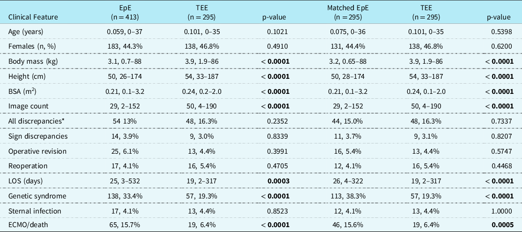
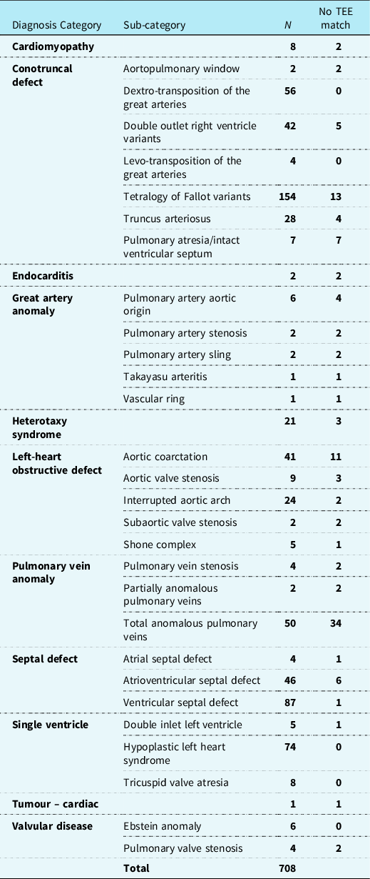
A total of 413 patients comprised the epicardial echocardiography group and 295 age- and diagnosis-matched patients comprised the transoesophageal echocardiography group, for a total of 708 patients (Table 1). The major indications for epicardial echocardiography are shown in Figure 1, with the most common involving patient size, inability of the cardiac anaesthesiologist to intubate the oesophagus (no more than two to three attempts are made), airway issues, and total anomalous pulmonary venous return. A subset of the epicardial echocardiography cohort contained rare diagnoses for which a comparable transoesophageal echocardiography patient of similar age could be not be located. Table 2 lists the cardiac diagnoses for which there were no transoesophageal echocardiography matches, most of which involved rare diagnoses or relative contraindications to transoesophageal echocardiography. For example, total anomalous pulmonary venous return is a relative contraindication to transoesophageal echocardiography at our institution, so there were very few comparable patients with this diagnosis in the transoesophageal echocardiography group during the study period. Another example involves infants with aortic coarctation requiring a median sternotomy because the proximal transverse aortic arch was too small to accomodatea clamp during aortic arch reconstruction. These two diagnoses represented the majority of patients without a comparative transoesophageal echocardiography study. Finally, transoesophageal echocardiogram probe placement was not performed in neonates smaller than 3 kg, as per manufacturer guidelines, so epicardial echocardiography patients in this size category did not have a transoesophageal echocardiography match.
Entire cohort analysis (includes unmatched EpEs).

BSA – body surface area, ECMO – extracorporeal membrane oxygenation, EpE – epicardial echocardiogram LOS – length of stay, Sign – significant, TEE – transoesophageal echocardiogram
Data expressed as median with range or number with percentage,
* include trivial patch leaks
The “Other” category in this diagram consisted of reasons that an epicardial echocardiogram (EpE) was performed instead of a tranoesophageal echo (TEE) including indications such as difficult intubation, oesophageal abnormalities, gastroinstestinal (GI) bleeding, interference of TEE probe with procedure, pre-operative instability, presence of a Nissen tube, TEE probe malfunction.

Diagnosis categories and missing TEE matches for entire cohort.

TEE – transoesophageal echocardiogram
Patient characteristics
Compared to the transoesophageal echocardiography group, the epicardial echocardiography group had lower body mass (median of 3.1 kg versus 3.9 kg; p < 0.0001), height (median 50 cm versus 54 cm; p < 0.0001), and body surface area (median 0.21 m2 versus 0.24 m2; p < 0.0001) (Table 1). Additionally, the epicardial echocardiography group was also more likely to have a genetic syndrome (33% versus 19%; p < 0.0001). With regard to genetic syndromes, the majority patients had a 22q11 microdeletion, trisomy 21, Charge syndrome (coloboma, heart defects, choanal atresia, growth restriction, genital abnormalities, and ear abnormalities), Vacterl association (vertebral defects, anal atresia, cardiac defects, tracheo-oesophageal fistula, renal anomalies, and limb abnormalities), or Williams syndrome.
Outcomes
Compared to the transoesophageal echocardiography group, the epicardial echocardiography group required longer hospitalisation (median 25 days versus 19 days; p = 0.0003), and they were more likely to require extracorporeal membrane oxygenator support or die (16% versus 6%; p < 0.0001). When only the epicardial echocardiography cases for which there was a direct transoesophageal echocardiography match were used for comparison, the differences in demographics and outcomes were similar. All other variables (gender, need for re-operation, or sternal wound infection) did not demonstrate a statistically significant difference between the two groups when comparing all of the epicardial echocardiography cases as well as only those with a match to the transoesophageal echocardiography cases. We did not find any documented instances of arrhythmia or hypotension (potential complications of epicardial imaging) and we found two instances of esophageal injury among the transoesophageal cohort, and we were unable to find documentation of any unintentional extubations resulting from transoesophageal imaging.
Imaging diagnostic accuracy
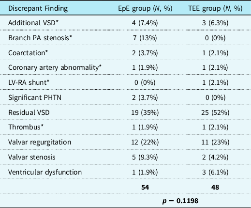
When comparing the diagnostic accuracy between the two groups, 111/708 patients (16%) had discrepant findings between the intraoperative imaging (epicardial echocardiography or transoesophageal echocardiography) and the postoperative discharge transthoracic echocardiogram. Following a secondary review of the images to determine whether the discrepant findings were truly discrepant, we determined that 102/708 (14%) had discrepant findings. The discrepant findings were classified into the following categories of major versus minor discrepancies. Major discrepancies included additional ventricular septal defects, left ventricular to right atrial shunt, branch pulmonary artery stenoses, aortic coarctation, coronary artery abnormalities, and thrombi. Minor discrepancies were classified as trivial or small residual ventricular septal defect patch leaks, mild valvar regurgitation, mild valvar stenosis, ventricular dysfunction, and significant pulmonary hypertension (Table 3).
Imaging discrepancies.

EpE – epicardial echocardiogram, LV – left ventricle, PA – pulmonary artery, PHTN – pulmonary hypertension, RA – right atrium, TEE – transoesophageal echocardiogram, VSD – ventricular septal defect,
* - classified as a clinically significant discrepancy
There were no statistically significant differences in the rates of overall imaging discrepancies between the epicardial echocardiography and transoesophageal echocardiography groups. When comparing all epicardial echocardiography versus all transoesophageal echocardiography patients, the rates of all imaging discrepancies (including trivial ventricular septal defect patch leaks) were 54/413 (13%) for the epicardial echocardiography group versus 48/295 (16%) for the transoesophageal echocardiography group (p = 0.2352). The rates were not significantly different when we evaluated only the clinically significant discrepancies for the entire cohort (14/413 [3.9%] for the epicardial echocardiography group versus 9/295 [3.0%] for the transoesophageal echocardiography group, p = 0.8339). These findings held true when comparing all discrepancies (including patch trivial septal leaks) among only the matched epicardial echocardiography and transoesophageal echocardiography cases (44/295 [15%] for the epicardial echocardiography group versus 48/295 [16%] for the transoesophageal echocardiography group, p = 0.7337.
Additionally, all imaging discrepancies were found to be false negative findings, indicating that the findings were likely present in the immediate post-operative period, but not identified by the intraoperative imaging and later defined on the pre-discharge transthoracic echocardiogram. This finding was confirmed by our secondary review of the images of the discrepant findings. During our secondary image review, we also confirmed that all the patients cardiac anatomic states were the same between the post-operative imaging and the pre-discharge imaging that we evaluated (no intervening cardiac surgeries were performed). In total, 37 total patients from both cohorts required an intra-operative revision (change or correction to the original surgery prior to leaving the operating room). Of these, 31 were initiated due to findings on the intra-operative imaging, 18 from the epicardial imaging group, and 13 from the transoesophageal imaging group.
Of note, two patients (0.78%) had an imaging discrepancy discovered during the transthoracic imaging prior to hospital discharge that warranted additional surgery during the same hospitalisation, one from the epicardial echocardiography group and one from the transoesophageal echocardiography group. One (epicardial imaging) was a mild coarctation that was initially not appreciated during the intraoperative imaging and progressed to require intervention, and the second (transoesophageal imaging) was an unrecognised anomalous right coronary artery origin that required a subsequent unroofing procedure.
Imaging studies performed via epicardial echocardiography tended to have overall fewer images captured than the transoesophageal echocardiography cohort (median 29 images, versus 50, p < 0.0001) and images tended to be more focused in nature than those acquired during a typical post-operative transoesophageal echocardiogram (Table 1).
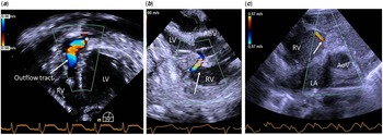
An example of intraoperative transoesophageal echocardiography imaging findings that might lead to revision of the intracardiac repair can be seen in Figure 2a and 2b. Similarly, examples of intraoperative epicardial echocardiography findings are seen in Figure 2c.
( a ): Intraoperative transoesophageal echocardiographic (TEE) view from the deep transgastric projection, showing residual post-operative right ventricular outflow tract flow obstruction (arrow) just below the pulmonary valve (*). ( b ): Intraoperative TEE image from the transgastric, long-axis projection, displaying the right ventricle (RV), left ventricle (LV) and aortic valve (AoV), showing a small, residual ventricular septal defect (VSD) patch leak (arrow). ( c ): Intraoperative epicardial echocardiographic (EpE) short-axis view from the base of the heart, demonstrating the left ventricle (LV), right ventricle (RV), aortic valve (AoV), and small, residual VSD patch leak (arrow) shunting left to right.

Figure 3 depicts the proportion of epicardial echocardiography, and transoesophageal echocardiography studies performed during each era over the 16-year period that we examined. Our institutional imaging technique and approach has remained comparable for both modalities over that time period with the exception of upgraded transducer equipment periodically.
Imaging studies performed by era for all EpE and matched TEE studies.
EpE – epicardial echocardiogram, TEE – transoesophageal echocardiogram. TEE as a percentage of the total, by era: 2003–2005 – 83%, 2006–2008 – 62%, 2009–2011 – 34%, 2021–2014 – 45%, 2015–2019 – 24%

Discussion
Initially, our hypothesis was that epicardial echocardiography would demonstrate a higher rate of imaging discrepancies due to the more limited nature of the imaging and the fact that at our institution, the experienced echocardiographer does not hold the probe or control the imaging, the surgeon does, with guidance from the echocardiographer. However, this study revealed similar diagnostic accuracy rates between epicardial echocardiography and transoesophageal echocardiography, therefore we are reassured that even without the direct involvement by an echocardiographer, comparable results are achievable. Additionally, we found higher rates of extra corporeal membrane oxygenation and death in the epicardial echocardiography cohort compared to transoesophageal echocardiography. In an effort to guide management and improve outcomes, intraoperative imaging allows surgeons to plan their operative approach, correct any significant residual lesions, and identify ventricular or valvar dysfunction. Reference Ozturk, Cansaran Tanidir and Ayyildiz5 Although prior studies have evaluated the diagnostic accuracy of transoesophageal echocardiography and epicardial echocardiography on a limited basis, these studies failed to directly compare the demographics and outcomes of patients who undergo intraoperative transoesophageal echocardiography and epicardial echocardiography. Our study fills this gap and helps to better understand the demographics of the patient populations undergoing either of the modalities. We excluded patients who had both types of intraoperative imaging to assure that our results more clearly delineated the differences in outcomes between the fragile, higher risk patient population with contraindications to transoesophageal echocardiography and the more mainstream transoesophageal echocardiography population.
In our large study of 708 patients, we found that epicardial echocardiography and transoesophageal echocardiography had very comparable diagnostic accuracies and rates of intra- and post-operative complications. Imaging discrepancies in our cohort were defined as discrepancies between the intraoperative imaging and the pre-discharge transthoracic echocardiogram. Small ventricular septal defect patch leaks accounted for the largest proportion of imaging discrepancies. When excluding small, haemodynamically insignificant ventricular septal defect patch leaks, valvar regurgitation accounted for most imaging discrepancies in both cohorts, followed by additional ventricular septal defects and branch pulmonary artery stenosis. It should be noted that only one case of a presumed missed thrombus was found in the imaging discrepancies for each modality. The cases with thrombi may not have represented true discrepancies, since it is possible for a thrombus to have formed post-operatively, although, upon secondary review of the images, the intraoperative imaging was not adequate to be entirely confident of the absence of thrombi. So, we included these as a discrepancy, given the significant potential for harm to the patient that a thrombus missed during intraoperative imaging could present. We aimed to establish a metric comparing the diagnostic accuracy of the two modalities; many studies have already evaluated their individual sensitivities, specificities, positive predictive values, and negative predictive values.
Patients with epicardial echocardiography or transoesophageal echocardiography as an imaging standard demonstrated similarly low rates of reoperation and intraoperative revisions. Patients requiring epicardial echocardiography were smaller in size than matched transoesophageal echocardiography patients, consistent with the relative contraindications for transoesophageal echocardiography in small neonates where transoesophageal echocardiogram probe placement may be more difficult and more likely to cause cardio-respiratory compromise. 3 In addition, premature neonates in the epicardial echocardiography group represent a more fragile and acutely ill population. The epicardial echocardiography population also had a higher incidence of genetic syndromes, likely due to associated cranio-facial abnormalities or compromised airways that might preclude intubation with a transoesophageal echocardiography probe. Interestingly, epicardial echocardiography did not confer additional infection risk, suggesting that sterile techniques employed in the operating room were adequate for prevention. All of these factors together paint a picture to indicate that the epicardial echocardiography patient population is an inherently higher risk group. While this study does not allow for us to control for these demographic factors and conclusively determine the reason for the worse outcomes, the seemingly higher risk composition of the epicardial echocardiography patient population likely accounts for the observation in our data of longer hospitalisations, more mechanical circulatory support, and higher rates of death among this population than their transoesophageal echocardiography counterparts. We speculate that is independent of the imaging modality employed.
This similarity in outcomes between the two imaging groups is very encouraging, especially when considering that epicardial echocardiography imaging tends to be less comprehensive. Transoesophageal echocardiography imaging allows for a more detailed, complete assessment of the repair since transoesophageal echocardiography imaging does not interfere with the surgical field and provides greater access to structures, more varied imaging planes and the opportunity to evaluate transitional physiologic changes associated with chest closure. Epicardial echocardiography by contrast, must balance bleeding, a beating heart, the surgeon’s time, and the need to move on to the final phases of the operation.
Literature review
Early in the history of intraoperative imaging and prior to the use of transoesophageal echocardiogram probes for paediatric interventions, Ungerleider et al described the value and accuracy of epicardial echocardiography. Over the course of 328 surgeries, they determined that epicardial echocardiography was superior to non-imaging methods for evaluating surgical repairs with a positive impact on surgical outcomes. Reference Ungerleider, Greeley and Sheikh1 More recently, Ozturk et al similarly determined epicardial echocardiography to be safe, effective, and accurate. Reference Ozturk, Cansaran Tanidir and Ayyildiz5 Both studies showed that epicardial echocardiography could detect residual lesions and complications in the operating room. Our study comparing epicardial echocardiography and transoesophageal echocardiography builds on this work by providing information related to post-operative patient outcomes.
Several studies evaluated the diagnostic accuracy of epicardial echocardiography and transoesophageal echocardiography and found good agreement between the two methods. Although our study demonstrated overall higher rates of discrepant findings, possibly due to differing standards for identifying discrepancies, we also found no difference between the diagnostic accuracies of the two modalities. Weineke et al, in a smaller cohort of 18 children undergoing ventricular septal defect surgery, found that both modalities demonstrated no false positives and only one false negative per modality. Reference Wienecke, Fyfe and Kline6 Manvi et al. studied 158 patients and reported good positive and negative predictive values for epicardial echocardiography, but they did not directly compare the predictive values of epicardial echocardiography to transoesophageal echocardiography as we did. Reference Manvi, Dixit and Srinivas7 Muhideen et al. studied 50 paediatric patients and found a diagnostic accuracy rate of 94 and 92% for epicardial echocardiography and transoesophageal echocardiography, respectively. Reference Muhiudeen, Roberson and Silverman8 More recently, Khumbarthi et al, prospectively studied 25 patients and found the image quality and Doppler values for both modalities to be comparable. Reference Kumbharathi, Taneja and Mehra9 Dragulescu et al and Stern et al found that epicardial echocardiography and transoesophageal echocardiography were more likely to miss residual lesions in different areas due to differences between the modalities in technique and access to structures. Transoesophageal echocardiography demonstrates advantages when imaging structures that lie more posteriorly, such as atrioventricular valves, and epicardial echocardiography generally has advantages when imaging more anterior structures such as the right ventricular outflow tract. We found a higher rate of some discrepant findings over others when epicardial echocardiography was employed in place of transoesophageal echocardiography, which harmonises with Stern’s recommendation for a hybrid imaging approach. Although, we speculate that in our data, the discrepancy rate in abnormal findings in the branch pulmonary arteries for the epicardial echocardiography group may be due in part to a higher incidence of complex pulmonary artery reconstructions at our institution which may skew our results somewhat rather than being intrinsic to the imaging modality itself. Similarly, Dragulescu et al found that overall detection of residual lesions in the operating room improved when both transoesophageal echocardiography and epicardial echocardiography were employed. Reference Dragulescu, Golding and Van Arsdell10,Reference Stern, Emani and Peek11
Limitations
The findings of this retrospective, single institution study may not apply to other institutions since indications and sterile method for epicardial echocardiography may not be standardised. We were unable to match a subset of the epicardial echocardiography patients with rare diagnoses, precluding a comparison to the transoesophageal echocardiography group for diagnostic accuracy or outcomes. Reoperations due to an imaging discrepancy were only tracked during the hospitalisation for the original surgery. Thus, reoperations during a subsequent hospitalisation would not be included in the analyses.
Only echocardiographic imaging findings were evaluated in this study, we did not compare the discrepant findings to any other imaging modalities.
Additionally, intraoperative complications such as exacerbated surgical bleeding or oesophageal injury were difficult to characterise and quantify for a comparison of these factors between epicardial echocardiography and transoesophageal echocardiography.
Conclusions
We conclude that epicardial echocardiography represents an effective alternative to transoesophageal echocardiography for intraoperative imaging when transoesophageal echocardiography is contraindicated. The two modalities yield similar diagnostic accuracies, although the patient population that receives epicardial echocardiography is often more fragile and acutely ill with the potential for worse outcomes, likely unrelated to the imaging modality employed.
Acknowledgements
None.
Financial support
This research received no specific grant from any funding agency, commercial, or not-for-profit sectors.
Conflicts of interest
None.