Introduction
Temporal bone surgical skills have traditionally been taught, acquired and refined via the repeated dissection of human cadaveric temporal bones. While this approach has successfully trained generations of otologists, it is being increasingly challenged by the availability of human cadaveric materials. Ethical, cultural, regulatory and financial issues have further added to the burdens of successfully running temporal bone skills courses. Moreover, paediatric temporal bones and bones with rare otic capsule anomalies are not easily available, but have become more important in training otologists who practice as cochlear implant surgeons.
Alternative animal temporal bones such as those from small rodents and sheep have limitations as their osseous anatomy differs significantly from that of humans. Computer-based virtual reality simulations have been developed, with haptic feedback capabilities. These show some benefits with specific task-based training; however, their ability to accurately convey subtle but important aspects of drill dissection such as drill ergonomics, drill tone, colour contrasts and accurate haptic feedback remain limited. Attempts to manufacture artificial temporal bones from ceramics and plastic have been suboptimal because they lack anatomical detail, and the materials’ properties are dissimilar to bone.
More recently, the use of advanced technologies, including high-resolution micro computed tomography (CT) scanning and three-dimensional (3D) printing technology, and greater attention to the mechanical properties of materials incorporated into synthetic temporal bones that best simulate those of natural bone, has produced more suitable temporal bones for training.Reference Hochman, Kraut, Kazmerik and Unger1 The standardised production of adult and paediatric bone, and anatomically normal and anomalous bone, for developing basic and advanced otological skills is an added advantage.
This study aimed to evaluate the face and content validity of a newer generation of 3D printed temporal bone manufactured by Phacon (Leipzig, Germany) for surgical skills training. Validity was assessed by asking trainees to compare their previous learning experiences using human cadaveric bones with their experiences using the artificial bone. It is hoped that measurement of the perceived value of this specific synthetic temporal bone model relative to the established standard will guide innovations in educational practice and available tools. The increasing use of newer, sophisticated manufactured temporal bones, for example, may complement and, in time, substitute for human cadaveric bones in skill development, while reducing cost and regulatory burdens.
Materials and methods
Simulated temporal bone
The temporal bone model used (model TFba) is manufactured by (and is available from) Phacon. It was developed based on data acquired by scanning normal human cadaveric temporal bone using micro CT with 12-μ resolution.
Based on these data, a simulated temporal bone was printed with a 3D printer (Z510; 4D Concepts, Groß-Gerau, Germany) using a cast powder and a bonding agent that has similar material properties to bone. The material properties were improved by varying the bonding agent and its concentration until the manufacturer considered them to be similar to bone. Layer by layer, the bonding agent was selectively added to the cast powder to create the solid structures, and colour was added to differentiate the relevant anatomical structures. The cast powder was then removed from the hollow structures such as mastoid air cells and the cochlear lumen. Additionally, anatomical structures such as the facial nerve and carotid artery, and the sigmoid sinus, were printed as hollow channels with coloured borders and filled with coloured wires. Finally, a polyurethane mixture was infiltrated into the model. A silicon sheet was used as dura and as the tympanic membrane. The cochlea and labyrinth, and their lumen, were reproduced in a detailed way. The soft tissue of the round window membrane could not be reproduced because of technical limitations, but the location and bony parts of the round window niche were clearly visible. The ossicular chain was cast as one rigid and continuous structure.Reference Roosli, Sim, Mockel, Mokosch and Probst2
Structured drilling exercises
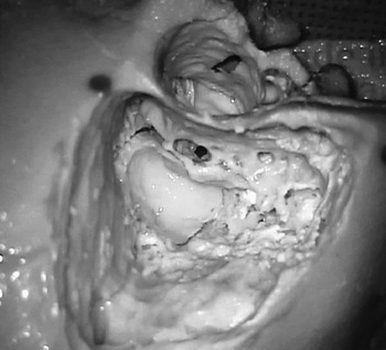
Nine late-year accredited otolaryngology (Surgical Education and Training) trainees were directed to perform structured temporal bone dissection exercises on three consecutive 3D simulated temporal bones. These exercises commenced with a cortical mastoidectomy, posterior epitympanectomy, posterior tympanotomy and round window dissection, progressing on to cochleostomy, and finally bony canalplasty.Reference Francis and Niparko3 If time permitted, they were then allowed to progress on to lateral temporal bone resection, wall down mastoidectomy and labyrinthectomy (Figure 1).
Photograph of the structured temporal bone following completion of the exercises and performance of canalplasty, demonstrating the anatomical realism of the cortical temporal bone, lateral ossicular chain, facial recess, cochleostomy and labyrinthectomy.

Each trainee was allowed 90 minutes to complete as many of the structured exercises as their skill development would allow. Each period of dissection was interleaved with a 90-minute discussion on various aspects of temporal bone anatomy and pathology, so as to allow maximal learning during the 1.5-day intensive teaching exercise. Immediate formative feedback from two experienced otologists (HWF and MdC) was given to each trainee during the course of the drilling so as to emulate the usual teaching method used during cadaveric-based skills courses. Preparation for the course consisted of reflecting on previous temporal bone experiences, both in the operating theatre and temporal bone laboratory, and the viewing of a structured temporal bone dissection video.4
Immediately following the dissection exercises, the trainee's experiences and reactions were surveyed using 23 semi-structured questions. Responses were made using five-point Likert-type scales varying from strongly agreeing (5), agreeing (4), neutral (3), disagreeing (2) to strongly disagreeing (1). Questions were grouped into four domains: anatomical realism, task-based usefulness, usefulness as a training tool and overall reactions. The first two domains were designed to assess face validity and the last two assessed content validity. Trainees were asked to use prior cadaver dissection experiences as a reference for their responses. A score of 3 indicated that the synthetic bone was not equivalent to human temporal bones, but was acceptable; scores of less than 3 indicated that the synthetic bone was not equivalent and was not acceptable. A score of 5 indicated that the synthetic bone was at least equivalent to human cadaveric temporal bones.
The study had been reviewed and approved by the local ethics committee, and was considered a low-to-negligible risk project.
Results
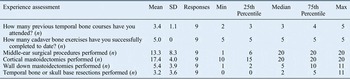
The range of trainees’ previous temporal bone experiences, both cadaveric and in the operating theatre, varied depending on the stage of training, but was generally greater in the Surgical Education and Training year five trainees. For the 9 trainees as a group, the mean number of previous temporal bone cadaveric courses attended was 3.4 (range = 2–5), and the mean number of previously successfully completed cadaveric bone exercises was 5 (Table I). This meant that all trainees had sufficient prior cadaveric dissection experience to use their pre-existing temporal bone knowledge as a reference point for their judgment of the artificial temporal bone experiences.
Trainees’ prior temporal bone operative experience*

* For the nine trainees involved in the validation exercise. SD = standard deviations; min = minimum; max = maximum
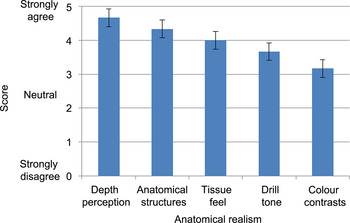
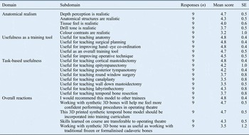
Anatomical realism was assessed in terms of five questions, which concerned depth perception, accuracy of anatomical structures, osseous tissue feel (haptic feedback), drill tone and colour contrasts (Table II). All trainees agreed that the anatomical realism of the 3D printed bone was comparable to cadaveric bones (Figure 2). In particular, the aspects of depth perception (score of 4.7), anatomical accuracy (4.3) and haptic feedback (4.0) were realistic. Drill tone and colour contrasts scored lower (achieving scores of 3.7 and 3.2 respectively), but were considered to be comparable or acceptable relative to cadaver experiences.
Mean ratings for the five semi-structured questions assessing anatomical realism of the synthetic temporal bone.

Trainees’ face and content validation questionnaire responses*

* For the nine trainees involved in the validation exercise. SE = standard error; 3D = three-dimensional
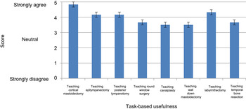
Task-based usefulness of the synthetic temporal bone was assessed by asking the trainees to compare the standardised temporal bone exercises performed on the 3D printed temporal bone with those performed on cadavers. For all dissection exercises, the trainees agreed that the 3D printed bone was at least equivalent to cadaveric bones. There was a stronger agreement regarding the usefulness of the synthetic model for tasks involving lateral aspects of the temporal bone (cortical mastoidectomy (score of 4.8), epitympanectomy (4.2) and posterior tympanotomy (4.2)) (Figure 3). The scoring was slightly weaker for more medial exercises: round window surgery (score of 3.7), bony canalplasty (3.5), wall down mastoidectomy (3.5) and temporal bone resection (3.7). However, these exercises were still scored as being acceptable compared to human cadaveric bones. In contrast to the other more medial exercises, the surgical labyrinthectomy exercise was scored highly (score of 4.3).
Mean ratings for the eight semi-structured questions assessing task-based usefulness of the synthetic temporal bone.

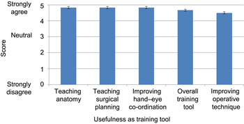
As compared to human cadaveric bones, trainees found the 3D temporal bone to be a useful training tool, particularly for teaching anatomy (score of 4.8), surgical task planning (4.8), improving hand–eye co-ordination (4.8), as an overall training tool (4.7) and for improving operative technique (4.5) (Figure 4). The feedback on this domain was of particular interest, as the responses indicated that the value of the 3D printed model was not just limited to the development of anatomical knowledge. The model was also useful for technical skills such as hand–eye co-ordination, more subtle cognitive skills such as surgical and task planning, which the trainees believed would lead to improvement in intra-operative efficiency, and dissection skills.
Mean rating for the five semi-structured questions assessing the synthetic temporal bone's usefulness as a training tool.

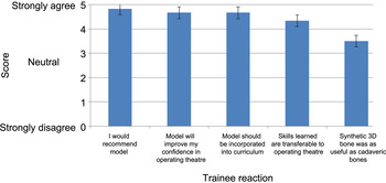
Overall trainees’ reactions also indicate a high degree of acceptance of the 3D printed temporal bone as a training tool comparable to cadavers (Figure 5). The trainees felt that the synthetic temporal bone was recommendable to other trainees (score of 4.8), the skills learned were transferable to the operating theatre (4.3) and its use improved confidence in the operating theatre (4.7). They universally agreed that these specimens should be incorporated into the training curriculum (score of 4.7). However, the use of 3D temporal bones as a replacement for human cadaveric bones in training programmes met with slightly lower acceptance ratings (score of 3.5). This lower scoring may be related to the novelty of using synthetic temporal bones in a traditional training course in which cadaver bones are the assumed ‘gold standard’.
Mean ratings for the five semi-structured questions assessing trainees’ overall reactions to the synthetic temporal bone as a teaching tool. 3D = three-dimensional

Face validation
Face validity was evaluated by asking trainees to assess whether the 3D printed model had a look, sound and feel comparable to cadaveric temporal bones, using simple measures. The domain assessing anatomical realism were all scored highly (mean = 4.0, range = 3.2–4.7). This suggests that the synthetic temporal bone provided a training experience close to that afforded by the cadaveric bone. In particular, the anatomical realism of fine structure detail (mean score = 4.2, range = 4.0–4.7) (which included pneumatised cortical temporal bone, and luminal structures such as the labyrinth and cochlea), bone dust production and handling, drill tone, and haptic feedback, were sufficiently represented to mirror the experiences of using human material. Colour contrasts were scored as slightly less acceptable (the sigmoid sinus and facial nerve were considered too bright, and the dura too dull) (score of 3.2). The task-based usefulness domain indicated that the bone generally provided satisfactory training experiences for the more lateral temporal bone exercises (cortical mastoidectomy, epitympanectomy and posterior tympanotomy). Labyrinthectomy was a notable medial dissection exercise, which scored highly (score of 4.3).
Content validation
Content validation of the temporal bone exercises was assessed in terms of the 3D bone's usefulness as a training tool and overall trainee reactions to the skills course. The trainees indicated that many aspects of their skill development were improved by the training experience, both technically (teaching anatomy, drill ergonomics and transferability to the operating theatre) and cognitively (task planning and improved confidence). There was strong agreement that the course was recommendable to other trainees (score of 4.8) and should be incorporated into the training curriculum (4.7).
Of particular note were the overall trainee responses indicating: acceptability of the manufactured temporal bone for inclusion in temporal bone skills development programmes (score of 4.7), recommendation to other trainees (4.8) and agreement that the course enhanced their operative skills (4.7).
Discussion
Osseous temporal bone dissection has traditionally been taught using repeated, structured dissection exercises within a purpose-built skills development facility. The decline in availability of human cadaveric temporal bones, coupled with increasing governmental restrictions, ethical and religious barriers, and the cost of running a licensed facility, have challenged this teaching paradigm. Other methods of skills development, such as virtual realityReference Arora, Khemani, Tolley, Singh, Budge and Varela5 and the use of animal alternatives,Reference Gocer, Eryilmaz, Genc, Dagli, Karabulut and Iriz6 have only partially satisfied the requirements for an alternative teaching model, as they lack human bone anatomical realism (e.g. fine structure detail, bone dust handling, drill tone and ergonomics, and haptic feedback).
The development and use of synthetic temporal bones has been attempted in the past with models manufactured from various plastics, ceramics and resins.Reference Suzuki, Hagiwara, Ogawa and Ono7, 8 However, their anatomical realism was poor and the materials used did not behave like bone. Fluid-filled structures such as the labyrinth and cochlea were solid, making their use as training tools suboptimal.
More recently, the company Phacon has used micro CT imaging, sophisticated printing technology and more appropriate materials to approximate cadaveric bone. This has resulted in a higher fidelity synthetic temporal bone. Preliminary experiences with this manufactured model suggested that many of the deficiencies of previous synthetic temporal bones had been sufficiently improved to warrant further investigation for its use as a cadaver substitute in otological training. Our aim was to further investigate the usefulness of this model as a substitute for temporal bone skills development by formally evaluating the face and content value of the manufactured bone, with a view to facilitating its use as a simulated training tool.
Semi-structured questionnaires aimed at both trainees and trainers have been used extensively in the past to validate potentially useful temporal bone training tools. The validation of virtual reality temporal bone training tools, including the Voxel-Man simulatorReference Arora, Khemani, Tolley, Singh, Budge and Varela5, Reference Khemani, Arora, Singh, Tolley and Darzi9 and University of Melbourne training simulator,Reference Zhao, Kennedy, Yukawa, Pyman and O'Leary10, Reference O'Leary, Hutchins, Stevenson, Gunn, Krumpholz and Kennedy11 demonstrates their usefulness as part of a skills development programme. Studies of learning have focused on the use of virtual reality as a complimentary tool to accelerate subsequent skills development practised on cadaveric bones.Reference Zhao, Kennedy, Yukawa, Pyman and O'Leary10, Reference Zirkle, Taplin, Anthony and Dubrowski12 However, despite the potential increase in the availability of simulated training opportunities and the demonstration of some measurable contribution to training, virtual reality tools are still hampered by high costs and limited face validity because of poor drill ergonomics and haptic realism. In their present state of development, these tools are best used as a compliment to the use of traditional cadaveric materials in skills development.
Prior attempts to manufacture synthetic temporal bones have had only modest success,8 and their use as training simulators has not been formally evaluated. Suzuki et al. manufactured synthetic temporal bones using 3D technology and partly evaluated their validity.Reference Suzuki, Hagiwara, Ogawa and Ono7, Reference Suzuki, Ogawa, Hagiwara, Yamaguchi and Ono13 However, their anatomical realism was hampered by the limited accuracy of some structures: the cochlea and labyrinth lacked a lumen, and haptic realism was limited because the behaviour of the synthetic bone was unlike that of cadaver specimens. These synthetic temporal bones have not been incorporated into the training curriculum.
Recent improvements in 3D printing technology and more careful manipulation of printing materials have led to improvements in the fidelity of manufactured bones. Semi-structured questionnaires, similar to those used in this study, have shown that these printed bones have higher face validity than previous generations of synthetic temporal bones. Mick et al. found a high degree of face validity for the lateral structures, and concluded that the models would be suitable for training residents earlier in their skills development programme (post-graduate years one to three).Reference Mick, Arnoldner, Mainprize, Symons and Chen14 Roosli et al. validated the Phacon temporal bone, specifically for training cochlear implant surgeons.Reference Roosli, Sim, Mockel, Mokosch and Probst2 The authors emphasised the presence of a cochlea lumen and the extended squamous temporal bone as necessary anatomical details for cochlear implant training. Experienced otologists rated the temporal bone as equivalent to human cadaver temporal bones for the purposes of training cochlear implant surgeons. The added advantage of printing paediatric bones and specimens with otic capsule anomalies was also highlighted.
Our validation findings were similar to those of Mick et al.Reference Mick, Arnoldner, Mainprize, Symons and Chen14 and Roosli et al.,Reference Roosli, Sim, Mockel, Mokosch and Probst2 particularly regarding the anatomical realism of the synthetic bones. This is most likely related to the choice of materials used to represent cortical and otic capsule derived bone, and the accurate representation of a realistic lumen within the labyrinth and cochlea. This shows substantial improvement over previous models, and is compelling evidence that a suitable alternative to cadaveric bones exists for teaching many of the osseous exercises required by trainees. Our validation study also showed that there is room for improvement in terms of: the fidelity of some of the structures, such as the tympanic and round window membrane; the mobility of the ossicular chain; the facial nerve and bony fallopian canal interface; and the dura and sigmoid sinus colour contrasts. Hopefully, this study will prompt manufacturers to make these improvements in subsequent generations of synthetic temporal bones.
• Advanced technologies and more suitable materials have enabled production of synthetic temporal bones with anatomical realism, suitable for skills training
• A formal validation study of a printed synthetic temporal bone was conducted using a semi-structured questionnaire
• Responses related to realism, teaching and task-based usefulness, and overall reaction, indicated a high degree of acceptance of the model
• Tympanic membrane and ossicular chain mobility could be improved to increase fidelity of the model
• The role of simulated temporal bones in skills development is discussed
The study involved surveying the responses of a small cohort of senior trainees in Australia and New Zealand who had pre-existing cadaveric dissection experience. The generalisability of findings to more junior trainees or novice learners in other countries is unclear. A study using similar methodologies in a North American training setting is planned, in order to obtain further data regarding the validity of using synthetic temporal bones for skills development in a different culture and training setting.
Conclusion
A synthetic temporal bone has been developed using composite materials and sophisticated 3D printing technology. Our study, which has a level of evidence 2b, suggests that the synthetic temporal bone has a high degree of face and content validity. This makes it a useful substitute for traditional cadaveric bones in terms of lateral temporal bone skills development for Australian otolaryngology trainees. The cost savings, low biohazard and the variety of locations where the synthetic temporal bones can be used make it likely that the models will be incorporated into traditional temporal bone skills development programmes in the near future.
Acknowledgement
The authors thank Dr Karen Byth for her kind assistance in the statistical analysis of this project.