



Non-technical Summary
This study describes the Serkinskaya faunal complex of the Siberian lower Cambrian Kessyusa Group, which includes many metazoan remains: a fragment of gnathobase, several scalids of priapulid-like organisms, various morphotypes of spines, organic films with outgrowths, and other organic remains. The presumed age of these remains is Cambrian Fortunian to Stage 2 (Vendian–Cambrian, possibly Tommotian; ca. 535–530 million years ago), which makes the described organic complex one the most diverse and ancient metazoan small carbonaceous fossil assemblages. Along with the description of the complex, we conducted Raman spectroscopy to reveal the composition of small carbonaceous fossils (SCFs). Raman spectroscopy allows the identification of molecules or materials; thus, for ancient organics, we aimed to identify types of materials. The results of the Raman spectroscopy analysis of the gnathobase fragment show heterogeneity on the surface of the microfossil and indicate differences in the preservation of the cuticular cover. Findings from the Serkinskaya faunal complex confirm the presence of filter-feeding, deposit-feeding (priapulids), vertical mixing of the sediment in the first centimeter, predation pressure (cuticle with outgrowths), and possible durophagy in the earliest Cambrian.
Introduction
The emergence of predatory lifestyles in the early Cambrian laid the foundation for modern ecosystems and accelerated the “arms race” between predators and prey (Cabej, Reference Cabej2019). Modern predatory invertebrates have specialized structures that facilitate the capture, retention, and ingestion of food (Manton, Reference Manton1964). In the early Cambrian, animals started to rework the sediment vertically, opening new ecological niches and expanding their feeding ecology. The emergence of durophagy (feeding on animals with hard skeletons) contributed to the diversification of biomineralized animals (Bicknell and Paterson, Reference Bicknell and Paterson2018).
Durophagous animals are characterized by rigid structures adapted for crushing or grinding hard food. One example of such structures is the gnathobase: masticatory spines (Cong et al., Reference Cong, Daley, Edgecombe and Hou2017; Yang et al., Reference Yang, Ortega-Hernández, Legg, Lan, Hou and Zhang2018), which can be located on mandibles, such as those found in extant copepod crustaceans (D’Agostino et al., Reference D’Agostino, Hoffmeyer and Degrati2020), or on walking appendages, such as those found in the horseshoe crab, Limulus polyphemus (Linnaeus, Reference Linnaeus1758) (Bicknell et al., Reference Bicknell, Ledogar, Wroe, Gutzler, Watson and Paterson2018a). Gnathobases have also been described in ancient chelicerates (e.g., Habelia optata Walcott, Reference Walcott1912, Sidneyia inexpectans Walcott, Reference Walcott1911; Zacaï et al., Reference Zacaï, Vannier and Lerosey-Aubril2016; Aria and Caron, Reference Aria and Caron2017), and in the radiodonts (Amplectobelua symbrachiata Hou, Bergström, and Ahlberg, Reference Hou, Bergström and Ahlberg1995; Cong et al., Reference Cong, Daley, Edgecombe and Hou2017).
Compression fossils of arthropods bearing gnathobases are known from the middle Cambrian (Zacaï et al., Reference Zacaï, Vannier and Lerosey-Aubril2016; Aria and Caron, Reference Aria and Caron2017; Bicknell et al., Reference Bicknell, Paterson, Caron and Skovsted2018b; Yang et al., Reference Yang, Ortega-Hernández, Legg, Lan, Hou and Zhang2018) and further occurred throughout the Phanerozoic. Molecular evidence suggests that panarthropods originated in the Ediacaran (Howard et al., Reference Howard, Giacomelli, Lozano-Fernandez, Edgecombe and Fleming2022), and it can be hypothesized that new ecological niches should have begun to develop with their appearance. In the paleontological record, remains of bilaterians and possibly arthropods occur before the Precambrian–Cambrian boundary (Fedonkin and Waggoner, Reference Fedonkin and Waggoner1997; Shahkarami et al., Reference Shahkarami, Mángano and Buatois2017; Kesidis et al., Reference Kesidis, Slater, Jensen and Budd2019; Evans et al., Reference Evans, Hughes, Gehling and Droser2020; Slater and Bohlin, Reference Slater and Bohlin2022). Bilaterian trace fossils (simple, horizontal, unbranched trails or burrows) are known in the Ediacaran and by the terminal Ediacaran their diversity and disparity increases (Mángano and Buatois, Reference Mángano and Buatois2020). Recently a new bilaterian, Uncus dzaugisi Hughes, Evans, and Droser, Reference Hughes, Evans and Droser2024, was described from the Ediacaran of South Australia indicating the appearance of nematoids (Hughes et al., Reference Hughes, Evans and Droser2024). Trace fossils increase in their number and complexity in the early Fortunian, and by the Cambrian Stage 2 trace fossils include scratch imprints, bilobate and branching trails indicating the appearance of arthropods, priapulids, nematodes, polychaetes, and enteropneusts (Mángano and Buatois, Reference Mángano and Buatois2017, Reference Mángano and Buatois2020; Laing et al., Reference Laing, Mángano, Buatois, Narbonne and Gougeon2019). Ecdysozoan remains preserved as small shelly fossils in the Cambrian Fortunian (SCFs) are represented by priapulid-like scalidophorans (Liu et al., Reference Liu, Xiao, Shao, Broce and Zhang2014), possible kinorhynchs (Zhang et al., Reference Zhang, Xiao, Liu, Yuan, Wan, Muscente, Shao, Gong and Cao2015; Shao et al., Reference Shao, Wang, Liu, Qin, Zhang, Liu, Shao, Zhao and Zhang2020), scalidophoran larvae (Liu et al., Reference Liu, Xian, Zhang, Eriksson, Liu and Shao2025), and problematic scalidophorans (Vannier, Reference Vannier2024; Wang et al., Reference Wang, Qiang, Guo, Vannier and Song2024). Other Fortunian SSFs include mollusks, problematic remains like anabaritids and chancelloriids (Parkhaev and Demidenko, Reference Parkhaev and Demidenko2010; Kouchinsky et al., Reference Kouchinsky, Bengtson, Landing, Steiner, Vendrasco and Ziegler2017, Reference Kouchinsky, Alexander, Bengtson, Bowyer and Clausen2022), and possible cnidarians (Liu et al., Reference Liu, Shao, Zhang, Wang, Zhang, Chen, Liang and Xue2017). By Cambrian Stage 2, tommotiids, brachiopods, lobopodians, paleoscolicids, and protoconodonts (possible chaetognaths) enter the stage of Cambrian Explosion (Gravestock et al., Reference Gravestock, Alexander, Demidenko, Esakova and Holmer2001; Kouchinsky et al., Reference Kouchinsky, Bengtson, Clausen and Vendrasco2015, Reference Kouchinsky, Bengtson, Landing, Steiner, Vendrasco and Ziegler2017, Reference Kouchinsky, Alexander, Bengtson, Bowyer and Clausen2022).
The very first small carbonaceous fossils (SCFs) (Butterfield and Harvey, Reference Butterfield and Harvey2012)—organic-walled fossils that are too small to be seen on bedding planes, but structures larger and more delicate than conventional palynomorphs—that possibly belong to ecdysozoans are described from the late Ediacaran (Moczydłowska et al., Reference Moczydłowska, Budd and Agić2015; Willman and Slater, Reference Willman and Slater2021). Late Terreneuvian SCFs from Baltica comprise several bilaterian remains, including total-group chaetognaths and cuticular spines characteristic of scalidophorans together with the possible hemichordate Sokoloviina (Slater et al., Reference Slater, Harvey and Butterfield2018a). By Cambrian Stage 2, organic remains of bilaterians become more abundant and ecdysozoans are commonly found (Slater and Bohlin, Reference Slater and Bohlin2022). By Stage 3, the suspension-feeders, deposit-feeders, and durophages become diverse (Mángano and Buatois, Reference Mángano and Buatois2014).
In this study, we present new organic microfossils of Cambrian Terreneuvian (Vendian–Cambrian) age from the Siberian Platform that include several spines characteristic of different feeding types. Despite their rarity, the discovered complex (Fig. 1) can help reconstruct the feeding ecologies present during this time interval.
Serkinskaya fauna complex from the core material of the Serkinskaya square, Muna Uplift, Siberian Platform. (1–5) Spines of Ceratophyton circufuntum, (1) №2124/26, (2) №2124/29, (3) №2124/28, (4) №2124/27, (5) №2124/34. (6–8) Spines of Ceratophyton groetlingboensis, (6) №2124/37, (7) №2124/36, (8) №2124/74. (9) Spine with the root, №2124/75. (10) Possible polychaete spine, №2124/76. (11) Two overlapping spines with hooks, №2124/77. (12–16) Tissues covered with small triangular processes, (12) №2124/78, (13) №2124/79, (14) №2124/80, (15) №2124/81, (16) №2124/82. (17) Small metazoan gnathobase-like fragment, №2124/83. (18, 22) fibrillar, porous spine, №2124/86. (19) large gnathobase fragment, №2124/84, (20) close-up squared area with interspersed spinules. (21) fragment with rounded spines, №2124/85, resembles fragments from Slater et al. (Reference Slater, Harvey and Butterfield2018a, fig. 6, L–P). (23–27) Priapulid-like scalids Baltiscalida njorda, (23, 26) type C teeth according to Smith et al. (Reference Smith, Harvey and Butterfield2015), №2124/87 and №2124/91, respectively; (24, 25, 27) type A teeth according to Smith et al. (Reference Smith, Harvey and Butterfield2015), №2124/88, №2124/89, and №2124/90, respectively. (28, 29) priapulid-like scalids Baltiscalida njorda, type 3 teeth, (28) №2124/92, (29) №2124/93; (30, 31) same specimens as in (28, 29), Baltiscalida njorda type 3 with spines outlined, not to scale; (32–34) Sokoloviina costata, (32) №2124/94, (33) №2124/95, (34) №2124/96. All numbers have the prefix Centre of Collective Use ‘Collection GEOCHRON’ of the Institute of Petroleum Geology and Geophysics SB RAS – CCU ‘GEOCHRON’ №2124/X.
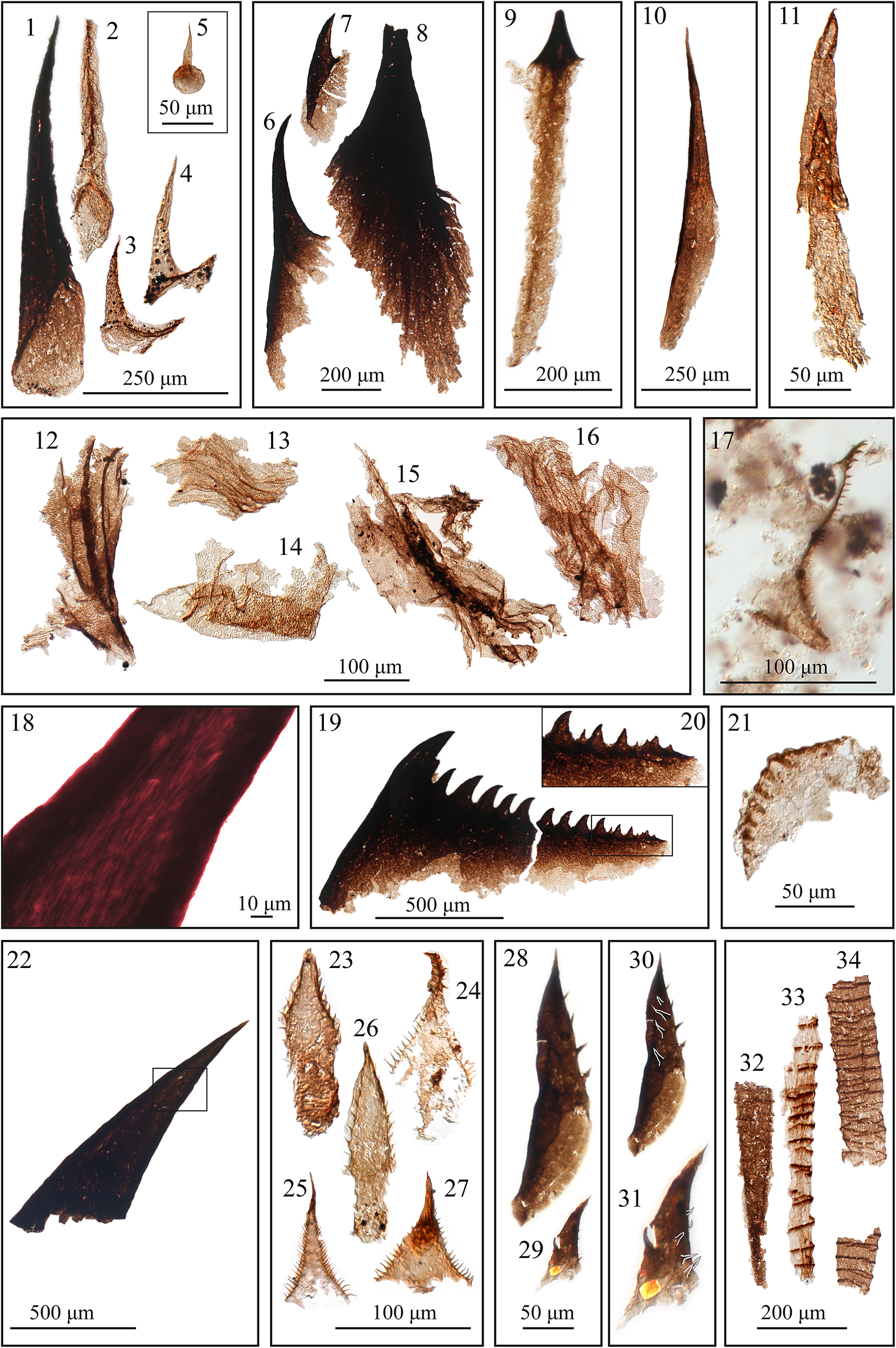
Figure 1. Long description
A multi-panel microscopic study organized into four main rows.
Top row: Panels 1 through 5 show elongated, dark, tapered spines of Ceratophyton circufuntum with a 250 micrometer scale bar; panel 5 is an inset with a 50 micrometer scale. Panels 6 through 8 display broader, serrated spines of Ceratophyton groetlingboensis with a 200 micrometer scale. Panel 9 shows a spine with a distinct root. Panel 10 is a slender, curved spine. Panel 11 shows two overlapping hooked spines with a 50 micrometer scale.
Second row: Panels 12 through 16 feature organic tissues covered in small triangular processes with a 100 micrometer scale. Panel 17 shows a small metazoan gnathobase-like fragment with a serrated edge.
Third row: Panel 18 is a high-magnification view of a fibrillar, porous spine at 10 micrometers. Panel 19 shows a large gnathobase fragment with a row of sharp teeth and a 500 micrometer scale; panel 20 is a close-up of its spinules. Panel 21 shows a fragment with rounded spines.
Bottom row: Panel 22 shows a large tapered spine. Panels 23 through 27 display various priapulid-like scalids of Baltiscalida njorda, including type A and C teeth with a 100 micrometer scale. Panels 28 and 29 show type 3 teeth, while 30 and 31 provide the same specimens with spines outlined. Panels 32 through 34 show the ribbed, elongated structures of Sokoloviina costata with a 200 micrometer scale.
Geological setting
We describe and analyze organic microfossils from drill-core material from the Serkinskaya area, Muna Uplift, Siberian Platform. Samples from drill cores from the Serkinskaya area (S-VII-1 and S-VIII-1) were transferred to us by the Amakinskaya geological exploration expedition. The macerated samples currently are deposited at the IPGG SB RAS, and the drill-core material is deposited at Novosibirsk, Russia, in the IPGG SB RAS drill-core storehouse.
In the Serkinskaya area, the oldest exposed sediments are identified as the Kessyusa Group, which is predominantly carbonate in composition (there is irregularity in vertical continuity, limestones are interbedded with clayey limestones, and brown, green, and gray-green mudstones) and has a thickness of up to 63 m; the top of this formation comprises a layer of green-gray mudstones (Fig. 2). The studied samples originated from these mudstones. Small carbonaceous microfossils from drill core VII-1 are from a depth of 106.8 m (the name of the sample is S-VII-1-106.8, as indicated in Fig. 2), and SCFs from drill core S-VIII-1 are from a depth of 182 m (the name of the sample is S-VIII-1-182, as indicated in Fig. 2). Above the mudstones, the Emyaksin Formation is identified, and its composition is predominantly carbonate (clayey, nodular limestones) with a thickness of 86 m. In the adjacent Anabar-Olenek region, 200 km to the west from the Serkinskaya bore holes (geological map sheets Q-51-VII, VIII), the Emyaksin Formation has a similar composition and is characterized by finds of the trilobites Triangulaspis meglitzkii (Toll, Reference Toll1899) and Pagetiellus lenaicus (Toll, Reference Toll1899) of the Atdabanian Stage (Stage 3) of the lower Cambrian (Pishchalnikov et al., Reference Pishchalnikov, Salikhov, Zarubin and Pyshko2020). Notably, according to the approved regional stratigraphic scheme of Cambrian sediments of the Siberian Platform (Sukhov et al., Reference Sukhov, Pegel and Shabanova2021), closest (200 km to the west) to the study area of the Muno–Olenek facies area in drill core 203, the Emyaksin Formation is predominantly composed of limestone, and overlies the limestone–dolomite Manykai Formation. The boundary between Precambrian and Cambrian sediments is defined in the lower part of the Emyaksin Formation by its small shelly fossil (SSF) complex, which transitions from the upper Vendian (the Purella antiqua zone of the Nemakit-Daldynian Stage) to the Tommotian Stage of the Cambrian (Sukhov et al., Reference Sukhov, Shabanov and Pegel2016). Thus, in the absence of additional paleontological data, the range of uncertainty in the age of the upper part of the Kessyusa Group of the Muna Uplift (extracted from drill cores of the Serkinskaya area) spans the Purella antiqua zone of the Nemakit-Daldynian Stage of the upper Vendian (Fortunian) to the Tommotian Stage (Stage 2) of the lower Cambrian.
Stratigraphic position of SCFs found from the Olenek and Muna uplifts of the Siberian Platform. SCF positions are indicated by the arrowheads next to the stratigraphic column. The names of the samples are S-VII-1-106.8 and VIII-1-182 (next to the arrowheads). S-VII-1 and VIII-1 above the stratigraphic columns are the names of the drill cores transferred to us by the Amakinskaya geological exploration expedition. Origin of sections: Olenek Uplift = composite section of the Olenek, Kersyuke, and Khorbusuonka rivers (Nagovitsin et al., Reference Nagovitsin, Rogov, Marusin, Karlova, Kolesnikov, Bykova and Grazhdankin2015); 203 borehole = from Sukhov et al. (Reference Sukhov, Shabanov and Pegel2016); Muna Uplift = data on the structure of the Serkinskaya borehole sections (S-VII-1 and VIII-1) provided to INGG SB RAS by Amga Exploration Expedition. Formation abbreviations: Ch = Chuskuna Formation, Em = Emyaksin Formation, Er = Erkeket Formation, Kn = Kuonamka Formation, Mn = Manykay Formation, Mt = Mattaia Formation, Ks = Kessyusa Formation, St = Staraya Rechka Formation, Sy = Syhargalakh Formation, Tr = Turkut Formation Zone abbreviations: At = Anabarites trisulcatus, Pa = Purella antiqua, Ns = Nochoroicyathus sunnanginicus, Dr = Dokidocyathus regularis, Dl = Dokidocyathus lenaicus, Pj = Profallotaspis jakutensis, R = Repinaella. Taxa shown on Figure 2 not mentioned in the text: Cambrotubulus decurvatus Missarzhevsky in Rozanov et al., Reference Missarzhevsky1969; Purella antiqua (Abaimova, Reference Abaimova1976); Watsonella crosbyi Grabau, Reference Grabau1900; Aldanella attleborensis (Shaler and Foerste, Reference Shaler and Foerste1888); Anabarites trisulcatus Missarzhevsky in Voronova and Missarzhevsky, Reference Voronova and Missarzhevsky1969; Anabarites korobovi Missarzhevsky in Rozanov and Missarzhevsky Reference Rozanov and Missarzhevsky1966; Treptichnus pedum (Seilacher, Reference Seilacher, Schindewolf and Seilacher1955)
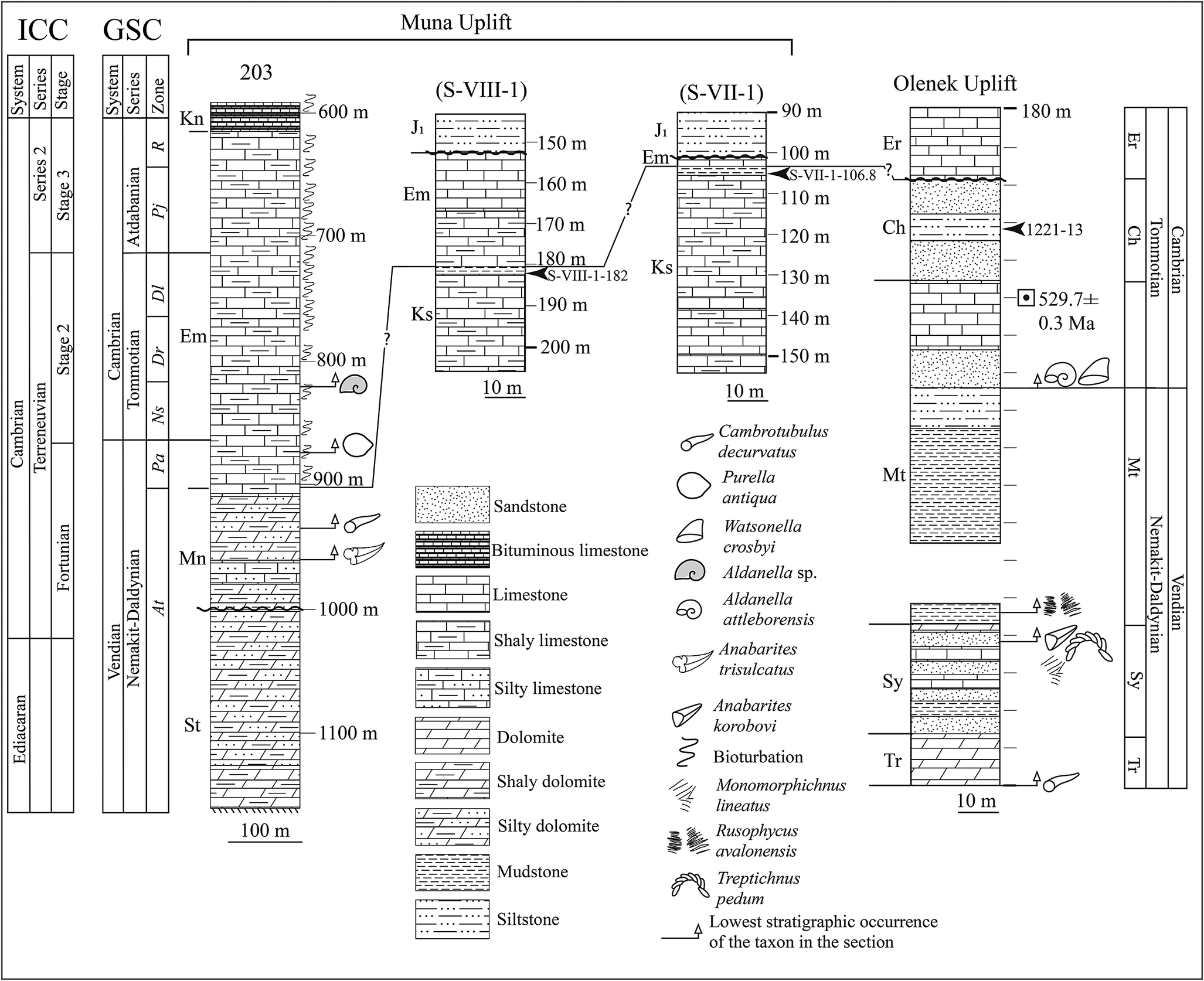
Figure 2. Long description
The diagram consists of four vertical stratigraphic columns. On the far left, two reference columns labeled I C C and G S C provide the temporal framework from the Ediacaran to Cambrian Series 2, Stage 3. The G S C column includes zones labeled At, Pa, Ns, Dr, Dl, Pj, and R.
Moving right, the Muna Uplift section contains three columns:
1. Borehole 203: A long column spanning from the St formation at 1100 meters up to the Kn formation at 600 meters. It shows a transition from silty dolomite and mudstone at the base to limestone and shaly limestone, with bituminous limestone at the top. Small shelly fossil icons and bioturbation symbols are marked along the right side.
2. Borehole S-VIII-1: Spans from the Ks formation (180 to 200 meters) to the J sub 1 formation (150 meters). An arrowhead marks sample S-VIII-1-182 at the boundary between Ks and Em.
3. Borehole S-VII-1: Spans from the Ks formation (110 to 150 meters) to the J sub 1 formation (90 meters). An arrowhead marks sample S-VII-1-106.8 within the Em formation.
On the far right, the Olenek Uplift composite section shows formations Tr, Sy, Mt, Ch, and Er. A radiometric date of 529.7 plus or minus 0.3 M a is noted in the Ch formation.
A central legend identifies lithologies including Sandstone, Bituminous limestone, Limestone, Shaly limestone, Silty limestone, Dolomite, Shaly dolomite, Silty dolomite, Mudstone, and Siltstone. Fossil taxa icons are defined for Cambrotubulus decurvatus, Purella antiqua, Watsonella crosbyi, Aldanella species, Aldanella attleborensis, Anabarites trisulcatus, Anabarites korobovi, Bioturbation, Monomorphichnus lineatus, Rusophycus avalonensis, and Treptichnus pedum. Horizontal lines and question marks indicate proposed correlations between the columns.
Materials and methods
For the extraction of organic microfossils from mudstones, the standard palynological extraction technique modified from T.N. Herman (Reference Hermann and Timofeev1974) was used. This method was previously described by Dantes et al. (Reference Dantes, Nagovitsin and Raevskaya2024). Large organic films and microfossils were extracted from the dissolved sediment without centrifugation. To optimize and speed up the work with the sediment, sieves with a meshs of 150–250 μm were used to extract the macerated coarse fraction. Afterwards, the fine fraction (less than 250 μm) was processed according to standard palynological methods, with separation of the organic part of the sediment from the mineral part in heavy liquid by centrifugation. After extraction, the organic material was preserved on slides with gelatin–glycerol adhesive or EOKITT polymer adhesive. The material was studied via a Zeiss AxioImager A1 microscope.
Raman spectroscopy
Raman spectra of the samples were obtained via an Automated Raman Imaging Microscope Apyron at the Analytical Center for Multielemental and Isotope Research SBRAS (Novosibirsk) with a 488-nm laser fiber coupled to the instrument. A 100× (NA 0.9 air) objective was used for excitation and detection, and the Rayleigh light was rejected via a RayShield filter. For all the measurements, a laser with a power of approximately 5 mW was employed. The spot size can be approximated by the full width at half maximum (FWHM) of the airy disc as 0.51×488/0.9 = 277 nm. Because of focus distortion, the spot inside the sample is expected to be slightly larger. A 50-μm core diameter multimode fiber served as the pinhole for confocality and as the entrance slit to the spectrometer. The spectrometer used was a UHTS600 (see Dieing and Hollricher, Reference Dieing and Hollricher2008, for more information) equipped with a back-illuminated CCD camera with peak quantum efficiencies of ~90% in the detection range (CCD camera stands for Charge Coupled Device camera). Two gratings (300 g/mm and 1800 g/mm) were used to disperse the light. The samples were scanned in X–Y using a highly precise piezoelectric scanner, while movement in the Z direction was performed by a stepper motor (single step 10 nm). Details of the experimental conditions for the 2D scans can be found in Table 1. The datasets were evaluated via WITec Project Plus software. The same equipment was used in another study (Korsakov et al., Reference Korsakov, Toporski, Dieing, Yang and Zelenovskiy2015).
Experimental parameters
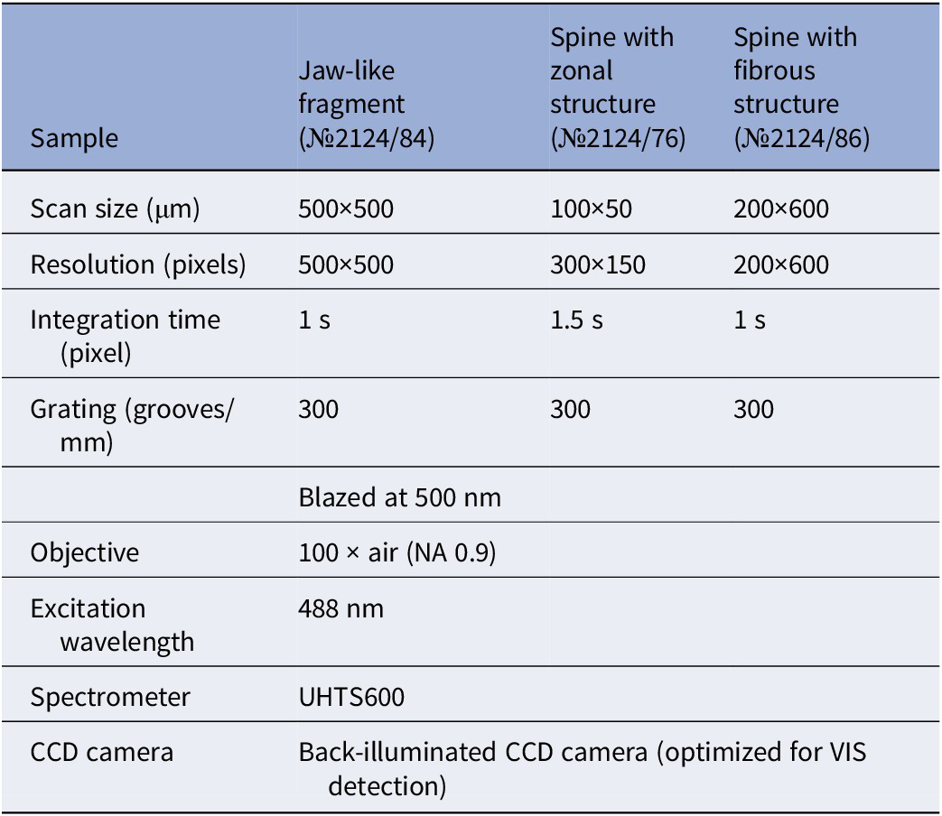
Table 1. Long description
The table consists of four columns. The first column lists the parameters, while the subsequent three columns represent the samples: Jaw-like fragment (number 2124/84), Spine with zonal structure (number 2124/76), and Spine with fibrous structure (number 2124/86).
* Scan size (mu m): 500 by 500 for the jaw-like fragment; 100 by 50 for the zonal spine; 200 by 600 for the fibrous spine.
* Resolution (pixels): 500 by 500 for the jaw-like fragment; 300 by 150 for the zonal spine; 200 by 600 for the fibrous spine.
* Integration time (pixel): 1 s for the jaw-like fragment; 1.5 s for the zonal spine; 1 s for the fibrous spine.
* Grating (grooves/mm): 300 for all samples. The jaw-like fragment specifies Blazed at 500 nm.
* Objective: 100 times air (N A 0.9) for the jaw-like fragment.
* Excitation wavelength: 488 nm for the jaw-like fragment.
* Spectrometer: U H T S 600 for all samples.
* C C D camera: Back-illuminated C C D camera (optimized for V I S detection) for all samples.
Repository and institutional abbreviations
The organic samples examined in this study were deposited at the Centre of Collective Use ‘Collection GEOCHRON’ of the Institute of Petroleum Geology and Geophysics SB RAS (CCU ‘GEOCHRON’, Novosibirsk, Russia). The collection number of the presented samples is №2124, the Ceratophyton circufuntum Zhang in Gravestock et al., Reference Zhang, Gravestock, Alexander, Demidenko, Esakova and Holmer2001, spines are №2124/26–29 and №2124/34, the C. groetlingboensis Hagenfeldt, Reference Hagenfeldt1989, spines are №2124/36, 37 and 74, the individual spines are registered under numbers №2124/75, 76, 77, and the spine with a fibrillar structure is №2124/86. Fragmentary cuticular tissues are numbered №2124/78–82. A small gnathobase fragment is registered under №2124/83, and a large gnathobase fragment is registered under №2124/84. The problematic fragment with rounded spines №2124/85. The priapulid scalids are registered under numbers №2124/87–93; Sokoloviina costata Kiryanov in Krandievsky et al., Reference Kiryanov, Krandievsky, Istchenko and Kiryanov1968, is registered under numbers №2124/94–96.
Microfossil complex of animal origin
The overall Serkinskaya faunal complex includes many metazoan remains (more than 30 remains for drill core S-VIII-1 and more than 20 for drill core S-VII-1), which are represented by molting invertebrates, possibly hemichordates, and many spines, most likely belonging to different taxa (Fig. 1).
Jaw-like fragment
The jaw-like fragment is the most interesting specimen among the whole complex (Fig. 1.19). The fragment is presented in lateral view with tooth-like outgrowths that cover the entire upper surface. The maximum length of the fragment is 1242 μm, the maximum width (the distance from the tip of the first tooth-like outgrowth to the end of the base) is 866 μm, length and width of the first outgrowth are 207 μm and 135 μm, respectively. The tooth-like outgrowths and their base have a denser structure; the base of the fragment is represented by a more transparent film. Two layers at the base of the jaw-like fragment are distinguishable, one superimposed on the other. It can be assumed that these layers correspond to the volumetric shape of the fragment in life. The tooth-like outgrowths decrease in size in the direction away from the first outgrowth. An additional smaller fragment was found together with the larger fragment (Fig. 1.17). One side of the smaller fragment is similar in structure to the larger jaw-like fragment, but most of it overlaps with debris, making it difficult to evaluate (Fig. 1.17). This specimen has a solid contour in the form of an arch, where the concave part is continuously covered with small tooth-like outgrowths. On one side of the fragment, the tooth-like outgrowths form a protruding structure.
Spines
There are several types of simple conical spines in the Serkinskaya fauna: spines with a rounded base (Ceratophyton circufuntum) (Fig. 1.1–1.5) and spines with flared uneven bases (Ceratophyton groetlingboensis) (Fig. 1.6–1.8). All simple conical spines are separated based on the morphology of the lower part: C. circufuntum (Fig. 1.1–1.5) has a rounded base, which could be a result of having an unsclerotized ring separating the spine from the rest of the cuticle (Richards and Richards, Reference Richards and Richards1979, Gorb, Reference Gorb2001). Other possible interpretations are claws of Onychophora that have even bases (Smith and Ortega-Hernández, Reference Smith and Ortega-Hernández2014) or spines with rounded bases covering the trunk of Cambrian kinorhynch representatives (Zhang et al., Reference Zhang, Xiao, Liu, Yuan, Wan, Muscente, Shao, Gong and Cao2015). Ceratophyton groetlingboensis (Fig. 1.6–1.8) with a flared uneven base was most likely tightly connected to adjacent tissues. Similar spines connected to the outer cuticle are described in insects (Gorb, Reference Gorb2001). With the exception of one specimen (Fig. 1.8), all the spines have a preserved long narrow tip that could not have been involved in coarse food processing (on the basis of a comparison with the anatomy of extant and Cambrian invertebrates) (Wu et al., Reference Wu, Liu, Wu, Zhang, Hao, Ma, Lin and Fu2025).
There are several types of conical spines with additional morphological structures: (1) a spine with a long anchor-like base (Fig. 1.9); (2) a spine with distinct zonal structures (Fig. 1.10); (3) overlapping spines with hooks at the tips (Fig. 1.11); and (4) a spine with a porous structure and thickened lateral walls (Fig. 1.18, 1.21).
The spine with a long anchor-like base (Fig. 1.9) likely had a sensory/tactile function. The presence of a long root suggests deep anchorage in the animal body and most likely a tactile function, such as a papillae anchor-like base (Giribet and Edgecombe, Reference Giribet and Edgecombe2020). The spine with a zonal structure (Fig. 1.10) is similar to the pharyngeal spines of polychaetes (Plyuscheva et al., Reference Plyuscheva, Martin and Britayev2004), which grow concentrically, and the number and spacing of growth lines reflecting age and growth rates (Plyuscheva et al., Reference Plyuscheva, Martin and Britayev2004). The comparison of spines with zonal structures and pharyngeal spines of polychaetes is only morphological, so the functions and growth rates of zonal structures in the early Cambrian spine are not considered in this paper.
Each of the overlapping spines (Fig. 1.11) exhibits a fine hook-like outgrowth at the tip. Since the hook on the lower spine and the body of the upper spine are in different optical planes at high magnifications, it can be concluded that these spines overlap, rather than forming a funnel-in-funnel structure. These spines were likely arranged in a row on one anatomical structure and served for fixation during the animal’s movement. Suitable analogues may be the hooks on the legs of tardigrades (Nelson et al., Reference Nelson, Guidetti, Rebecchi, Kaczmarek, McInnes, Rogers, Damborenea and Thorp2020).
The spine with a porous structure (Fig. 1.18, 1.22) is the only spine from the Chuskuna Formation of the Kessyusa group of the Olenek Uplift presented in this study (arrowhead on Fig. 2). The spine has a porous structure, and the pores are represented by tubes (Fig. 1.18); the pores on the tip are better preserved than those on the base. The lateral walls of the spine are thickened and pierced with pores. This spine is important in the analysis of jaw-like fragments and is important for understanding the structure of gnathobase fragments (see discussion).
Priapulid scalids
Some of the most recognizable remains are scalids of priapulid-like organisms. Similar scalids, described as SCFs by Smith et al. (Reference Smith, Harvey and Butterfield2015), were attributed to the Burgess Shale (Miaolingian) priapulid Ottoia. In that study, the sclerites were organized into four types: (1) Type A teeth: accessory denticles similar in shape on both sides of the sclerite; (2) Type B teeth: three broad spines with wide bases; (3) Type C teeth: basal part without spines and accessory denticles in the upper part of the sclerite; and (4) Type D: comb-like sclerites with longer accessory spines (Smith et al., Reference Smith, Harvey and Butterfield2015). Singular scalids were described in Slater et al. (Reference Slater, Harvey and Butterfield2018a) as Baltiscalida njorda Slater et al., Reference Slater, Harvey and Butterfield2018a, and organized into three types: (1) Type 1: sclerites with numerous short denticles; (2) Type 2: sclerites with medium to long denticles with a size gradient; and (3) Type 3: elongate sclerites with sparse denticles and no size gradient or medium-sized denticles.
Based on the classification of Smith et al. (Reference Smith, Harvey and Butterfield2015), the sclerites found in the Serkinskaya faunal complex are most similar to Type A teeth (Fig. 1.24, 1.25, 1.27) and Type C teeth (Fig. 1.23, 1.26); however, we cannot claim that they belong to Ottoia. Ottoia sclerites usually have ornamentation on the central pad and fine elongate denticles (Smith et al., Reference Smith, Harvey and Butterfield2015). Baltiscalida sclerites, on the other hand, have shorter, robust, triangular denticles and no ornamentation on the central pad, which is consistent with the Serkinskaya sclerites; thus, we describe them within the Baltiscalida group.
We also found sclerites similar to those of Baltiscalida njorda, Type 3 sclerites (Fig. 1.28–1.31) (Slater et al., Reference Slater, Harvey, Guilbaud and Butterfield2017). Unlike the originally described Baltiscalida njorda, Type 3, the base of the Serkinskaya samples is preserved, and the spines were hollow in life, as evinced by the varying length of the lateral walls at the base. Sclerites are covered with small nonbranching outgrowths, which are arranged in two rows (Fig. 1.28, 1.30).
Sokoloviina
Among the faunal complex, tubular fossils were found and assigned to Sokoloviina costata based on their fusellar construction with oblique sutures (Fig. 1.32–1.34). Sokoloviina is interpreted as a hemichordate (Maletz, Reference Maletz2019), and the tubular organisms found in the Serkinskaya assemblage could be among the earliest representatives. However, within the Sokoloviina group, different combinations of common features have been described (Slater et al., Reference Slater, Harvey and Butterfield2018a, Reference Slater, Willman, Budd and Peelb; Golubkova et al., Reference Golubkova, Kuzmenkova, Laptsevich, Kushim, Vaskaboinikava and Silivanov2022; Mussini and Butterfield, Reference Mussini and Butterfield2025b), which complicates the understanding of and morphological variation of these organisms. In the present study, these remains are described only in the faunal complex without a detailed analysis.
Problematica
The most common microfossils among the Serkinskaya fauna are organic films covered with small outgrowths; some large specimens also have stiffened ribs, indicating a rather complex structure of the cuticle (Fig. 1.12–1.16). The lack of characteristic features makes it impossible to assign these microfossils to specific groups of animals, but it can be assumed that they belong to molting invertebrates. Together with cuticle fragments, a fragment with tooth-like outgrowths with rounded ends was found (Fig. 1.21). Relatively similar but less convex elements have been previously described in Slater et al. (Reference Slater, Harvey and Butterfield2018a, fig. 6, L–P).
Raman spectroscopy analysis
Organic microfossils are generally composed of kerogen (ancient organic matter), which is not homogeneous and undergoes changes in chemical composition and disruption of molecular structure during burial (Hodge, Reference Hodge1953; Wiemann et al., Reference Wiemann, Fabbri and Yang2018). Reconstructing the primary structure of chemical compounds in fossil material is extremely difficult, but has been demonstrated in the case of chitin, where the authors used many different methods, including application of the enzyme chitinase directly to identify chitin organic material (Ehrlich et al., Reference Ehrlich, Rigby, Botting, Tsurkan and Werner2013). In another paper, chitin was identified via FTIR (Fourier transform infrared spectroscopy) with additional Raman spectroscopy (Loron et al., Reference Loron, François, Rainbird, Turner, Borensztajn and Javaux2019).
Previously, ancient organic matter and bone tissues were studied via Raman spectroscopy to determine their molecular compositions (Wiemann et al., Reference Wiemann, Fabbri and Yang2018, Reference Wiemann, Crawford and Briggs2020, Reference Wiemann, Menéndez, Crawford, Fabbri, Gauthier, Hull, Norell and Briggs2022; McCoy et al., Reference McCoy, Wiemann, Lamsdell, Whalen, Lidgard, Mayer, Petermann and Briggs2020). However, the results of these studies have been questioned, and the interpretations presented in the aforementioned papers were described as controversial (Alleon et al., Reference Alleon, Montagnac, Reynard, Brulé, Thoury and Gueriau2021), although see (Wiemann and Briggs, Reference Wiemann and Briggs2022).
In the present study, phase distribution maps for a jaw-like fragment, a porous spine, and a spine with a zonal structure were constructed via Raman spectroscopy (Fig. 3.4, 3.9, 3.14). Phase distribution maps were obtained via true component analyses using WITec Five Plus software. Minimal heterogeneity in the composition of organic microfossils is sufficient for Raman scattering to emit at different intensities and to show compositional heterogeneities. The construction of maps by means of Raman scattering allowed us to identify different phases of kerogen, which reflect differences in the spectrum or intensity level of Raman scattering due to the structure of the object (related to different compositions within one fragment or the presence of more sclerotized parts relative to others). Unfocused parts of the object can also cause the separation of “phases” because microfossils are not flat and the medium in which they are preserved (in this case, EOKITT) contributes more to the resulting spectrum of unfocused areas. This situation is observed in Figure 3.7.
Results of Raman imaging. The original Raman spectra for each graph are provided in Supplementary 1. (1–4) Part of the jaw-like gnathobase fragment (№2124/84); (1) optical image with the study area indicated; (2) Raman image for the fragment with the phase distribution; (3) map with the phase distribution; (4) Raman spectra for the jaw-like gnathobase fragment; (5–9) possible polychaete spine with zonal structures (№2124/76); (5) optical image; (6) enlarged image, rectangle indicates the area of Raman image; (7) map with phase distribution; (8) Raman image for the fragment with phase distribution; (9) Raman spectra; (10–14) spine with porous structure (№2124/86); (10) optical image, rectangle indicates the area of Raman image; (11) phase distribution map for the spine fragment; (12) Raman image for the area with phase distribution; (13) enlarged fragment of the Raman image with the dot-like structure, possibly showing pores; (14) Raman spectra.
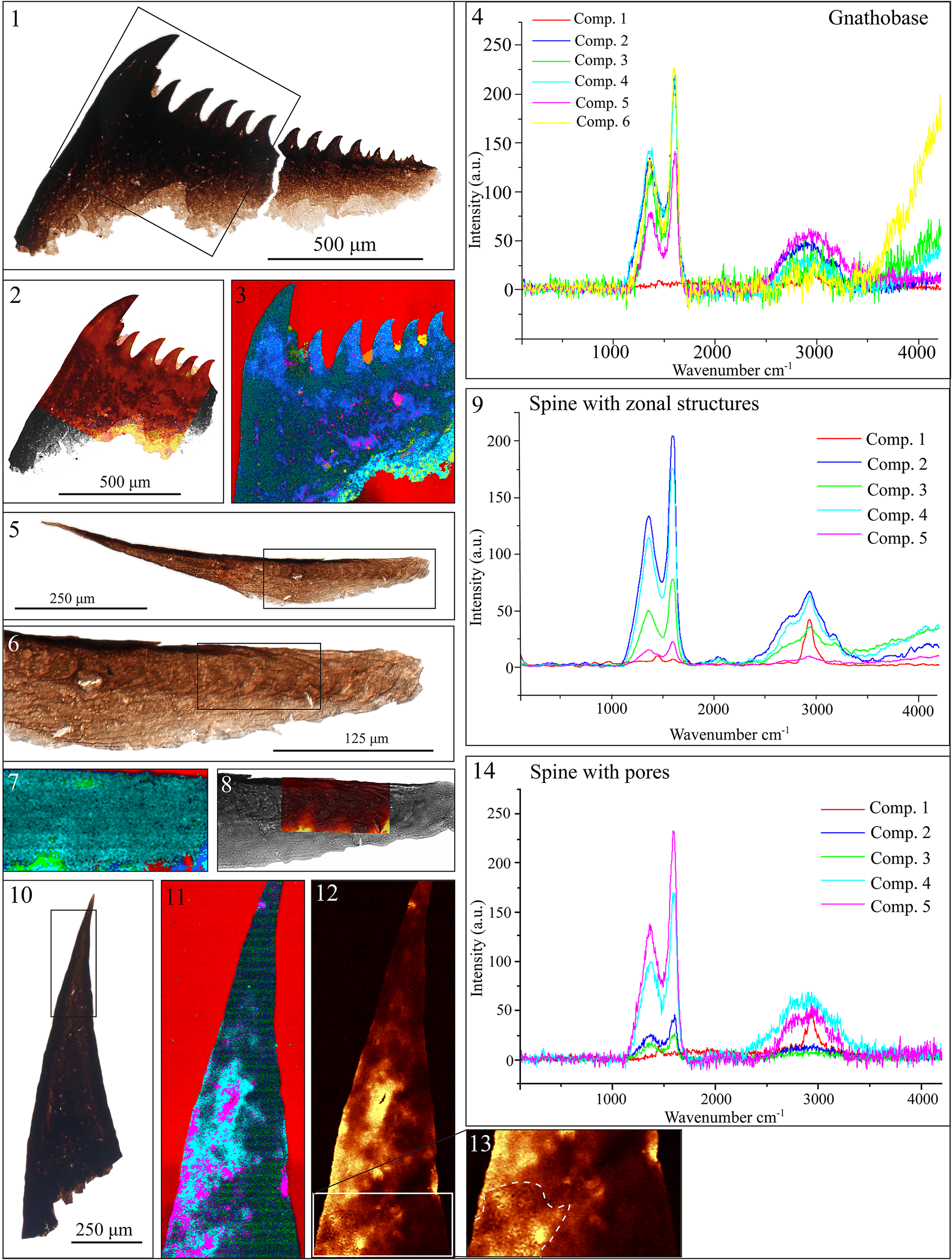
Figure 3. Long description
The figure is organized into three rows.
Top row (Panels 1 to 4) focuses on a jaw-like gnathobase fragment. Panel 1 is a brown optical micrograph with a 500 micrometer scale bar. Panel 2 and 3 show Raman phase distribution maps with vibrant red, blue, and green false colors. Panel 4 is a line graph titled Gnathobase, plotting Intensity in a u dot against Wavenumber cm super minus 1 from 0 to 4000. It shows two major peaks between 1000 and 2000 and a broad peak near 3000 for six components.
Middle row (Panels 5 to 9) displays a possible polychaete spine with zonal structures. Panels 5 and 6 are optical micrographs at different magnifications (250 and 125 micrometer scales). Panels 7 and 8 show the Raman imaging of the zonal area. Panel 9 is a line graph titled Spine with zonal structures, showing five components with distinct peaks at approximately 1350 and 1600 Wavenumber cm super minus 1.
Bottom row (Panels 10 to 14) illustrates a spine with a porous structure. Panel 10 is a dark optical image with a 250 micrometer scale. Panels 11, 12, and 13 provide high-resolution Raman maps showing dot-like porous textures. Panel 14 is a line graph titled Spine with pores, showing five components with a dominant sharp peak at 1600 Wavenumber cm super minus 1 and a secondary broad peak around 2900.
Different phases are automatically assigned the color of the mapped phase in the program (in the same sequence for all objects), so we use a single protocol to unify the results. True component analysis was used, with manual selection of regions. As the first phase, we selected the medium in which the microfossils were preserved (EOKITT), which is red in color everywhere. Next, we specified regions of the object itself, starting with the areas with the least intense Raman scattering and ending with brighter areas until the entire object area was filled. Mineral grains were not included in the analysis. Individual Raman spectra (Supplementary 1, Excel table) were analyzed in OriginPro, where the graphs for the phase distribution maps were constructed.
Raman spectroscopy results for small carbonaceous fossils
The Raman spectroscopy results for the jaw-like fragment in the phase distribution map revealed heterogeneity in the structure: the transparent base near the line of detachment differed from the denser part, which contained structures of two main phases (green and blue) (Figs. 1.19, 3.3). The dense part of the base consists mainly of the green phase, which shows linear zones enriched with fragments of the blue phase (Fig. 3.3). The spines of the jaw-like fragment are also not homogeneous in kerogen composition, and the first two large spines (green phase) differ from the smaller spines (blue phase). In addition, against the background of the first spine, Raman spectroscopy revealed another large spine of the blue phase. The two superimposed spine contours on the first large spine may represent the physical superposition of spines with different kerogen compositions, reflecting two generations of spines.
A second possible explanation concerns different compositions within a single spine. It was previously shown that the cuticular structure of Limulus polyphemus gnathobase is not homogeneous and includes the endocuticle, inner exocuticle, hyaline exocuticle, and epicuticle, with the hyaline layer exhibiting strong fluorescence and differing in composition (Rubin et al., Reference Rubin, Lamsdell, Prendini and Hopkins2017). If we assume that the composition of the Serkinskaya jaw-like fragment is similar to the structure of the cuticle of the extant horseshoe crab (Limulus polyphemus), we can suggest that the heterogeneity on the surface of the microfossil on the phase distribution map is due to different preservation of the cuticular cover, which reflects two different layers of the cuticle. However, this assumption can be made only as a hypothesis.
The spine with porous/fibrillar structure was also analyzed via Raman spectroscopy (Fig. 3). Although a porous structure was visible throughout the spine via light microscopy, heterogeneity in the structure in the Raman image was visible at only one location (Fig. 3.12 and 3.13). At the tip of the spine, where small tubes were most clearly visible under light microscopy, they were not visible in the Raman image. A small fragment with a heterogeneous dot structure, possibly corresponding to the structure of the tubes, is visible only on one side of the image (Fig. 3.13). Whether this is due to a peculiarity of the structure of the spine, heterogeneity in the preservation of tissues or parts of the spine, a difference in composition, or an error in the construction of the Raman image is currently an open question. The fragment with a dot-like structure is not a marginal effect (because it is not on the edge of the Raman image), and the presence of the structure most likely resulted from a difference in composition.
Raman images of spines with growth lines visible via light microscopy (Fig. 3.5–3.9) did not provide additional information. The initial assumption was that the composition of the growth lines might differ, and that zonation would be visible in the Raman image; however, when the phase distribution map was plotted, the composition of the kerogen did not differ in any way, and the growth lines did not stand out. Two fields (green color) with compositions different from those of the main kerogen (blue color) were identified, but the pattern of their locations is still unclear (Fig. 3.5–3.9). We interpret these green areas as unfocused images because of the different heights of object fixation. On the basis of the Raman spectroscopy results, we can assume that the composition of the spine did not change with changes in living conditions (e.g., seasons, temperature fluctuations, food availability, etc.). However, possible taphonomic changes, during which a small difference in composition could have been homogenized, cannot be excluded either.
Discussion
Age of the studied microfossils
The Serkinskaya faunal complex comes from the upper layers of the Kessyusa Group in the Serkinskaya area, and according to its stratigraphic position (which is correlated with the nearest drill core (S-203) and a section in the Olenek Uplift; Fig. 2), it may be of early Tommotian (Stage 2) or even Nemakit–Daldynian (Fortunian) age. The overlying Emyaksin Formation in the adjacent territory was characterized by the trilobites Triangulaspis meglitzkii (Toll) and Pagetiellus lenaicus (Toll), which are characteristic of Atdabanian (Stage 3) age (Pishchalnikov et al., Reference Pishchalnikov, Salikhov, Zarubin and Pyshko2020), thus indicating that the Serkinskaya fauna complex is older. Previously, trace fossils of possible arthropods, Monomorphichnus lineatus Crimes et al., Reference Crimes, Legg, Marcos and Arboleya1977, and Rusophycus avalonensis Crimes and Anderson, Reference Crimes and Anderson1985, were found in the lower Cambrian (lower Fortunian) sediments of the Siberian Platform (Nagovitsin et al., Reference Nagovitsin, Rogov, Marusin, Karlova, Kolesnikov, Bykova and Grazhdankin2015), whereas other studies have described sclerites (Moczydłowska et al., Reference Moczydłowska, Budd and Agić2015) and spines (Willman and Slater, Reference Willman and Slater2021) of possible late Ediacaran-aged metazoans. By Fortunian times, rich metazoan diversity was already present (Mángano and Buatois, Reference Mángano and Buatois2014, Reference Mángano, Buatois, Alderton and Elias2021). Despite the uncertainty interval of the Serkinskaya complex, the studied faunal complex reflects one of the oldest assemblages of SCFs preserving multiple animal phyla (taking into account the rare and potentially less diverse Terreneuvian instances from Baltica) (Slater et al., Reference Slater, Harvey, Guilbaud and Butterfield2017, Reference Slater, Harvey and Butterfield2018a). The diversity of the assemblage is consistent with molecular and other fossil data for early ecdysozoans (Liu et al., Reference Liu, Xiao, Shao, Broce and Zhang2014; Shao et al., Reference Shao, Wang, Liu, Qin, Zhang, Liu, Shao, Zhao and Zhang2020; Howard et al., Reference Howard, Giacomelli, Lozano-Fernandez, Edgecombe and Fleming2022; Hughes et al., Reference Hughes, Evans and Droser2024; Turk et al., Reference Turk, Pulsipher, Mocke, Laflamme and Darroch2024).
Adaptations indicative of durophagy
In the Serkinskaya complex we described a jaw-like fragment with tooth-like outgrowths that decrease in size in the direction away from the first tooth (Fig. 1.19, 1.17). Several interpretations can be proposed. A radular identity for the specimen (Fig. 1.19) most likely can be excluded based on the lack of a visible microvillar construction, thick continuous base, and irregular dentation of the specimen. Since priapulid teeth also occur in the assemblage, the jaw-like fragment could be a part of a scalidophoran tooth or introvert hook. Recently described specimens from the Pika Formation (Mussini et al., Reference Mussini, Veenma and Butterfield2025b, figs. 6A–D, 9H) are heavily sclerotized and optically dense, similar to the Serkinskaya SCFs, and show similar serrated margins. Some (similarly hollow) introvert hooks can even bear a subterminal “notch” below the prong (Mussini et al., Reference Mussini, Veenma and Butterfield2025b, fig. 5U), resembling that of the Serkinskaya specimen.
Another possible correlation can be made with the Cambrian spiralian Amiskwia. The jaw apparatus of Amiskwia is interpreted to be similar to the Gnathifera-like jaw, which consists of symmetrical plates that bear a series of curved rod-like elements (Caron and Cheung, Reference Caron and Cheung2019; Bekkouche and Gąsiorowski, Reference Bekkouche and Gąsiorowski2022; Mussini and Butterfield, Reference Mussini and Butterfield2025b). The wide base, irregular tooth size (emphasized by the small interspersed spinules found at one end of the tooth row; Fig. 1.20), and the sharp angle defined by the two solid edges of the Serkinskaya specimen are more indicative of gnathobase identity than a scalidophoran tooth or spiralian jaw apparatus (Caron and Cheung, Reference Caron and Cheung2019; Luo et al., Reference Luo, Parry, Boudinot, Wang, Jarzembowski, Zhang and Wang2025, fig. 2; Mussini et al., Reference Mussini, Hagadorn, Miller, Karlstrom, Evans, Dehler, Bastien and Butterfield2025a, 3AG, fig. 2P). Similar gnathobases have been described from imprints of the Cambrian chelicerates Habelia optata and Sidneyia inexpectans (Zacaï et al., Reference Zacaï, Vannier and Lerosey-Aubril2016; Aria and Caron, Reference Aria and Caron2017), the radiodont Amplectobelua symbrachiata (Cong et al., Reference Cong, Daley, Edgecombe and Hou2017), and the recently described trilobite-like Acanthomeridion serratum Hou, Chen, and Lu, Reference Hou, Chen and Lu1989 (Wu et al., Reference Wu, Liu, Wu, Zhang, Hao, Ma, Lin and Fu2025). A similar structure has also been shown in the Silurian eurypterid Eurypterus tetragonophthalmus Fischer, Reference Fischer1839 (Bicknell et al., Reference Bicknell, Paterson, Caron and Skovsted2018b). The sizes of the gnathobases of other Cambrian representatives correlate with the size of the Serkinskaya specimen (Aria and Caron, Reference Aria and Caron2017; Mussini and Butterfield, Reference Mussini and Butterfield2025b).
If the Serkinskaya specimen (Fig. 1.19) is interpreted as a gnathobase fragment, the durophagous habit needs further justification, since not all animals that possess a gnathobase are necessarily durophagous and consume hard-shelled prey. The gnathobase teeth of durophagous animals that consume hard-shelled prey tend to exhibit fibrous constructions (Bicknell et al., Reference Bicknell, Paterson, Caron and Skovsted2018b), evident in a recently described fragment from the Hess River Biota in Mussini and Butterfield (Reference Mussini and Butterfield2025b, fig. 3AG) and in extant Limulus polyphemus (Bicknell et al., Reference Bicknell, Paterson, Caron and Skovsted2018b). The Serkinskaya gnathobase fragment does not possess a fibrous structure, and its acuminate tips and arcuate shape are comparable to those of Eurypterus, whose durophagous habits are less compelling (Bicknell et al., Reference Bicknell, Paterson, Caron and Skovsted2018b). The gnathobase of Olenoides has slender and longer teeth with acuminate tips, and these animals did not crush hard prey (Bicknell et al., Reference Bicknell, Holmes, Edgecombe, Losso, Ortega-Hernández, Wroe and Paterson2021). The Serkinskaya specimen has less slender and more robust teeth than Olenoides but is not as blunt and subconical as the teeth of Sidneyia (Bicknell et al., Reference Bicknell, Paterson, Caron and Skovsted2018b). This may suggest that the animal bearing the Serkinskaya gnathobase most likely consumed prey with intermediate sclerotization (nonbiomineralized but heavily cuticularized animals), which implies a degree of durophagy in the Serkinskaya fauna.
In the present work, we used the gnathobase from the Vth appendage of Limulus polyphemus for Raman spectroscopy and to draw possible analogies with the described jaw-like fragment from the Serkinskaya fauna. Owing to their similar organization, Limulus polyphemus was previously used to study the biomechanics of Sidneyia inexpectans (Bicknell et al., Reference Bicknell, Ledogar, Wroe, Gutzler, Watson and Paterson2018a). When the modern material of Limulus polyphemus was analyzed by Raman spectroscopy, strong luminescence was observed in the examined range, and the construction of maps with phase distributions was impossible. Since the hyaline layer has previously been shown to have strong fluorescence and different compositions in extant Limulus polyphemus (Rubin et al., Reference Rubin, Lamsdell, Prendini and Hopkins2017), it was hypothesized that the heterogeneity in the phase distribution map of the Serkinskaya fragment may correspond to the multilayered nature of the fragment during life followed by patchy decomposition (Fig. 3.3).
As shown previously (Bicknell et al., Reference Bicknell, Paterson, Caron and Skovsted2018b), the structure of the gnathobase cuticle in Limulus polyphemus and Sidneyia inexpectans has a fibrous cuticle and is likely a plesiomorphic trait, whereas in Eurypterus tetragonophthalmus, the cuticle is laminate and is likely an apomorphic trait. The Serkinskaya gnathobase fragment does not possess any indication of fibrous structure, and, having been embedded in the plastic medium (EOKIT), it is not possible to investigate the internal structure. However, the gnathobase fragment presented in Mussini and Butterfield (Reference Mussini and Butterfield2025b, fig. 3AG) also has a possible longitudinal structure. The spines shown in Figure 1.2 and 1.18 have a dense outline and a fibrillar, porous structure, suggesting that the structure of the spine may be similar to that of the growth on the gnathobase in Sidneyia inexpectans. The described spine with thick lateral walls and a porous structure from the Olenek Uplift (Fig. 1.22) could thus be a part of the gnathobase fragment; however, it is unlikely to have been involved in grinding coarse food particles because the tip is rather thin and, consequently, not strong enough. The nonpreserved base also does not reveal how this spine was attached. On the basis of morphology, the spine may have been located closer to the mouth opening. The presence of pores suggests a tactile or other sensory or conductive function (Gorb, Reference Gorb2001).
Other microfossils with specific trophic adaptations
Priapulid-like organisms are known to occur near the base of the Cambrian (Smith et al., Reference Smith, Harvey and Butterfield2015; Shao et al., Reference Shao, Liu, Wang, Zhang, Tang and Li2016; Kesidis et al., Reference Kesidis, Slater, Jensen and Budd2019; Turk et al., Reference Turk, Pulsipher, Mocke, Laflamme and Darroch2024) and have been interpreted to combine carnivory with deposit-feeding (Vannier, Reference Vannier2012). Cambrian priapulid teeth from the recently described Hess River Formation (Mussini and Butterfield, Reference Mussini and Butterfield2025a), from the middle Cambrian Bright Angel Formation (Mussini et al., Reference Mussini, Hagadorn, Miller, Karlstrom, Evans, Dehler, Bastien and Butterfield2025a), and from the latest Cambrian/Tremadocian Deadwood Formation (Mussini and Butterfield, Reference Mussini and Butterfield2025c) are compatible with deposit-feeding or filter-feeding. Serkinskaya samples of priapulid teeth (Fig. 1.23–1.27) lack large, cuspidate denticles and instead show similar hair-like or minute spinulose projections, which are characteristic of filter-feeding.
The spine with a zonal structure from the Serkinskaya complex (Fig. 1.10) resembles the pharyngeal teeth of polychaetes (Plyuscheva et al., Reference Plyuscheva, Martin and Britayev2004); it can be assumed that the animal bearing the spine might have had a predatory lifestyle. For comparison, an attempt was made to study the Raman spectra of the pharyngeal teeth of the annelid Lepidonotus squamatus (Linnaeus, Reference Linnaeus1758) (comparative material for the spine with zonal structure) and the scalids of the extant priapulid Priapulus caudatus Lamarck, Reference Lamarck1816 (comparative material for Cambrian scalids). When the pharyngeal teeth and scalids of extant priapulids were analyzed, strong luminescence was observed, and the construction of maps with phase distributions was not possible.
An attempt was also made to analyze the scalids of Cambrian priapulids via Raman spectroscopy. Priapulid scalids were fixed in glycerol-gelatin media, and under laser heating during Raman spectroscopy strong luminescence was also observed for this type of optical medium, which made it impossible to distinguish separate kerogen spectra in the organic material. Despite the lack of Raman data for priapulid remains and possible pharyngeal teeth, morphological characteristics suggest that filter-feeding was already present. The large number of organic films covered with small outgrowths (Fig. 1.12–1.16) may also indicate predation pressure and the need to develop protective structures on the surface of the organisms.
Based on interpretation of the described remains, we suggest that the Serkinskaya faunal complex contains evidence for possible predators (spine with zonal structure), potential durophagy (fragments of gnathobase) with consumption of heavily sclerotized prey but probably not hard-shelled, defensive (cuticle fragments covered with small outgrowths) and tactile (spine with anchor-like base, spine with pores) structures, and possibly sessile planktivore hemichordates (Sokoloviina) and filter-feeding priapulids. This early appearance of diverse feeding strategies and behaviors occurred early in the Cambrian before the diversification of modern animal classes and orders.
Previously, Mángano and Buatois (Reference Mángano and Buatois2014) proposed the hypothesis that the appearance of Phanerozoic-type body plans in the Fortunian (e.g., deposit-feeders, large suspension-feeders) and the formation of Phanerozoic-type ecological structure in Cambrian Stage 2 (diverse trophic webs) were decoupled. The Fortunian appears to contain more animals than the Ediacaran, yet the eco-space remained underexploited, and vertical mixing of the sediment was restricted to <1 cm depth. The appearance of deposit-feeders facilitated the appearance of suspension-feeding infauna, benthos and plankton were coupled, and animal-dominated trophic webs started to form by Cambrian Stage 2, thus establishing the Phanerozoic-type ecology (Mángano and Buatois, Reference Mángano and Buatois2014, Reference Mángano and Buatois2017). Our findings, together with those of previously described Precambrian sclerites (Moczydłowska et al., Reference Moczydłowska, Budd and Agić2015; Willman and Slater, Reference Willman and Slater2021), support the hypothesis that complex metazoan behaviors preceded the establishment of diverse infaunal ecology and trophic webs.
Acknowledgments
We thank A. Yu. Zhuravlev (PIN RAS) for constructive discussion of the material and assistance in the analysis, D.E. Shcherbakov and A.P. Rasnitsyn (PIN RAS) for providing appendage of the horseshoe crab (Limulus polyphemus), and R.A. Rakitov for their comments and friendly support. We would also like to thank the anonymous reviewer and the associate editor A. Liu for their valuable comments and suggestions, and the paper greatly benefited from it!
Author contribution
Conceptualization O. Dantes (OD), K.E. Nagovitsin (KEN); data curation OD, KEN; formal analysis OD, KEN, A.V. Korsakov (AVK); funding acquisition OD, KEN, AVK; investigation OD, AVK; methodology OD, KEN, AVK; project administration OD; resources KEN, AVK; supervision OD, KEN; validation OD, KEN, AVK; visualization OD; writing: original draft preparation OD, KEN, AVK; writing: review and editing OD, KEN, AVK.
Competing interests
We declare that we have no competing interests.
Data availability statement
Data available from the Zenodo Digital Repository: https://doi.org/10.5281/zenodo.18713452.
Funding
Data acquisition was supported by Government Contract FWZZ-2026-0042 (IPGG SB RAS). Paleobiological work was financed by the Russian Science Foundation (project 23-17-00202). A.V. Korsakov (Raman spectroscopy work) was supported by Government Contract FWZN-2026-0002 (IGM SB RAS).



