Introduction
Ear arteriovenous malformation (AVM) represents a therapeutic challenge owing to its location in aesthetically sensitive areas. An AVM is an abnormal connection between the arteries and veins, with an absent capillary network.Reference Whitty, Murray, Null, Elwood and Jones1 In 1971, Doppman described this anomalous connection as the nidus. Although congenital, AVMs may not become clinically apparent until later in life.Reference Doppman2
Among extracranial AVMs, the ear is the second most commonly affected site after the cheek (16 per cent vs. 31 per cent).Reference Kohout, Hansen, Pribaz and Mulliken3 Due to the rarity of this condition and limited literature, there remains no consensus on optimal management. We propose an algorithmic approach to the management of auricular AVMs based on our institutional experience, representing one of the largest reported series to date.
Method
Approval was obtained from the Institutional Review Board Committee (IRB No: S2406160). Patients presenting with auricular AVMs between 2013 and 2022 were retrospectively reviewed. Patients with incomplete data or insufficient follow-up were excluded.
After the initial clinical examination in the Outpatient Clinic, a multi-disciplinary team (MDT) meeting was held with an otologist, interventional radiologists, vascular surgeons, and plastic surgeons. Decisions regarding embolisation and surgery were reached via consensus. Patients undergoing embolisation underwent surgical excision of the nidus within 24 hours. Post-operative follow-up was conducted via clinical review and telephone interviews.
Results
Nineteen patients (10 male, 9 female) were included, all presenting with unilateral AVMs (11 left-sided, 8 right-sided). Clinical features included pulsation (100 per cent), discolouration (80 per cent), fremitus (80 per cent), bleeding (67 per cent) and pain.
All patients underwent magnetic resonance imaging (MRI) of the lesions. Two-thirds of patients received pre-operative embolisation, followed by surgical resection within 24 hours (Table 1). None of the patients required blood transfusion. The patients were discharged on the fifth post-operative day. Outpatient department follow-up was performed once every three months for the first year. Subsequently, teleconsultation was performed every six months. Patients were offered secondary reconstruction of the ear, but none had undergone it at the time of writing this paper.
Patient details; STA = Superficial temporal Artery; PA = Postauricular artery; OA = Occipital artery; P = Peripheral lesion; C = Central lesion. Size if given in reference with ear; STSG = Split thickness skin grafting; MRI = Magnetic resonance imaging

Feeding vessels included the superficial temporal, postauricular, and occipital arteries. Surgical approaches varied based on disease extent:
AVM excision with skin excision and primary closure (n = 4)
AVM excision with skin grafting (n = 2)
Flap coverage over cartilage (n = 4)
Excision of deformed cartilage with AVM (n = 4)
Total auricular amputation (n = 2)
Three patients (15.8 per cent) developed recurrence within one year. Representative cases are summarised in Figures 1–2.
Case 1 – Excision and temporoparietal flap, Case 2 – Excision and primary closure, Case 3 – Occipital flap, Case 4 – Total amputation of Ear.

Case 5 – Partial amputation of Ear, Case 6 –Total amputation + Skin grafting, Case 7 – Total amputation of ear + Scalp flap.

In cases where AVM was excised and the underlying cartilage was found to be healthy, the residual cartilage was covered using a temporoparietal fascia (TPF) flap. A split-thickness skin graft, harvested from the thigh, was applied over the TPF flap. Case 1 (Figure 1).
When the AVM was small and the skin remained adequate following excision, primary closure over the cartilage was performed. Case 2 (Figure 1).
An occipital fascia flap was utilised for healthy underlying cartilage coverage in cases where the superficial temporal artery was either involved in the disease process or had to be ligated during surgery, rendering the TPF flap unusable. Case 3 (Figure 1).
In cases of progressive and extensive auricular AVMs associated with recurrent haemorrhagic episodes, total auricular amputation was required due to the severity and extent of disease. Case 4 (Figure 1).
When both skin and cartilage were involved in a portion of the auricle, partial ear amputation was undertaken to ensure complete removal of the nidus and achieve disease control. Case 5 (Figure 2).
In a case presenting with profuse bleeding, auricular amputation was necessary. However, the skin defect could not be closed primarily. A skin graft was used for wound coverage, but progressive graft loss occurred, and the wound healed by secondary intention over five months. Case 6 (Figure 2).
In another case where ear amputation was performed, the resultant skin defect was reconstructed using a random scalp flap with an intact vascular supply. Case 7 (Figure 2).
Discussion
In this series, patients commonly presented a decade after symptom onset. This delay in presentation was attributed to a lack of awareness among both patients and primary care providers. Pulsation and fremitus were the most frequently reported symptoms, with their resolution correlating strongly with post-operative patient satisfaction. Most patients were referred to tertiary care centres only after an episode of bleeding or ulceration.Reference Sisk, Kerr and King4
Magnetic resonance imaging remains the imaging modality of choice, offering detailed assessment of soft tissue and cartilage involvement and permitting exclusion of intra-cranial extension. Digital subtraction angiography (DSA) can be performed if embolisation is planned, as it has diagnostic and therapeutic value. Routine DSA is not warranted in lesions involving less than 20 per cent or greater than 90 per cent of the auricle, as it is unlikely to alter clinical management.
The primary aim of management is complete eradication of the AVM nidus, which requires comprehensive planning encompassing soft tissue, cartilage and wound considerations, as outlined in Table 2.
Treatment planning

AVM soft tissue management
Clinically, AVMs are staged based on the Schobinger classification: stage I (quiescence), stage II (expansion), stage III (destruction) and stage IV (decompensating) malformations. Although early-stage lesions (Stages I–II) are often asymptomatic, 80 per cent in our cohort demonstrated disease progression. Consequently, early surgical intervention is advocated where feasible, in preference to expectant management.Reference Liu, Mulliken, Zurakowski, Fishman and Greene5
Smaller lesions, particularly those located along the helical rim, where intra-operative haemorrhage is anticipated to be controllable with direct pressure, may be managed with primary surgical excision without the need for pre-operative embolisation. In our series, approximately one-third of patients underwent excision without prior embolisation.
‘Superselective’ embolisation, employing glue or particulate agents, was followed by definitive surgical excision within 24 hours in all applicable cases.Reference Potts, Zumofen, Raz, Nelson and Riina6 Non-selective embolisation or proximal vessel ligation represent outdated techniques that confer no additional benefit and may complicate the management of recurrence.Reference Ayahao7 At our institution, resection was consistently performed within 24 hours of embolisation, facilitating accurate localisation of the nidus and significantly reducing intra-operative blood loss.Reference Rane, Lamba, Gohil and Gupta8 The literature supports surgical excision within 48–72 hours, following embolisation to optimise outcomes.Reference Bhandari, Sadhotra, Bhargava, Bath, Mukherjee and Maurya9–Reference Deng, Huang, Chen, Zhang, Li and Li11 Additionally, intra-lesional embolisation may be employed as an adjunct to superselective embolisation to further devascularise the nidus in selected cases.Reference Zou, Qiao, Hua, Yang, Wang and Jin12
Although embolisation alone may appear attractive for managing lesions in cosmetically sensitive regions, it is associated with a high recurrence rate and the risk of disease progression into adjacent angiosomes.Reference Liu, Mulliken, Zurakowski, Fishman and Greene5, Reference Strassmair and Wilhelm13 The auricle receives vascular supply from two principal angiosomes: the anterosuperior segment, perfused by the superficial temporal artery, and the posteroinferior segment, supplied by the postauricular and occipital arteries.Reference Mitchell, Taylor, Houseman, Mitchell, Breidahl and Ribuffo14
Debulking or partial excision of AVMs carries a significant risk of recurrence and is associated with increased intra-operative haemorrhage.Reference Maftei, Howard, Brown, Gunning and Standfield15 In our series, recurrence was suspected in cases where complete nidus excision was not achieved. Hence, debulking or subtotal excision should be avoided in favour of definitive resection.
Cartilage management
Retention of native auricular cartilage, even when hypoplastic or distorted, is advocated wherever feasible, as it contributes significantly to maintaining the structural contour and definition of the ear.
In the presence of multiple fenestrations and significant cartilage destruction, excision of deformed or non-viable cartilage is warranted to facilitate effective soft tissue reconstruction and reduce the risk of post-operative complications.
Wound closure
Primary closure is the easiest method of wound closure. Its use is limited to small AVMs and partial ear amputations when tissue approximation can be easily achieved. Minor wound dehiscence or marginal skin necrosis was noted in several cases and was successfully managed with conservative measures. Delayed suture removal was employed to promote optimal healing.
Where primary wound closure was not feasible, local flap reconstruction was undertaken. Delayed healing at the interface between flap and native skin was commonly observed but responded well to conservative management, including routine dressings and delayed suture removal. No significant haemorrhage was encountered from these areas during the healing period.
Local flaps can be of the following two types
Fascia with skin graft is favoured when preservation of the native cartilage framework is achievable. As demonstrated in Cases 1 and 4, a thin fascial flap provides effective coverage of residual cartilage, thereby maintaining auricular contour. The TPF flap was the preferred choice in our series.
A random-pattern scalp flap was utilised when fascia flap was not available due to disease involvement.
Skin graft may appear an easy and fast solution for significant defects, or when the patient/surgeon hesitates to raise the flap to avoid incision over a non-affected area. However, it is associated with complications, such as early post-operative bleeding, graft loss and delayed wound healing.
If the defect is allowed to heal with secondary intention, it is associated with maximum complications, which include recurrence of AVM, multiple episodes of bleeding, wound infection and delayed wound healing. This method should be avoided.
Recurrence remains a recognised complication in the management of AVMs. In comparison with previously published series, our cohort included a reasonable number of patients with adequate follow-up. Only two patients exhibited recurrence following excision of lesions involving the helical region, which was likely attributable to incomplete resection of the nidus. (Table 3).
Comparison with other studies

Proposed treatment protocol
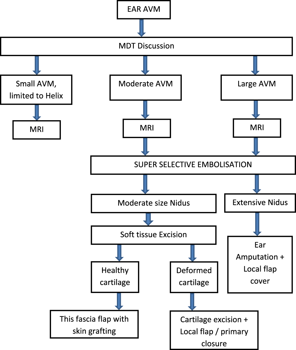
We propose a management algorithm for AVMs. All cases should be reviewed by an MDT, following thorough clinical assessment and imaging with MRI. The MDT should comprise an otologist, plastic surgeon, interventional radiologist, and, in cases of advanced or decompensated disease, an additional physician for optimisation of the patient’s clinical status.
When the lesion is confined to the auricular helix, it is classified as a small AVM. Surgical intervention is planned without the need for pre-operative embolisation. Typically, excision followed by primary closure is undertaken in these cases. Intra-operative haemorrhage may be controlled using proximal digital pressure or a soft clamp, as illustrated in Case 5.
For moderate to large auricular AVMs, pre-operative embolisation is recommended, followed by surgical excision of the nidus together with the involved skin and deformed cartilage. The choice between primary closure and cartilage coverage with a flap is determined intra-operatively. In this series, local fascial flaps, such as the TPF or occipital flap, along with skin graft, were employed to preserve auricular contour. Although free fascial flaps may theoretically be utilised, our centre lacks experience with their application in ear AVM excision.
In cases of extensive auricular AVMs where ear amputation provides optimal disease control, surgical intervention may proceed without pre-operative embolisation. Nevertheless, in the absence of adequate surgical expertise, embolisation can be employed pre-operatively to enhance safety. When primary closure following ear amputation is unfeasible, wound coverage can be achieved using random-pattern local scalp flap.
Conclusion
The initial intervention offers the optimal opportunity for successful treatment of auricular AVMs. An MDT approach is essential to ensure complete excision of the nidus. Adherence to a standardised treatment protocol facilitates optimal patient outcomes (Figure 3). Conservative management should be discouraged during the early stages of ear AVM, as curative excision is achievable at this point. DSA-guided pre-operative embolisation significantly reduces intra-operative haemorrhage. All surgical procedures in this series were performed within 24 hours of embolisation, consistent with literature advocating surgery within 48 to 72 hours. Following excision of auricular AVMs, primary skin closure, with or without flap coverage, is recommended. The use of skin grafting alone post-excision is not advisable. Secondary intention healing should be avoided due to the elevated risks of bleeding and lesion recurrence.
Treatment algorithm; AVM = arteriovenous malformation; MDT = multi-disciplinary team; MRI = magnetic resonance imaging

Financial support
This research received no specific grants from any fundraising agency, commercial or not-for-profit actors.
Competing interests
The authors declare none.
Ethical statement and informed consent
The duty was conducted in accordance with the Declaration of Helsinki and was approved by the Institutional Ethics Committee (IRB No. S2406160). The requirement for informed consent was waived because of the retrospective design.