Introduction
There exist more than 30 different morphological amacrine cell types (MacNeil & Masland, Reference MacNeil and Masland1998; Masland, Reference Masland2001), but recent physiological studies in ganglion cells suggest that there may be significantly fewer functional types based upon the limited forms of inhibition elicited by flashed spots in ganglion cells (Chen et al., Reference Chen, Hsueh, Greenberg and Werblin2010). Four prominent narrow-field ganglion cell types, the ON and OFF beta and parasol cells, appear to receive common but limited spatiotemporal patterns of inhibition: a transient broad and a sustained local inphase GABAergic inhibition and a local “crossover” form of glycinergic inhibition (Werblin, Reference Werblin2010).
Here, we extended our study from ganglion cells (Chen et al., Reference Chen, Hsueh, Greenberg and Werblin2010) to amacrine cells. We investigated whether different amacrine cell types might also receive common and limited forms of inhibition and whether the forms of inhibition found in amacrine cells resembled the forms of inhibition found in ganglion cells.
We parsed amacrine cells into two general morphological classes: 1) narrow-field amacrine cells with lateral spread of processes less than 200 μm and 2) wide-field amacrine cells with spread of processes greater than 300 μm. Each amacrine cell class received distinct but limited spatiotemporal forms of GABAergic and glycinergic inhibition, similar to, but representing only a subset of the forms of inhibition found in ganglion cells. Although there are more than 30 different amacrine cell morphologies, we measured much more limited forms of inhibition, suggesting that many morphologically diverse amacrine may generate similar forms of inhibition.
Methods
Surgery
New Zealand white rabbits (2.5 kg) were anesthetized and sacrificed in accordance with protocols approved by the Office of Laboratory Animal Care at University of California, Berkeley, CA. The procedure has been described previously (Roska et al., Reference Roska, Molnar and Werblin2006; Hsueh et al., Reference Hsueh, Molnar and Werblin2008). Briefly, rabbits are anaesthetized with 2 ml Ketamine/Xylazine cocktail (35 and 5 mg/kg) subcutaneously. Subsequently, the animals are euthanized with 1.5 ml Euthasol that is administered intracardially or intravenously. Enucleation does not begin until death is confirmed by cessation of respiration, the absence of heartbeat, and lack of blink reflex. The eyes were quickly enucleated. Each eye was dissected in dim red light by opening the eyeball removing the lens, then removing the vitreous, and finally cutting away the retina periphery to preserve the visual streak, a region about 3 × 4 mm in dimension. Before recording, the retina was peeled from the sclera and flat mounted on a Millipore filter having a precut 4 mm hole at its center. The retina with Millipore filter was then transported into a recording chamber. The flat-mounted retinas in the chamber were perfused with Ames solution at 32°C. The Ames solution for perfusion and for storing, the extra visual streak pieces was saturated with a mixture of O2 (95%) and CO2 (5%) and buffered with Na2CO3 to a pH of 7.4.
Patch clamp and recording
The images were viewed in a video monitor through a charge-coupled device camera attached to one of the microscope ports. A Nikon upright microscope with 60× water immersion lens was used to observe the shadow of the targeted cell body in the inner nuclear layer (INL). The aperture of the condenser and the contrast of the monitor were adjusted to produce the best contrast for viewing the cells.
To reach the amacrine cells beneath the inner limiting membrane (ILM), an empty glass pipette burrowed a channel at about 20 deg from horizontal, penetrating through ILM, the ganglion cell layer (GCL), and inner plexiform layer (IPL), finally reaching the INL. The preparation was illuminated with a red light source during the penetration process. After retracting the empty penetrating pipette, a second electrode filled with intracellular solution was aligned with the burrow to penetrate the retina through the previously created channel. When the pipette approached the desired cell, a small positive pressure was applied to remove debris near the tip. Then the pressure was quickly released. A negative pressure was often applied allowing the pipette to adhere to the cell. An adequate seal was ascertained when the measured tip resistance exceeded 1 Gohm. Bipolar cells and displaced ganglion cells were identified and avoided by viewing their morphology and location within the depth of the retina.
The intracellular solution contained in mM: 113 CsMeSO4 (Sigma, St Louis, MO), 1 MgSO4 (Fisher Scientific, Waltham, MA), 0.0078 CaCl2 (Fisher Scientific), 0.5 1,2-bis(o-aminophenoxy)ethane-N,N,N,′N′-tetraacetic acid (Fisher Scientific), 10 HEPES (Sigma, St. Louis, MO), 4 ATP Na2 (Sigma), 0.5 GTP Na3 (Sigma), 5 N-(2,6-Dimethylphenylcarbamoylmethyl)triethylammonium bromide (Sigma), and 7.5 Neurobiotin-Cl (Vector Labs, Burlingame, CA), pH 7.2.
Excitatory currents were measured by voltage clamping the cell at the calculated reversal potential for chloride (−60 mV). Inhibitory currents were recorded by voltage clamping the cell at the cation reversal potential (0 mV). The currents in the distal axonal regions of the wide-field cells were likely left unclamped because of the large space constant. But the signals in the dendritic regions of the broad cells (Volgyi et al., Reference Volgyi, Xin, Amarillo and Bloomfield2001) were probably better clamped. Signals were filtered at 60 Hz for analysis.
Stimuli
A variety of stimuli were presented against a gray background set at 2.3 μW/cm2. The retina was light adapted to this background level for 20 min before beginning recording. The image from a standard digital light processor projector was focused first on a diffusing plate and then focused onto the photoreceptor layer via a prism, projecting the image through the microscope condenser. We first flashed spots at 10.2 μW/cm2 to identify the receptive field center followed by annuli of the same intensity. Spot diameters were 30, 50, 100, 200, 400, 600, 1000, and 1500 μm presented in order of increasing diameter. The outside diameter of all annuli was fixed at 1500 μm; the inside diameter increased in sequence of 100, 200, 400, and 600 μm. The stimuli were systematically presented at least two times: first, with diameters from smallest to largest and then from largest to smallest. The data show the average from the pair of stimulus presentations. For Figs. 6, 7, 8, and 9, flashed bright spots of 10.2 W/cm2 or dark spots removing background of 400 μm were used.
Pharmacology
To infer the pharmacological origins of inhibitory components, experiments were performed in the presence and absence of pharmacological blockers of inhibitory receptors. Usually, only one cell was recorded in each preparation. The retina was then fixed and prepared for staining. Receptor blockers were added to Ames medium and perfused across the preparation. Four different receptor blockers were used: 20 μM 2-amino-4-phosphonobutyric acid (L-AP4; Tocris Bioscience, Ellsville, MO) to block metabotropic glutamate receptors and thereby selectively inactivate the ON system, 5 μM SR95531 (Sigma) to block GABAA receptors, 100 μM TPMPA 1,2,5,6-Tetrahydropyridin-4-yl)methylphosphinic acid (Tocris Bioscience) to block GABAc receptors, and 1 μM strychnine (Sigma) to block ionotropic glycinergic receptors. Recordings were repeated under Ames as a control when possible to confirm washout; although in some cases, it was impossible to fully wash the drugs and return to control conditions.
Morphology
Retinas were fixed in 4% paraformaldehyde for 1 h and then incubated in phosphate-buffered saline solution overnight with TritonX 100 (0.5%, Sigma) increasing the permeability of membrane. The Fluor 488-streptavidin conjugate (0.005 mg/ml, Invitrogen S11223; Invitrogen, Camarillo, CA) reacted with neurobiotin to stain the soma and dendrites. 4′,6-diamidino-2-phenylindole (0.01 mg/ml, Molecular Probes, Eugene, OR) was used to stain the nuclear layers. Retinas were then washed and sealed with Vectashield. Images were viewed and recorded using an Olympus FV1000 confocal microscope. The dendritic size was estimated by taking the average of longest and shortest diameter processes from a single optical section.
Results
Morphology of wide- and narrow-field amacrine cells
We studied 28 amacrine cells in the flat-mount retina. These amacrine cells were parsed into two nonoverlapping populations based upon the extent of the lateral spread of their dendrites: 1) 21 narrow-field neurons with lateral spread from 25 to 143 μm averaging 65.5 ± 7.2 μm (s.e., n = 19) (Two AII amacrine cells, described later, are not included in this count.) 2) 7 wide-field neurons with lateral spread from 300 to more than 1000 μm morphologically similar to the polyaxonal amacrine cells (Volgyi et al., Reference Volgyi, Xin, Amarillo and Bloomfield2001; Wright & Vaney, Reference Wright and Vaney2004). Consistent with earlier work (MacNeil & Masland, Reference MacNeil and Masland1998; MacNeil et al., Reference MacNeil, Heussy, Dacheux, Raviola and Masland1999), we found no amacrine cells with lateral spreads between 200 and 300 μm. But we could not clearly separate those amacrine cells with processes less than 200 μm into narrow- and medium-field groups because our staining of processes (see Methods) was not as accurate as those of earlier studies (MacNeil & Masland, Reference MacNeil and Masland1998; MacNeil et al., Reference MacNeil, Heussy, Dacheux, Raviola and Masland1999). The soma of all cells was in the INL to distinguish them from displaced amacrine cells or ganglion cells.
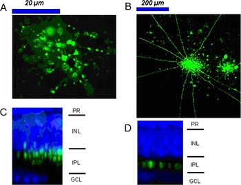
Examples of a narrow-field and a wide-field amacrine are shown in Fig. 1. All of the narrow-field amacrine cells were diffuse or multiply stratified in the IPL. The wide-field amacrine cells were broad and monostratified. Similar morphological forms were reported in previous studies (MacNeil & Masland, Reference MacNeil and Masland1998; MacNeil et al., Reference MacNeil, Heussy, Dacheux, Raviola and Masland1999). Throughout this paper, we refer the morphology of the recorded amacrine cells as “narrow field” and “wide field.” To distinguish morphology from physiology, we refer to the spatial extents over which inhibition was elicited as “local” and “broad.”
Morphologies of typical narrow- and broad-field amacrine cells. (A) Flat-mount confocal image of a narrow-field amacrine cell. The image shows the processes that remained after the electrode was removed from the preparation, pulling the soma with it. An approximate indication of where the soma would have been is by the transparent green oval at the center of the image. (B) Flat-mount confocal image of a wide-field amacrine cell. (C) Stratification of the narrow-field amacrine cell derived from a z confocal stack and (D) stratification of the wide-field amacrine cell with processes near the center of the IPL. Blue indicates DAPI staining to show the boundaries of the IPL. PR, photoreceptor; INL, inner nuclear layer; IPL, inner plexiform layer; GCL, ganglion cell layer.

Spatial and temporal and pharmacological characteristics of inhibition to narrow-field amacrine cells
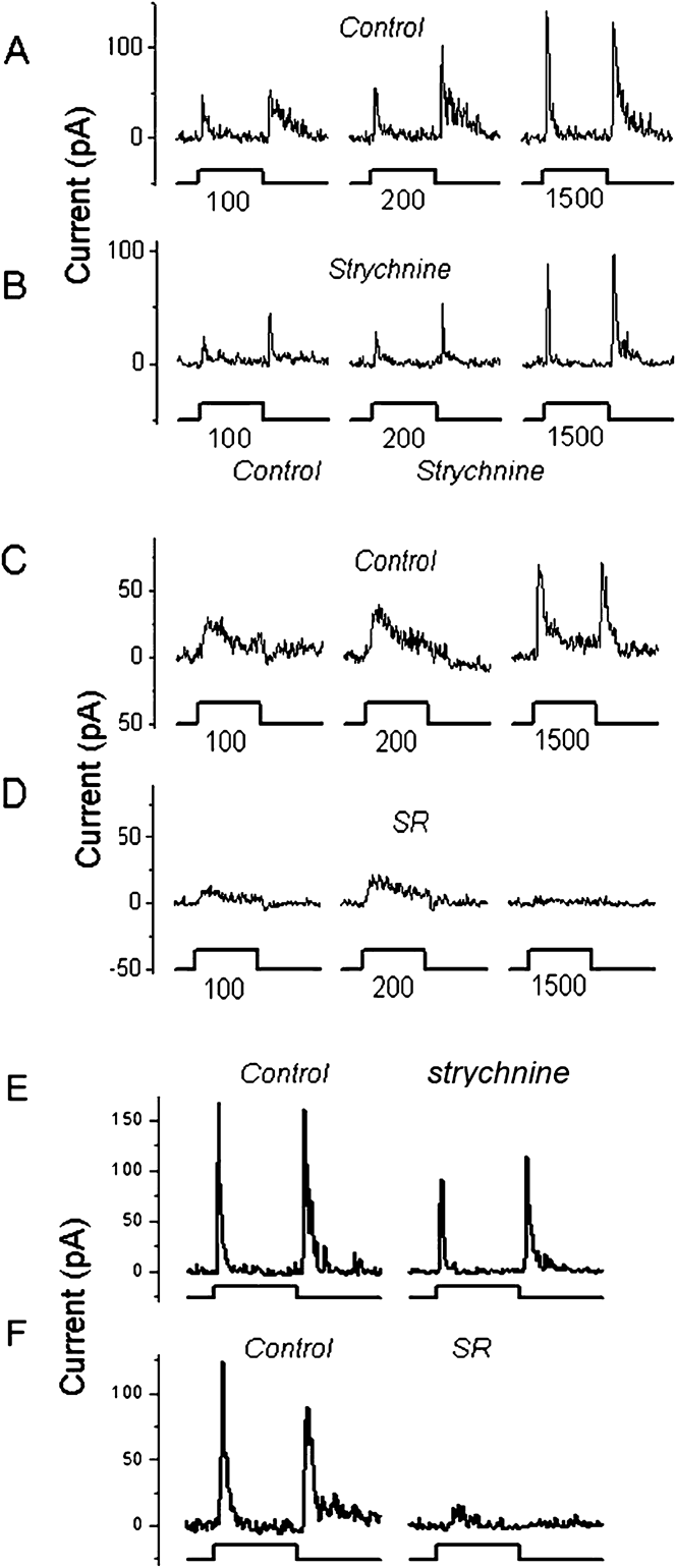
Narrow-field amacrine cells were stimulated with bright spots and with annuli of different dimensions (see Methods). The cells responded with a sustained inward (excitatory) current. Seventeen of these cells received a transient inhibition reaching a sharp peak in less than 200 ms. The peak responses grew with increasing spot diameter up to 1500 μm as shown in Fig. 2A. We did not measure changes in the timing of the peak response with increasing spot diameter. A broad annulus, falling outside the region of the dendrites of the narrow-field amacrine cell, evoked a similar transient inhibitory current as shown in Fig. 2E and 2F. These transient peaks could occur at ON and OFF as shown here or at either ON or OFF in other recordings. Because the response is transient and the integration area is so large, we refer to this inhibition as “broad transient inhibition.”
Transient GABAergic inhibitory currents are elicited over broad areas in narrow-field amacrine cells. (A) and (B) Dendritic extent of this amacrine cell was 45 μm. Larger spots evoked larger transient inhibitory currents, increasing up to 1500 μm. The transient components of these currents were insensitive to strychnine. (C) and (D) Different cell. Extent of this amacrine cell was 81 μm. The transient currents in this cell were blocked by SR95531. (E) and (F) Annulus-evoked outward currents in two different narrow-field amacrine cells. Annulus outer diameter, 1500 μm and inner diameter, 400 μm. Left panels: (control). Right panels: Transient currents are unaffected by strychnine (E) but blocked by SR95531 (F). Stimulus traces represent 1.5 s flash duration. Receptor blockers are indicated above each set of responses.

Four other narrow-field amacrine cells did not receive this broad transient inhibition. Two of them were AII amacrine cells. A GABAergic inhibition of AII activity has been reported (Bloomfield & Xin, Reference Bloomfield and Xin2000) but that is thought to result from feed back to bipolar cell terminals, possibly explaining why we did not measure this current directly in these four amacrine cells.
The broad transient inhibition in narrow-field amacrine cells was GABAergic
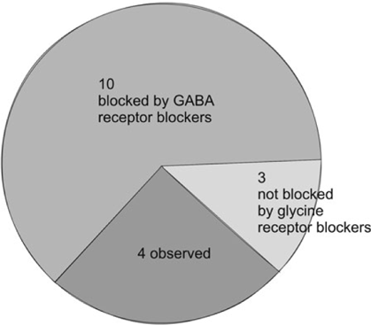
Seventeen narrow-field amacrine cells received broad transient inhibition. For spots of different diameters, the transient inhibition was virtually unaffected by strychnine in three cells as shown in Fig. 2A and 2B. The residual transient current in the presence of strychnine was 62 ± 18% (n = 3, s.e.) of the control. SR95531 or SR95531 + TPMPA blocked this inhibition in 10 cells as shown in Fig. 2C and 2D. The residual transient current in the presence of GABAergic receptor blockers was 5.1 ± 2.4% (n = 10, s.e.) of control.
No blockers were used four other cells but the largest spot (1500 μm) always evoked the largest transient inhibitory current, and the average peak transient inhibitory current was 134 ± 21 pA.
The results using annular stimuli were similar: Annuli elicited only transient currents that were blocked by GABA receptor blockers but unaffected by the glycine receptor blocker strychnine.
The sustained inhibition in narrow-field amacrine cells was glycinergic
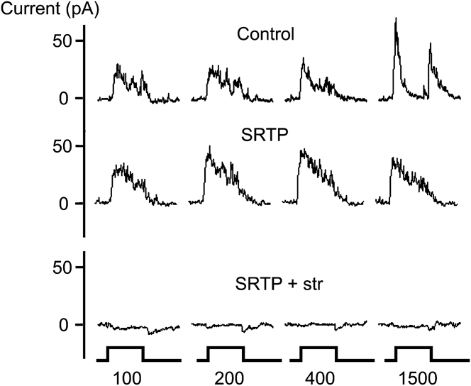
We used GABA antagonists to isolate glycinergic inhibition in 10 narrow-field amacrine cells. These measurements are taken from the same group of amacrine cells in Table 1, but in these cells, the predominant current was sustained rather than transient. Whereas the GABA receptor blockers SR95531 and TPMA decreased the transient responses in Fig. 2, these drugs increased the sustained component of the responses as shown in Fig. 3 (compare the first and second rows). For example, the 400-μm stimulus spot in the third column, inhibition increased from 18 pA in control to 24 pA averaged from 200 to 1000 ms after stimulus onset. This suggests that GABA might normally inhibit the generation of glycine inhibition. Fig. 3C shows that the remaining sustained inhibition was completely blocked by the addition of strychnine. In another three narrow-field amacrine cells, strychnine when added first blocked the sustained part of the control current. In all 13 narrow-field amacrine cells, the dominant sustained current was blocked by strychnine.
Strychnine blocked sustained inhibitory currents in narrow-field amacrine cells. (A) Control: Sustained and transient inhibitory currents. (B) SRTP: Transient inhibitory currents are blocked in the presence of SR95531 and TPMPA. (C) SRTP + str: Sustained inhibitory currents in the presence of SR95531 and 1,2,5,6-Tetrahydropyridin-4-yl)methylphosphinic acid are blocked by the further addition of strychnine. GABA receptor blockers (SRTP) eliminated the transient current elicited by the 1500-μm spot. Strychnine eliminated the sustained currents.

Broad-field transient inhibition in 17 narrow-field amacrine cells. GABA receptor blockers blocked inhibition in 10, strychnine had no effect in 3, and 4 more showed broad-field inhibition but were not studied pharmacologically

Inhibition can be broader than the dendrites of narrow-field cells
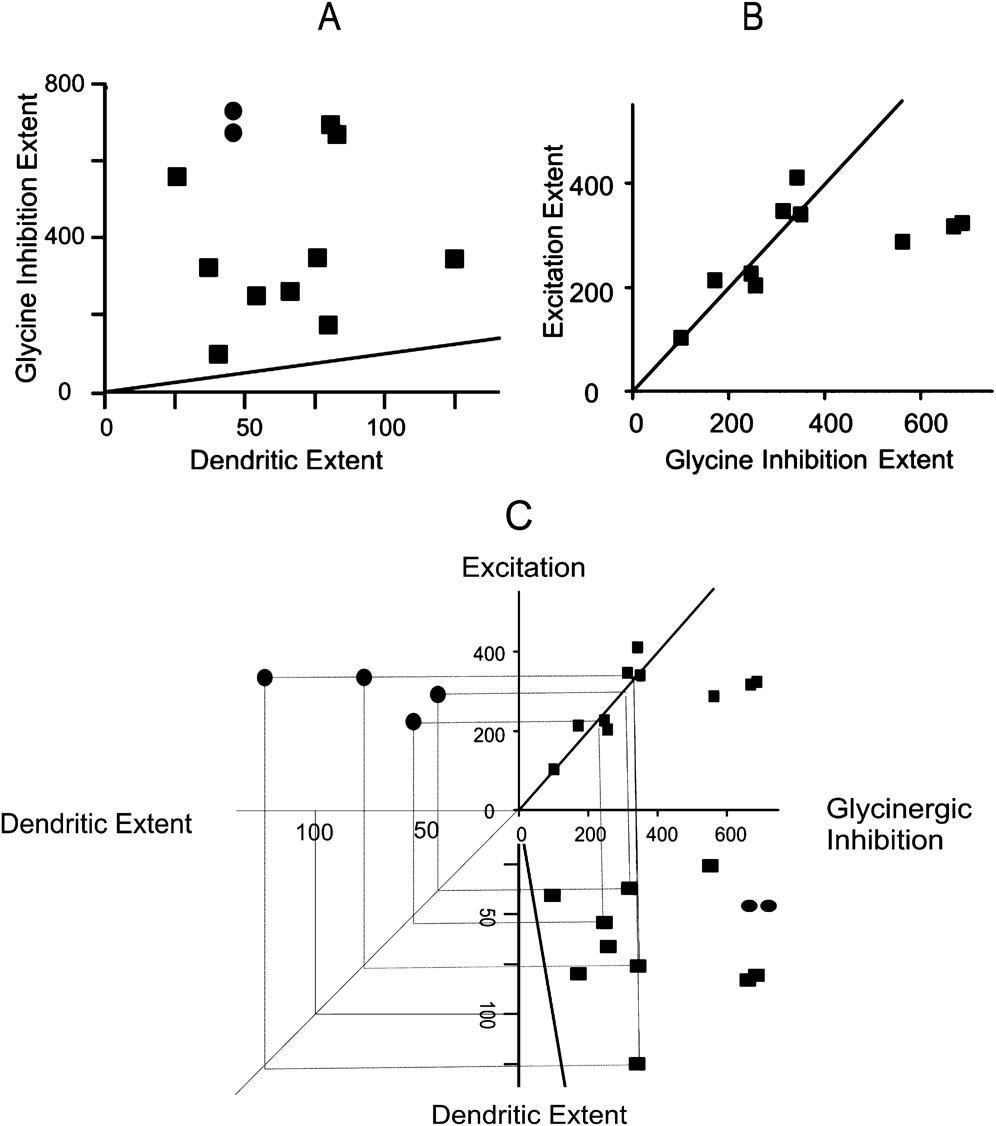
We compared the maximum spatial extent of glycinergic inhibition with the cell’s dendritic extent by measuring inhibition in response to a flashed disc of increasing diameter. In these measurements, we blocked GABA inhibition with SR95531. Sometimes we added TPMPA to SR95531 with similar results. Fig. 4A shows that in 10 narrow-field amacrine cells average size of the disc generating maximum sustained glycinergic inhibition was 371.2 μm, significantly broader than the extent of the dendrites that typically spanned less than 200 μm. We also included two AII amacrine cells (solid circles) showing that extent of glycinergic inhibition in AII cells was also significantly larger than the extent of the dendrites. Strong electric coupling between AII amacrine cells (Bloomfield & Volgyi, Reference Bloomfield and Volgyi2004) may extend glycinergic inhibition beyond the dendrites of any single narrow-field amacrine cell.
(A) Glycinergic inhibition is integrated over a much more broad area than the dendritic extents of narrow-field amacrine cells. Solid line dendritic spread equals inhibitory breadth. Circles represent two AII amacrine cells. Squares represent all other narrow-field amacrine cells. All of the data points fall well above the equal diameter line showing that inhibition is always significantly broader than the dendritic extent of any narrow-field amacrine cell. (B) Comparison of the spatial extent of glycinergic inhibition versus the spatial extent of the excitatory input to a population of 10 narrow-field amacrine cells. Glycine inhibition is integrated over areas as broad or broader than excitation in these narrow-field amacrine cells. Diagonal line: extent of excitation equals extent of inhibition. In most narrow-field amacrine cells (7 of 10), the spatial extent of excitatory input is similar as that of glycinergic inhibition. The spatial extent of all excitatory inputs was less than 450 μm, but some of the glycine inhibitory inputs extended up to 600 μm. Dimensions are in microns. (C) Excitation is plotted against dendritic extent by reflecting excitation and dendritic extent around the common axis of glycinergic inhibition taken from (A) and (B). Excitation is always broader than dendritic extent suggesting that strong coupling in these amacrine cells extends both excitation and inhibition beyond the dimensions of the dendrites.

Glycine inhibition can be broader than excitation
We measured the maximum extent of excitation in the same way; we measured the extent of inhibition using the magnitude of the response to spots of increasing diameter as plotted in Fig. 4B. Excitation extended about as broadly as inhibition for spots up to 400 μm in diameter, but in some cells, inhibition extended to 800 μm, much broader than either excitation or the span of the processes of the cell. It is likely that electrical coupling extends the collecting area of individual narrow-field amacrine cells beyond their dendritic extent as shown by Bloomfield and Volgyi (Reference Bloomfield and Volgyi2004) for AII amacrine cells. Additional electrical coupling of the glycinergic inhibitory interneurons may provide the extended inhibition to these cells beyond 400 μm.
Excitation can be broader than the dendritic extent of the cell
For spot diameters up to 400 μm, excitation and inhibition are roughly equally broad. We can use this equivalence to reflect the measurement of dendritic spread and excitation around the axis for inhibition as shown in Fig. 4C. For four of the measured points taken as examples, excitation is shown to be broader than the dendritic spread of the cells.
Wide-field amacrine cells receive glycinergic but not GABAergic inhibition
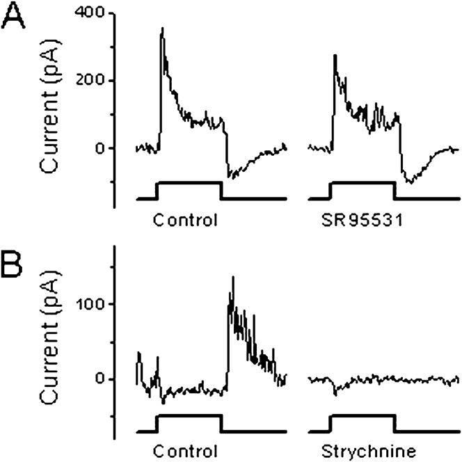
The GABA receptor blocker SR95531 had no effect the inhibitory current in three wide-field amacrine cells as shown in Fig. 5A. The currents, in the presence of SR95531 when averaged over the first 1000 ms following the onset of the spot stimulus, were 88.2 and 206% of the control currents. Conversely, strychnine completely blocked the inhibitory current-blocked inhibition in three other wide-field cells as shown in Fig. 5B. The residual currents in these cells, averaged from 0 to 1000 ms following the start of the spot stimulus, were −1.6, 5.6, and 29.5% of the control level. The high value in the last case (10.5 pA in control and 3.1 pA in glycine blocker) may be due to a noisy measurement of a weak inhibitory current in both the control and blocked conditions.
Inhibitory current in wide-field amacrine cells is glycinergic. (A) SR95531 did not block inhibition in this wide-field cell. (B) Strychnine completely blocked inhibition in this wide-field cell. These results are representative of the six wide-field cells studied. Spot size, 400 μm. Perfused receptor blockers are indicated below each recording.

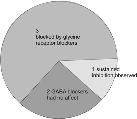
Table 2 parses each of the six wide-field amacrine cells and the experimental conditions under which they were studied.
Experimental conditions under which the wide-field amacrine cells were studied. Sustained inhibition was blocked by strychnine in three cells, not blocked by GABA receptor blockers in two cells, and observed in one other wide-field amacrine cell

The peak latency of broad transient GABAergic inhibition is much shorter than that of glycinergic inhibition
The peak latency of transient GABAergic currents, recorded in all amacrine cells studied here, was 140 ± 16 ms (n = 5, s.e.). The transient GABAergic current decayed to 30% of peak level in 208 ± 27 ms (n = 5, s.e.). The peak latency of glycinergic inhibitory currents recorded in amacrine cells under the same stimulus conditions was 286 ± 23 ms (n = 15, s.e.). By the time the glycinergic currents reach their peak level, the transient GABAergic had already decayed to levels below 30% of the peak value. The difference in latency of transient GABAergic inhibition and glycinergic inhibition was a significant (t-test, P = 0.001). The distribution of the peak latencies of the transient GABAergic and glycinergic inhibition is shown graphically in the Discussion in Fig. 10.
Glycinergic inhibition and crossover
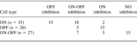
Earlier studies suggested that most of the glycinergic inhibition measured in bipolar and ganglion cells was of the crossover type where ON inhibition combines with OFF excitation or OFF inhibition combines with ON excitation to enhance the voltage response of the cell (Molnar & Werblin, Reference Molnar and Werblin2007; Hsueh et al., Reference Hsueh, Molnar and Werblin2008; Molnar et al., Reference Molnar, Hsueh, Roska and Werblin2009; Werblin, Reference Werblin2010). This appeared to be consistent with an earlier study of crossover in amacrine cells, measured in the retinal slice. Here, in a larger population of amacrine cells in flat-mount retina, we looked specifically at the relationship between the phase (ON vs. OFF) of excitation and inhibition, but we were not able to identify the morphology or process spread of this population. Of the 96 cells studied here, we found that the majority of glycinergic ON inhibition to OFF cells was crossover, about half of the OFF inhibition to ON cells was crossover, some ON-OFF cells received no inhibition, and that there was also considerable glycinergic ON-OFF inhibition to ON-OFF amacrine cells.
ON amacrine cells receive OFF and ON-OFF glycinergic inhibition
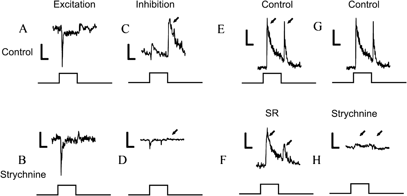
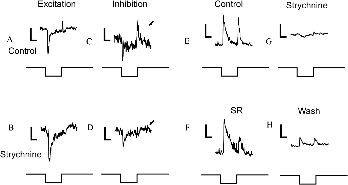
Of the 35 ON amacrine cells studied, 15 received inhibitory input at light OFF, 18 cells received inhibitory input at both light ON and OFF, and 2 cells received inhibitory input at light ON. The synaptic currents are shown in Fig. 6. Excitatory currents at ON are shown in Fig. 6A and 6B. The inhibitory current at OFF was blocked by strychnine as shown in Fig. 6D. Strychnine also blocked the ON-OFF inhibitory current as shown in Fig. 6H, but SR had little effect on ON-OFF inhibition as shown in Fig. 6F. ON inhibition occurred too rarely to analyze pharmacologically. These results show that the majority of inhibitory input to the ON amacrine cells is mediated by glycine.
Inhibition to ON amacrine cells is primarily glycinergic. (A) and (B) ON inward current. (C) OFF crossover outward current (occurring at OFF). (D) Outward current blocked by strychnine leaving a small residual inward current. (E) and (G) transient outward currents representing ON-OFF inhibition in control. (F) Outward currents were unaffected by SR; smaller responses represent rundown during the introduction of SR. (H) Outward currents almost completely blocked by strychnine (arrows). In this and in Figs. 7–9, the vertical bar indicates 25 pA. Stimulus traces represent 1 s flashes.

OFF amacrine cells receive ON and ON-OFF glycinergic inhibition
Of the 20 OFF cells, 15 received ON inhibition as shown in Fig. 7C and 7D and 5 OFF amacrine cells received ON-OFF inhibition as shown in Fig. 7E and 7F. In both cases, inhibition was blocked by strychnine, as shown in Fig. 7D and 7G.
Inhibition to OFF amacrine cells is glycinergic. (A) and (B) Excitatory currents at onset of a dark flash. (C) Arrow: Crossover inhibitory current at offset of the dark flash. (D) Arrow: Strychnine blocked the ON inhibitory current. (E) ON-OFF outward current. (G) Strychnine blocked the ON-OFF outward current. (F) outward current was unaffected by SR. (H) Inhibition begins to return after strychnine (from G) was washed out. Washout with strychnine is never complete.

Some ON-OFF cells receive no inhibition
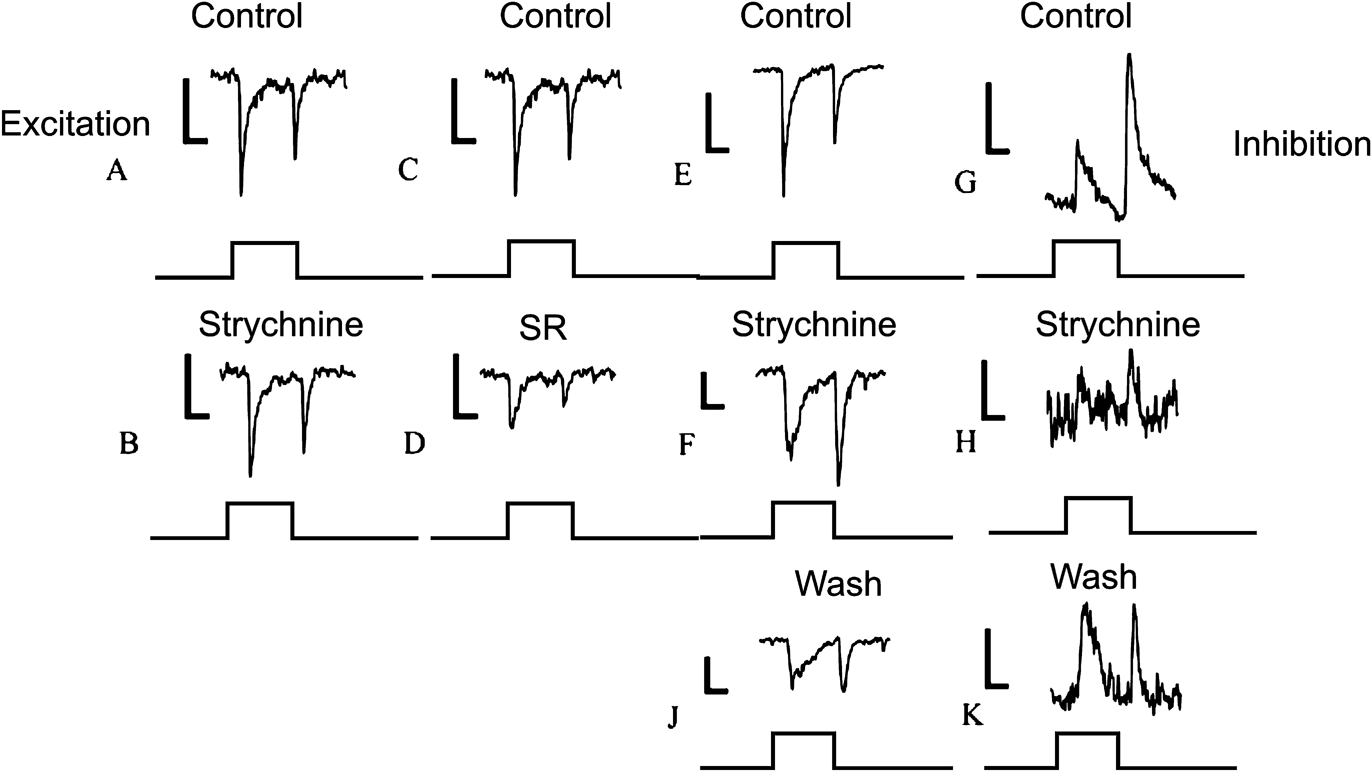
Of the 27 amacrine cells that received ON-OFF excitation, 15 retained inward currents even at 0 mV that were smaller than those measured at −60 mV. This suggests that the cells were not well space clamped, allowing some excitation to be generated at unclamped regions of the cell. If so, the anticipated outward current at 0 mV might have been overwhelmed by the unclamped inward current. To test for this, we blocked the inhibitory input pharmacologically to determine if the inward current contained an outward current component. In 9 of the 15 cells tested, application of SR or strychnine did not increase the magnitude of the inward current as shown in Fig. 8A–8D. This suggests that any inhibitory input to these cells was insignificant. We measured a notable decrease in the magnitude of the inward current in both strychnine and SR95531. This decrease was most likely due to the gradual run-down of the light response and not a result of the blockers. We saw similar rates of run-down in the native excitatory input at −60 mV that did not recover in the wash. These results suggest that this type of ON-OFF amacrine cell, likely a wide-field cell because it was not space clamped, does not receive any significant direct inhibitory input.
ON-OFF cells receive predominant ON-OFF glycinergic inhibition. Traces (A) through (D) represent cells that receive no inhibition; Traces (E) through (K) represent cells that receive glycinergic inhibition. (A) Excitatory ON-OFF inward current in control. (B) ON-OFF inward current is unaffected by strychnine. (C) Same ON-OFF inward current as in (A) under control. (D) ON-OFF inward current persists in SR (with some rundown). (E) ON-OFF inward current in control. (F) ON-OFF inward current is not affected by strychnine. (G) ON-OFF outward current under control. (H) ON-OFF outward current blocked by strychnine. (J) and (K) recovery of excitation and inhibition after wash.

Some ON-OFF amacrine cells receive ON-OFF glycinergic inhibition
In seven cells, of these ON-OFF cells, strychnine strongly suppressed ON-OFF inhibition as shown in Fig. 8H. In SR95531, the ON-OFF inhibitory responses become more sustained and lost any transient components that were present in control conditions. We found that SR95531 blocked wide-field responses, while leaving local responses relatively unaffected consistent with the results shown in Fig. 2. Taken together, the results suggest that local ON-OFF inhibition is not mediated by GABA, but when present, it is likely mediated primarily by glycine. Broad transient ON-OFF inhibition may be mediated primarily by GABA.
ON-OFF amacrine cells receive ON glycinergic inhibition
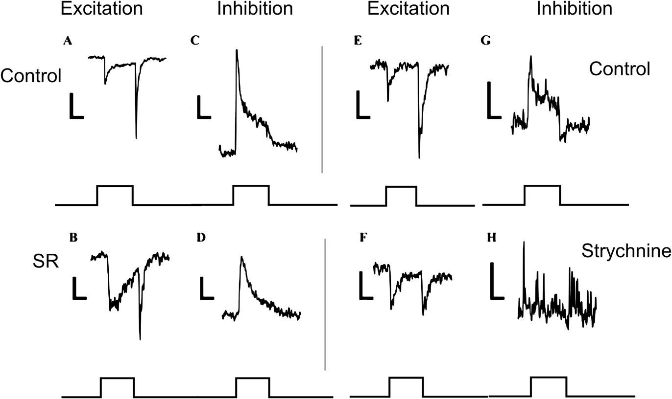
Five of the ON-OFF cells received ON inhibition that was significantly blocked by strychnine as shown in Fig. 9.
ON-OFF cells that receive glycinergic ON inhibition. (A–D) were measured from one amacrine cell, and parts (E–H) were measured from a second amacrine cell. (A) ON-OFF excitatory current. (B) In the presence of SR95531, the ON phase of the excitatory input became more sluggish, but the response was relatively unchanged. (C) The ON inhibitory input under control conditions. (D) The ON inhibition persisted in the presence of SR95531. (E) ON-OFF excitatory current in a different amacrine cell. (F) In the presence of strychnine, the ON-OFF excitation was somewhat smaller but largely unaffected. (G) ON inhibitory input under control conditions. (H) In the presence of strychnine, ON inhibition was blocked. Taken together, these experiments indicate that ON inhibition to ON-OFF cells was glycinergic and not GABAergic.

Table 3 below summarizes the number of cells studied and the forms of inhibition that they received.
Summary of cell types and the forms of inhibition that they receive

Discussion
Amacrine cells receive only a subset of the three main inhibitory signals generated in the IPL
Three main forms of inhibition emerge from studies in bipolar (Molnar & Werblin, 2007) and ganglion cells (Roska & Werblin, Reference Roska and Werblin2003): These three are: 1) broad transient GABA inhibition, 2) local sustained crossover glycine inhibition, and 3) local sustained noncrossover GABA inhibition. Some of the most prevalent ganglion cell types, the ON and OFF beta and parasol cells, receive all three components (Chen et al., Reference Chen, Hsueh, Greenberg and Werblin2010). However, in amacrine cells, we found only two forms of inhibition: local GABA inhibition was missing in all amacrine cells and broad transient GABA inhibition was missing in wide-field amacrine cells. The peak latencies of the transient GABA and sustained glycinergic inhibitory currents found in amacrine cells match closely two currents of similar spread, kinetics, and pharmacology found in ganglion cells as shown as comparison in Fig. 10.
Comparison of the peak latencies of the two inhibitory components found in amacrine cells in the present study, with the peak latencies of the three inhibitions found different ganglion cell types in Chen et al. (2010). The peak latencies for the broad GABAergic inhibition and the local glycine inhibition are quite similar. Light bars: transient GABA inhibition; dark bars, glycine inhibition; medium bars, sustained local GABA inhibition recorded only in ganglion cells.

Broad transient GABA inhibition
Both narrow-field amacrine cells (Fig. 2) and beta and parasol ganglion cells (Chen et al., Reference Chen, Hsueh, Greenberg and Werblin2010) receive a broad (up to 1 mm in breadth), fast (140 ms latency), transient ON, OFF, or ON-OFF GABAergic inhibition. This form of inhibition is likely carried by amacrine cells similar to the polyaxonal amacrine cells (Famiglietti, Reference Famiglietti1992a,Reference Famigliettib,Reference Famigliettic; MacNeil & Masland, Reference MacNeil and Masland1998); a group of neurons with very widely ramifying processes that appear to propagate information with brief transient signals at ON-OFF or ON-OFF over long lateral distances that can be blocked by tetrodotoxin or SR95531 (Taylor, Reference Taylor1999; Volgyi et al., Reference Volgyi, Xin and Bloomfield2002; Roska & Werblin, Reference Roska and Werblin2003).
Very broad short latency brief GABAergic inhibition has been associated with saccadic suppression (Roska & Werblin, Reference Roska and Werblin2003) as well as the object motion sensitive response (Olveczky et al., Reference Olveczky, Baccus and Meister2003; Baccus et al., Reference Baccus, Olveczky, Manu and Meister2008). It is striking that this form of inhibition is not measured in the wide-field amacrine cells themselves, the same neurons that appear to generate this form of inhibition. These studies and those of Wright and Vaney (Reference Wright and Vaney2004) suggest that the wide-field amacrine cells that carry broad transient inhibition are GABAergic.
Glycinergic inhibition
Both narrow- and wide-field amacrine cells (Hsueh et al., Reference Hsueh, Molnar and Werblin2008, the current study) and beta and parasol ganglion cells (Chen et al., Reference Chen, Hsueh, Greenberg and Werblin2010) receive a slower (peak latency 267 ms) glycinergic inhibition, but glycinergic inhibition has different spatial properties in ganglion and amacrine cells: In ganglion cells, glycinergic inhibition is spatially quite narrow, usually falling within the dendritic collecting area of the ganglion cell (Chen et al., Reference Chen, Hsueh, Greenberg and Werblin2010; Russell & Werblin, Reference Russell and Werblin2010). However, in narrow-field amacrine cells, a glycinergic inhibition of similar latency (267 ms) was elicited over a much broader region than the dendritic area of the recorded cells (Fig. 4), and inhibition can sometimes be elicited over regions even broader than excitation. Wide-field amacrine cells also receive glycinergic inhibition, but because of the large collecting area of these cells, it was not possible to determine whether this inhibition originates from a broadly coupled or narrow population of glycinergic amacrine cells.
These measurements suggest that there exist at least two classes of narrow-field glycinergic amacrine cells: one class is uncoupled, found in most ganglion cells. It falls locally within their dendritic field and acts either as crossover or to modify the response kinetics (Chen et al., Reference Chen, Hsueh, Greenberg and Werblin2010; Russell & Werblin, Reference Russell and Werblin2010). The other class of narrow-field amacrine cells integrates excitation over areas broader than their dendrites (Fig. 4) and generates inhibition in some narrow-field amacrine cells that is much broader than their dendrites, suggesting that they are electrically coupled. These coupled amacrine cells may represent a separate class of glycinergic interneurons from those that generate local inhibition in ganglion cells
Sustained local GABA inhibition
We found no local sustained GABA inhibition to amacrine cells in this study. However, a local, sustained, GABAergic inphase inhibition (i.e., ON inhibition to ON cells or OFF inhibition to OFF cells) is a common inhibition found in most ganglion cells (Chen et al., Reference Chen, Hsueh, Greenberg and Werblin2010). No crossover GABAergic inhibition (ON to OFF or OFF to ON) was measured in those ganglion cells. In earlier studies, local sustained GABA inhibition has been associated with edge enhancement, and traditional antagonistic surround activity usually found to be GABAergic (Cook & McReynolds, Reference Cook and McReynolds1998; Lukasiewicz et al., Reference Lukasiewicz, Eggers, Sagdullaev and McCall2004; Ichinose & Lukasiewicz, Reference Ichinose and Lukasiewicz2005; Eggers & Lukasiewicz, Reference Eggers and Lukasiewicz2006, Reference Eggers and Lukasiewicz2010; Zhang & Wu, Reference Zhang and Wu2009, Reference Zhang and Wu2010). This GABA inhibition is likely carried by the medium-field amacrine cells (MacNeil & Masland, Reference MacNeil and Masland1998) that have been shown to be GABAergic (Chen et al., Reference Chen, Hsueh, Greenberg and Werblin2010), and unlike the narrow amacrine cells, the medium-field amacrine cells have a broad enough spatial spread to carry inhibition laterally. This local GABAergic inhibition is not likely to be carried by the wide-field amacrine cells, although they are GABAergic (Wright & Vaney, Reference Wright and Vaney2004), because these wide-field cells propagate activity over broad rather than local regions, their response is transient and with much shorter latency. These studies taken together suggest that some medium-field amacrine cells generate GABAergic inhibition.
The missing pharmacological pathways
Surprisingly, we found no sustained local GABAergic inhibition acting upon any amacrine cell type in response to flashed spots. These narrow- and medium-field GABAergic amacrine cells are thought to generate local antagonistic surround. In that role, it may be counterproductive for the medium amacrine cells themselves to receive a local antagonistic surround. Consistent with our finding, recordings in salamander amacrine cells showed center-surround antagonism in ganglion cells but not in amacrine cells (Zhang & Wu, Reference Zhang and Wu2010). In an earlier study, Molnar and Werblin (Reference Molnar and Werblin2007) found sustained GABAergic inphase (ON to ON) inhibition only between ON cone bipolar cells and between ON cone to rod bipolar cells mediated in both cases by ON local GABAergic amacrine cells. In that study, we found no OFF to OFF or ON to OFF GABAergic inhibition to bipolar cells.
Summary of connectivity in the rabbit retina
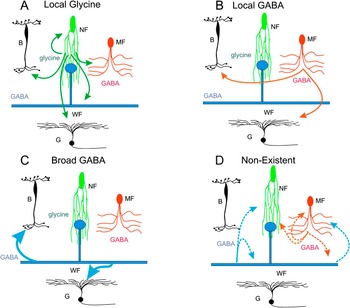
There are likely to be species differences among mammalian retinas, so this work refers only to the rabbit. Fig. 11 attempts to summarize the results of pharmacological studies in rabbit bipolar, amacrine, and ganglion cells. The figure relies upon associations between the three morphological amacrine cell types, their inferred transmitter content, and their inferred role in local and broad GABAergic and glycinergic inhibition as described in the previous paragraphs.
Summary of the known glycinergic and GABAergic pathways to amacrine and ganglion cells. (A) Local glycinergic inhibition affects all cell types. Text above infers that this local inhibition arises from narrow-field amacrine cells. (B) Local GABA inhibition only affects bipolar and ganglion cells. Text above infers that this inhibition arises from narrow- and medium-field amacrine cells. (C). Broad GABA inhibition affects all cell types except wide-field amacrine cells (wide-field amacrine cells don’t inhibit each other). (D) Pathways that have not been measured. Local GABA inhibition is not found in narrow- and medium-field amacrine cells and broad-field inhibition is not found in wide-field amacrine cells. Arrows signify measured pathways from narrow-field amacrine cells (NF, green), medium-field amacrine cells (MF, orange), and wide-field amacrine cells (WF, blue) to bipolar (B), ganglion (G), and other amacrine cells. All of the known pathways are described in the text.

Panel A shows narrow glycinergic inhibition to all cell types. This is based upon the studies of Molnar and Werblin (Reference Molnar and Werblin2007) describing glycinergic crossover feedback to bipolar cells where most OFF bipolars received ON glycinergic inhibition, but only about half of the ON bipolar cells received OFF glycinergic inhibition. The study of Hsueh et al. (Reference Hsueh, Molnar and Werblin2008) described glycinergic feedback to amacrine cells. In that study, most of the ON amacrine cells received OFF inhibition, but only about half of the OFF amacrine cells received ON inhibition. The study of Chen et al. (Reference Chen, Hsueh, Greenberg and Werblin2010) described glycinergic feedforward to four classes of ganglion cells. This paper describes additional pathways for glycine inhibition to narrow- and wide-field amacrine cells.
Panel B shows local GABA inhibition to bipolar cells but only between ON bipolars either to other ON bipolars with a brief delay and feedback to rod bipolars (Molnar & Werblin, Reference Molnar and Werblin2007). Chen et al., (Reference Chen, Hsueh, Greenberg and Werblin2010) described local GABA feedforward inhibition to ON and OFF beta and parasol cells. Neither narrow- nor wide-field amacrine cells received local GABAergic inhibition.
Panel C shows broad GABA inhibition as described by Roska and Werblin (Reference Roska and Werblin2003). This broad inhibition is fed back to bipolar cells and forward to ganglion cells. This is consistent with the measurements of Chen et al., (Reference Chen, Hsueh, Greenberg and Werblin2010) describing broad-field feed forward inhibition to ganglion cells. This paper also shows broad inhibition to narrow amacrine cells, but wide-field amacrine cells did not receive broad inhibition.
Panel D shows two significant pathways were not measured in any of these studies: 1) We measured no broad transient GABA inhibition to other wide-field cells and 2) We measured no local GABA inhibition to narrow-field amacrine cells. This suggests that narrow GABAergic amacrine cells do not inhibit other narrow-field GABAergic or glycinergic amacrine cells. This finding is consistent with the findings of Volgyi et al. (Reference Volgyi, Xin and Bloomfield2002) who showed that GABAergic surround inhibition to AII amacrine cells was not direct but was fed back to rod bipolar terminals. Eggers and Lukasiewicz (Reference Eggers and Lukasiewicz2006) describe GABA inhibition between amacrine cells, but this may also be indirect via bipolar cell terminals. It is the absence of these wide and narrow pathways that may be the most significant result of this study.
There are, of course, exceptions to the “rules” that we outline in Fig. 11. For example, some narrow-field amacrine cells affect timing rather than compensate for rectification (van Wyk et al., Reference van Wyk, Taylor and Vaney2006; Russell & Werblin, Reference Russell and Werblin2010). Some medium-field amacrine cells do much more than simply provide a GABAergic antagonistic surround such as the starburst cells (Fried et al., Reference Fried, Munch and Werblin2002; Zhou & Lee, Reference Zhou and Lee2008) and the A17 amacrine cell (Grimes et al., Reference Grimes, Zhang, Graydon, Kachar and Diamond2010). One medium-field amacrine cell, the DAPI-3 cell is glycinergic rather than GABAergic. Some wide-field amacrine cells serve as neuromodulators rather than mediate saccadic suppression such as the serotonin-accumulating cells (Vaney, Reference Vaney1986) or the dopamine cells (Dowling, Reference Dowling1986; Keeley & Reese, Reference Keeley and Reese2010). But for the majority of our measurements, the description of pathways outlined in Fig. 11 seems a reasonable summary of the general functional properties and interactive pathways of the three main morphological amacrine cell types.