Introduction
Multiple sclerosis (MS) is a chronic inflammatory and demyelinating disease affecting the central nervous system (CNS) by autoimmune-mediated myelin injury (Sospedra & Martin, Reference Sospedra and Martin2005). Currently, MS is considered a Th1–Th17-mediated organ-specific inflammatory autoimmune disorder (Panitch et al., Reference Panitch, Hirsch, Schindler and Johnson1987; Rocken et al., Reference Rocken, Racke and Shevach1996), caused by both genetic and environmental factors. The incidence of autoimmune disorders like MS and allergies has increased sharply in developed countries over recent decades (Hirst et al., Reference Hirst, Ingram, Pickersgill, Swingler, Compston and Robertson2009). Several epidemiological studies show an inverse global distribution of such disorders in developed countries and infectious diseases, especially parasitic infections in developing countries. An explanation of this phenomenon was first proposed in 1989 with Strachan's ‘hygiene hypothesis’, which suggested that repeated contact with infectious agents during childhood may protect against allergy and autoimmunity development (Strachan, Reference Strachan1989). The incidence of allergy has been found to be reduced in children infected with intestinal helminths, in particular hookworms or Schistosoma spp. (Strachan, Reference Strachan1989; Araujo et al., Reference Araujo, Lopes, Medeiros, Cruz, Sousa-Atta, Sole and Carvalho2000; Scrivener et al., Reference Scrivener, Yemaneberhan, Zebenigus, Tilahun, Girma, Ali, McElroy, Custovic, Woodcock, Pritchard, Venn and Britton2001; Yazdanbakhsh et al., Reference Yazdanbakhsh, van den Biggelaar and Maizels2001). These observations have also been confirmed in multiple experimental animal models of autoimmune conditions, such as type 1 diabetes, collagen-induced arthritis, experimental autoimmune encephalomyelitis (EAE) and colitis, and in allergic skin, intestine or airway conditions (David et al., Reference David, Thomas, Zaccone, Dunne and Cooke2004). Interestingly, in EAE, the mouse model most closely resembling human MS, treatment with Schistosoma mansoni eggs has been reported to be protective (La Flamme et al., Reference La Flamme, Ruddenklau and Backstrom2003; Sewell et al., Reference Sewell, Qing, Reinke, Elliot, Weinstock, Sandor and Fabry2003). Additionally, a negative correlation has been demonstrated between the incidence of MS and naturally acquired helminth infections (Fleming & Cook, Reference Fleming and Cook2006). One study of 12 MS patients with parasitic worm infection examined the clinical course of the disease by magnetic resonance imaging (MRI) and selected parameters, such as level of eosinophilia in peripheral blood. Over a follow-up period of 4.6 years, the patients showed significantly lower disease exacerbation and drastically reduced MRI changes as compared to uninfected MS patients (Correale & Farez, Reference Correale and Farez2007).
Helminths are known to use various immune regulatory strategies to induce tolerance in the host. For example, they are strong inducers of Th2 responses, characterized by up-regulated production of anti-inflammatory cytokines such as interleukin (IL)-4, IL-5, IL-10 and IL-13. This suggests that a change in Th responses from Th1/17 to Th2 patterns might be one underlying mechanism for the protective effect of helminths against autoimmune disorders. Moreover, the immunomodulatory effects of worm infections have also been associated with an increase of CD4+CD25+FoxP3+ regulatory T cells in peripheral blood (Yazdanbakhsh et al., Reference Yazdanbakhsh, van den Biggelaar and Maizels2001; Maizels et al., Reference Maizels, Balic, Gomez-Escobar, Nair, Taylor and Allen2004; Belkaid et al., Reference Belkaid, Sun and Bouladoux2006; Correale & Farez, Reference Correale and Farez2007). Thus, overall, the literature shows that increased production of IL-10 by regulatory T cells, in particular, as well as by T-helper cells (Satoguina et al., Reference Satoguina, Mempel, Larbi, Badusche, Loliger, Adjei, Gachelin, Fleischer and Hoerauf2002; Kamradt et al., Reference Kamradt, Goggel and Erb2005), B cells and also innate immunity cells such as macrophages (Araujo et al., Reference Araujo, Lopes, Medeiros, Cruz, Sousa-Atta, Sole and Carvalho2000; Hartmann et al., Reference Hartmann, Schonemeyer, Sonnenburg, Vray and Lucius2002; Satoguina et al., Reference Satoguina, Mempel, Larbi, Badusche, Loliger, Adjei, Gachelin, Fleischer and Hoerauf2002; Mangan et al., Reference Mangan, van Rooijen, McKenzie and Fallon2006; Smits et al., Reference Smits, Hammad, van Nimwegen, Soullie, Willart, Lievers, Kadouch, Kool, Kos-van Oosterhoud, Deelder, Lambrecht and Yazdanbakhsh2007) are involved in helminth-mediated suppression of inflammatory disorders.
In recent animal studies, different groups have attempted to identify and utilize the protective effect of helminth-derived single components in order to modulate the immune response in autoimmune diseases (Harnett & Harnett, Reference Harnett and Harnett2010). Therapy with Trichuris suis ova (TSO) uses vital eggs of the non-human pathogen parasite T. suis and has already been demonstrated to be effective in the treatment of inflammatory bowel disease (Summers et al., Reference Summers, Elliott, Qadir, Urban, Thompson and Weinstock2003, Reference Summers, Elliott, Urban, Thompson and Weinstock2005).
Taking into account these preliminary data, a non-randomized, pilot study was designed and four MS patients were treated with TSO. Over a treatment period of 6 months, immunomonitoring was used to detect a possible Th1–Th2 shift, basic changes in innate and adaptive immunology and changes in lymphocyte and natural killer (NK) subsets.
Materials and methods
Patients
Four patients with secondary progressive MS, some with overlapping relapses, were treated for 6 months with TSO. Patients were recruited from the Department of Neurology, Charité – Universitätsmedizin Berlin, and fulfilled the 2005 McDonald criteria (Polman et al., Reference Polman, Reingold, Edan, Filippi, Hartung, Kappos, Lublin, Metz, McFarland, O'Connor, Sandberg-Wollheim, Thompson, Weinshenker and Wolinsky2005). The study was approved by the local ethics committee and all subjects provided informed consent.
TSO were produced and provided by Ovamed GmbH, Barsbüttel, Germany, as a non-sterile aqueous suspension containing 2500 embryonated TSO in 15 ml of sulphate-stabilizer (0.05 mmol/l H2SO4). The patients were administered with TSO every 14 days over 6 months. The same dose was used in previous clinical trails for Crohn's disease and allergic rhinitis and is supposed to have immunomodulatory effects (Summers et al., Reference Summers, Elliott, Urban, Thompson and Weinstock2005; Bager et al., Reference Bager, Arnved, Ronborg, Wohlfahrt, Poulsen, Westergaard, Petersen, Kristensen, Thamsborg, Roepstorff, Kapel and Melbye2010).
Immunological and clinical parameters were surveyed 1 month before ( − 1), at the beginning (0) and in months 1, 2, 3 and 6 during therapy, as well as 1 and 4 months after therapy (defined as time points 7 and 10). Routine blood tests, including differential blood count, liver (γ-glutamyl transferase (GGT), alanine transaminase (ALAT), aspartate aminotransferase (ASAT), alkaline phosphatase (AP)) and renal (creatinine, urea) function, and electrolytes were performed by the laboratory of the Charité hospital, Berlin. Furthermore, we monitored immune cells and their subsets, including CD4+, CD8+ lymphocytes, B cells, NK cells, monocytes, and IgE and IgG subclasses. CD4+T cells were analysed quantitatively, qualitatively and in functional assays to identify cytokine secretion patterns. Samples were obtained by taking peripheral blood from patients using vacuum collection tubes anticoagulated with heparin (227 ml), and ethylenediaminetetraacetic acid (EDTA; 4 ml), as well as serum tubes (10 ml) (all Vacutainer Systems BD, Plymouth, UK). Patients were clinically monitored at the beginning of the observation period and in months 3, 6 and 10, including assessment according to the expanded disability status scale (EDSS).
Whole-blood stimulation of CD4+T cells
For whole-blood stimulation of CD4+T cells, sixteen 1 ml samples of heparinized blood were stimulated with 1 μl anti-CD28 antibody (Becton Dickinson, BD Pharmingen, Germany). A total of four control samples were treated with anti-CD28 alone, while four other samples were activated with either 1.5 μg/ml staphylococcus enterotoxin A (SEA), 1.5 μg/ml staphylococcus enterotoxin B (SEB) or 1.0 μg/ml toxic shock syndrome toxin (TSST). Samples were incubated for 6 h at 37°C in an atmosphere containing 100% air humidity and 5.2% carbon dioxide. At 2 h after the start of stimulation 2 μg/ml brefeldin A was added to block cytokine excretion (all from Sigma, St. Louis, Missouri, USA).
Flow cytometry
For whole-blood staining of stimulated CD4+T cells, samples were incubated for 10 min with phosphate-buffered saline (PBS) containing 20 mM EDTA at room temperature (RT). Erythrocytes were lysed by treatment with FACS-lysing solution (BD Pharmingen, Heidelberg, Germany). Leucocytes were permeabilized with FACS-Perm (BD Pharmingen) for 10 min at RT. After washing with PBS containing 2% fetal calf serum, fixed cells were stained for 20 min at RT in the dark with the following antibodies: anti-CD4-PerCP, anti-CD154-PE, anti-IL-2-FITC, anti-IL-4-APC, anti-IL-10-APC or anti-IFN-γ-FITC (all BD Pharmingen). Octagam (OCTAPHARMA GmbH, Langenfeld, Germany) was added to block unspecific binding. After washing a minimum of three times, 105 CD4+T cells per sample were analysed using a FACS Calibur cytometer.
For quantitative and qualitative analysis of immune cells, whole-blood staining in EDTA blood was performed in Trucount tubes (BD Pharmingen). The following anti-human antibodies were added to analyse the CD4+ lymphocyte subsets: anti-CD4 APC, anti-CD27 PE, anti-CD45RA biotin and anti-CD62L FITC. For analysis of CD8+ lymphocytes, anti-CD3 FITC, anti-CD4 PE, anti-CD8 APC and anti-CD45RA biotin were added. SA-PerCP was used as a secondary conjugate. For analysis of NK cell subsets, anti-CD3 APC, anti-CD56 PE and anti-CD62L FITC were added. Monocytes and B cells were analysed by staining with anti-CD4 PE, anti-CD14 PerCP, anti-CD23 PE and anti-CD62L FITC (all BD Pharmingen), and anti-CD19-APC (Beckman Coulter Immunotech, Marseille, France), respectively. All stains contained 2 μl Octagam. Afterwards, all samples were lysed and washed. Cytometric analysis was performed using CellQuest on a FACS Calibur cytometer.
To quantify the proportion of CD56++ NK cells, whole-blood staining was performed in 1.5 ml Eppendorf tubes by adding anti-human CD3 APC, CD44 FITC, CD56 PE (BD Pharmingen) and Octagam. IgE and IgG subclasses in serum samples taken from patients were analysed using an enzyme-linked immunosorbent assay (ELISA) kit from Euroimmune (Lübeck, Germany) and ELISA components from the hospital laboratory of Charité, Berlin, respectively.
Statistical analysis
The FACS staining was analysed using CellQuest. These and all other results were then analysed statistically by means of a paired two-sided t-test, ANOVA, Kruskal–Wallis test and Dunn's post-hoc test using GraphPad Prism 5. P values < 0.05 were considered significant.
Results
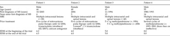
Four patients suffering from secondary progressive multiple sclerosis were treated with TSO for 6 months and subsequently monitored for another 4 months. Clinical characteristics are summarized in table 1.
Clinical characteristics of the subjects.

EDSS, expanded disability status scale; i.v., intravenous; MRI, magnetic resonance imaging; MS, multiple sclerosis.
Clinical course and adverse clinical events
All patients had previously been diagnosed with secondary progressive MS. Frequent clinical monitoring, including EDSS, was performed and the results remained stable both during therapy and during the follow-up period in three out of four patients. Patient 1 exhibited mild clinical impairment during therapy and progression of EDSS from 6.0 to 6.5. A cerebral MRI had been obtained for each patient within the past 2 years prior to the treatment. No gadolinium-enhancing lesions were detected in patients 1, 2 and 4, while patient 3 had refused gadolinium application. No further MRIs were performed during the therapy or thereafter.
TSO was well tolerated by all patients. No side-effects were reported, except mild gastrointestinal symptoms in one patient, possibly related to the TSO therapy. None of the patients ceased the therapy prematurely.
Routine laboratory parameters
Routine blood parameters such as differential blood count, liver (GGT, ALAT, ASAT, AP) and kidney (creatinine, urea) parameters and electrolytes obtained by the Charité's central hospital laboratory were all within the reference range (data not shown).
Lymphocyte subsets during TSO therapy
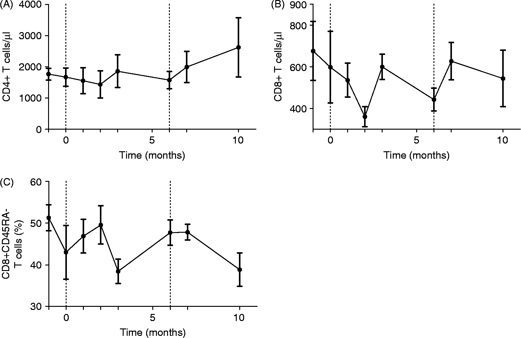
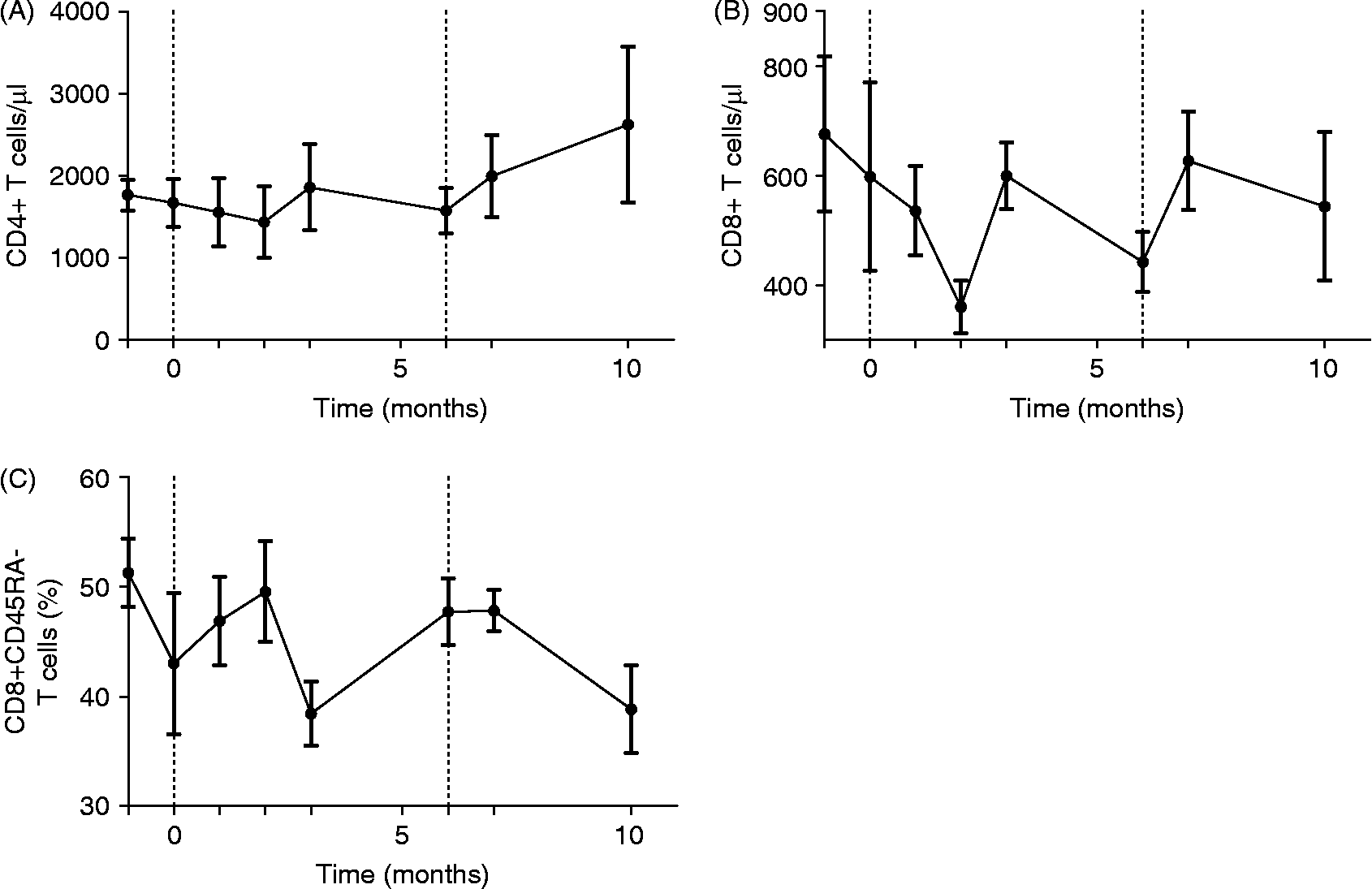
A slight decrease of CD4+ (fig. 1A) and a more relevant decline in CD8+T cell numbers (fig. 1B) as compared to baseline within the reference range could be observed after 2 months of therapy, although none reached statistically significant differences. A transient decrease in the percentage of memory or effector CD8+T cells (CD8+CD45RA − ) among CD8+T cells was detected after 3 months of therapy (fig. 1C).
Number of (A) CD4+T cells; (B) CD8+T cells; and (C) proportion of CD8 +/ CD45RA − cells of gated lymphocytes in the whole blood of patients, given as mean ± SEM. Dotted lines mark the duration of treatment.
Regarding CD4+T cells, during therapy with TSO a slight decrease in naïve CD4+T cells (CD3+/CD4+/CD45RA+/CD27+) was observed, while memory (CD3+/CD4+/CD45RA − /CD27 − ) and effector (CD3+/CD4+/CD45RA − /CD62L − ) CD4 T cell counts remained stable (data not shown).
Influence of TSO on the Th1–Th2 immune response
To determine whether TSO limits the CD4+ Th1 response or instead increases the general Th2 response, we stimulated whole-blood cells with different superantigens (SEA, SEB and TSST) before, during and after therapy with TSO in vitro and subsequently stained them for CD154, CD4, interferon (IFN)-γ, IL-2, IL-4 and IL-10.
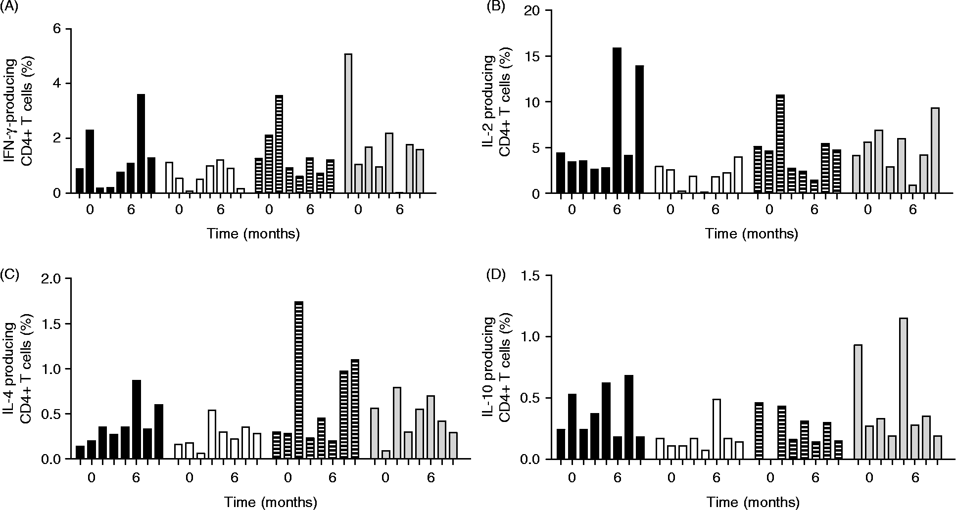
During TSO treatment, we detected a trend towards decreased IFN-γ responses to all T-cell stimuli, although this did not reach statistical significance (fig. 2A). A clear decrease in IL-2-producing T cells was detected in month 2 after the start of therapy (fig. 2B). For SEA stimulation, changes in IL-2 in month 2 compared to baseline levels were significant (Student's t-test; month − 1 versus month 2: P = 0.01; month 0 versus month 2: P = 0.05). For SEB and TSST stimulation, there was a tendency towards lower IL-2 values, although P values did not reach statistical significance. Surprisingly, IFN-γ and IL-2+T cells showed an increase at 1 month after the start of therapy.
Number of cytokine-producing CD4+CD154+T cells after whole-blood stimulation with staphylococcus enterotoxin A (SEA) plus anti-CD28-antibody in the course of TSO therapy. Cytokine production – (A) IFN-γ; (B) IL-2; (C) IL-4; (D) IL-10 – as percentages of all gated CD4+T cells in all four patients. The treatment period lasted from months 0 to 6. Black bars, patient 1; white bars, patient 2; striped bars, patient 3; grey bars, patient 4.
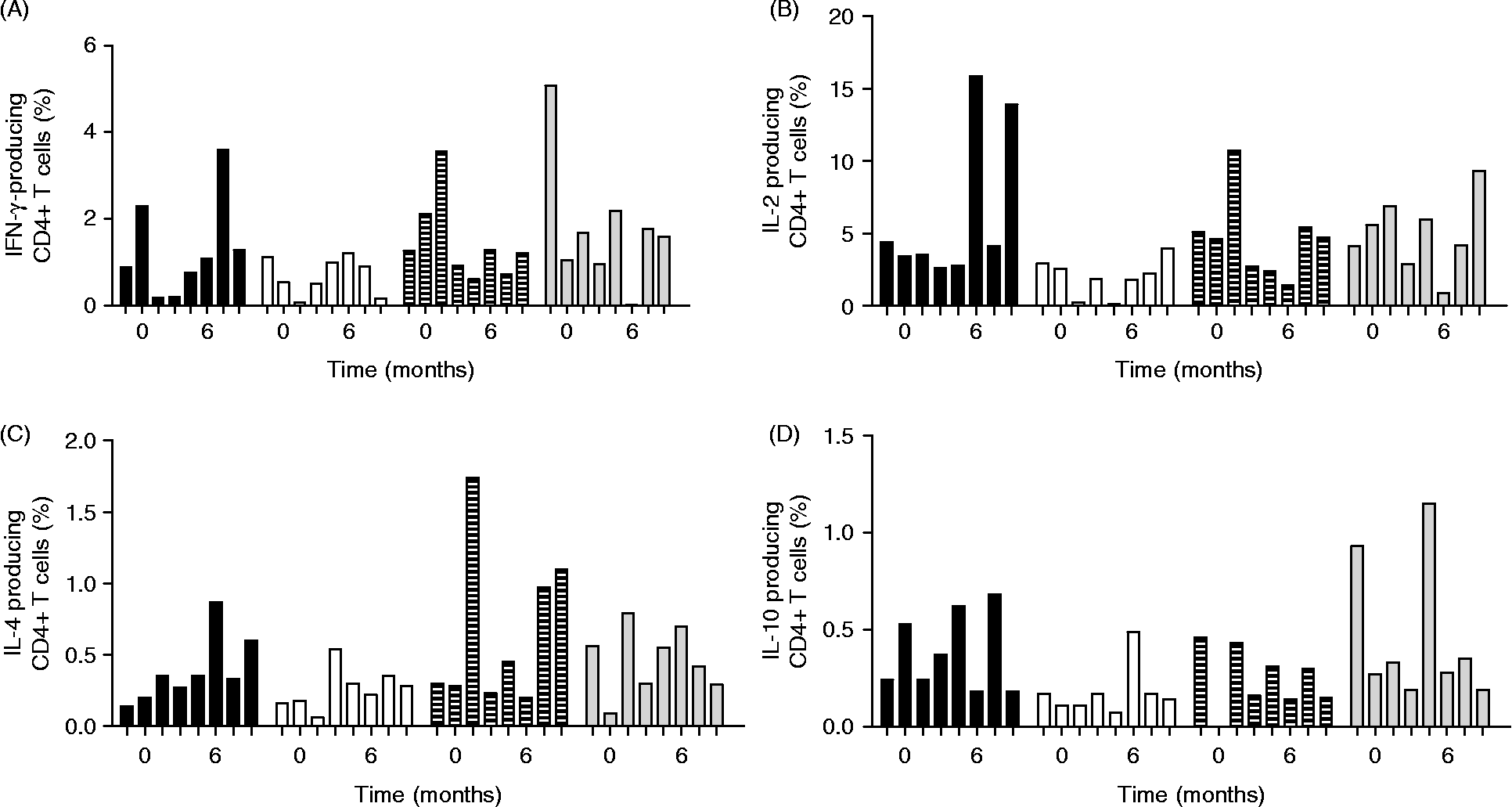
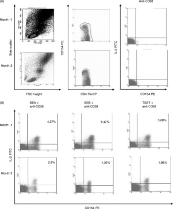
In contrast, an increase of IL-4-producing CD4+T cells was detected at the first month, followed by a decrease towards baseline levels at month 2 (fig. 2C). IL-10-producing CD4+T cells remained relatively unaffected by the treatment (fig. 2D). Representatively, FACS selection and analysis of IL-2-producing cells in one patient are shown as dot plots (fig. 3A and B).
(A) FACS dot plot of lymphocytes from one patient at time points − 1 and 2 (1 month before and 2 months after the start of therapy). The left panel shows lymphocytes gated in FSC/SSC (R1). In the middle panel CD4+ and CD154+/ − lymphocytes are gated in R2. The right panel shows CD4+ lymphocytes after stimulation with anti-CD28 antibody only. (B) CD4+ lymphocytes after stimulation with anti-CD28 Ab and SEA or SEB or TSST and IL-2-producing cells as percentages of all gated lymphocytes from R2 (upper right quadrant).

In summary, we detected a trend toward decreased Th1 responses in patients during TSO therapy, while Th2 and IL-10 responses remained relatively unaffected. No further statistical significance was detected in the comparison of cell counts and cytokine patterns with baseline values during and after therapy.
Immunoglobulin levels during TSO therapy
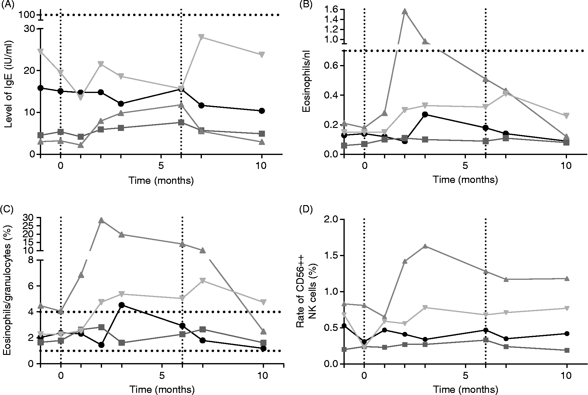
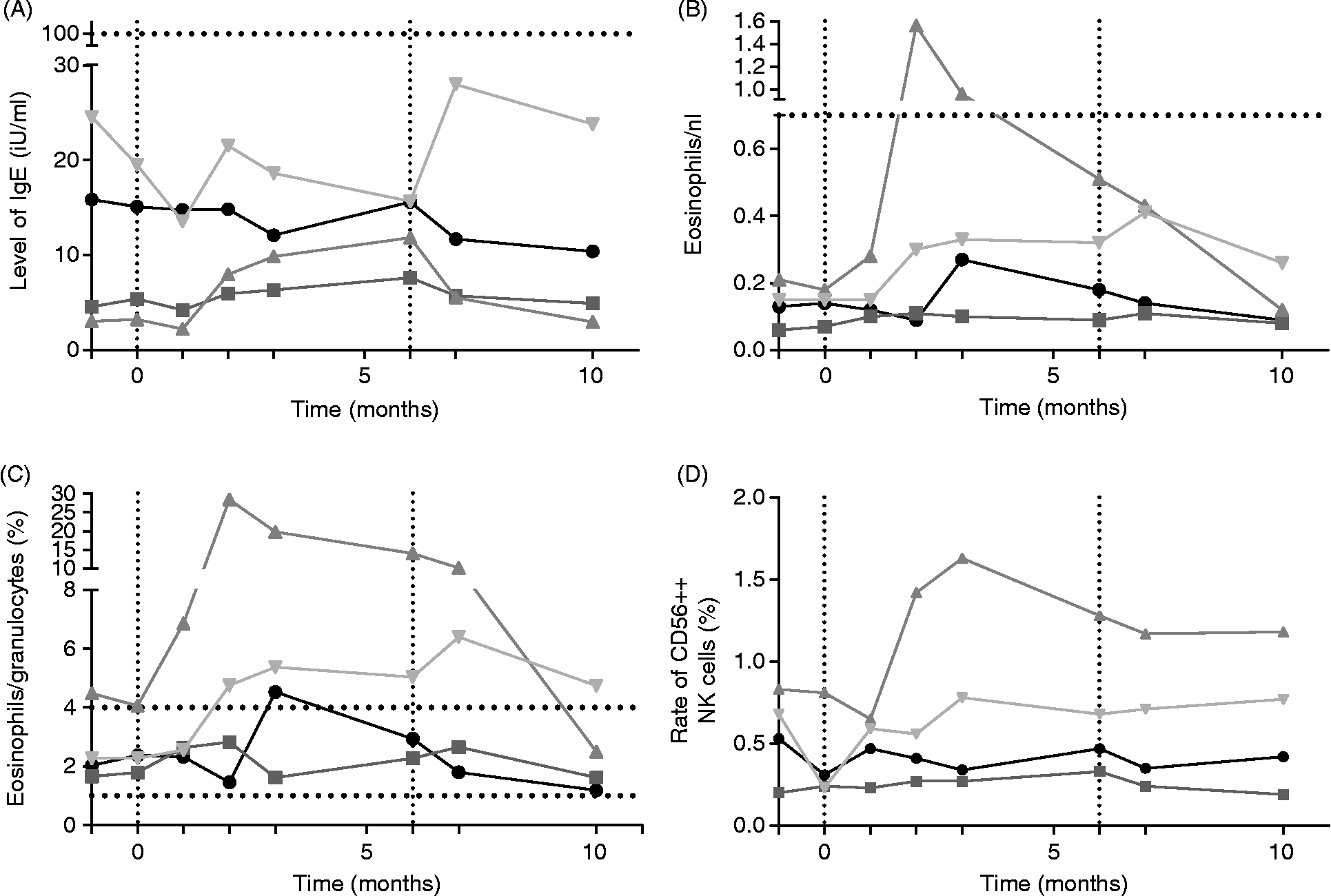
Minor variation in IgE-levels within the reference range was detected in three out of the four patients (fig. 4A). IgG subclasses remained unchanged except for an increase in IgG4 within the reference range in one patient (data not shown).
(A) Serum level of IgE in iU/ml. (B) Eosinophil counts per nanolitre of peripheral blood. (C) Eosinophils as percentage of all granulocytes. (D) CD56++ NK cells as percentage of total NK cells. All figures show data from four patients over the course of TSO therapy. Horizontal dotted lines mark upper and lower limit of the laboratory's reference range, vertical dotted lines mark the duration of treatment. ●, Patient 1;  , patient 2;
 , patient 3;
 , patient 4.
Leucocyte subsets under TSO therapy
The total leucocyte count and neutrophils did not vary significantly under therapy (data not shown). Eosinophil counts slightly increased after the beginning of therapy and only reached pathological values in one patient (fig. 4B). The proportion of eosinophils in granulocytes clearly exceeded the reference range in two patients (fig. 4C). During therapy, basophils increased in two patients and decreased in the other two, returning to baseline levels after the end of therapy (data not shown).
The total number of CD19+B cells varied significantly between patients, increasing slightly during the treatment in three patients but remaining within the reference range (80–500/μl). Prior to the start of treatment patient 4 had already had elevated B-cell numbers (approximately 1000/μl), which then decreased to values within the reference range over the course of the treatment. Approximately 80% of B cells were CD23+ in all patients with no significant changes during the treatment course (data not shown).
CD14+ monocyte/macrophage counts remained unchanged over the course of the therapy. A mean level of 400/μl within the reference range (200–1000/μl) was maintained. Accordingly, no significant changes were observed in the subgroups of CD62-L − and CD62-L+ CD14+ monocytes.
NK cells during TSO therapy
The number of NK cells (defined as both CD3 − /CD56+ and CD44+CD56++ NK cells) remained stable during the TSO treatment. One patient showed a transient increase in the proportion of CD56++ NK cells, followed by a decrease after termination of therapy. In the other patients, a stable level was maintained (fig. 4D). During the observation period, 0.67–5.46% of NK cells were CD56++, based on an absolute cell count varying between 3 and 37/μl of peripheral blood.
Discussion
Recent reports suggest that parasitic infections, especially worm infections, may protect against the development of inflammatory reactions associated with allergic and autoimmune diseases (Yazdanbakhsh et al., Reference Yazdanbakhsh, van den Biggelaar and Maizels2001; Maizels et al., Reference Maizels, Balic, Gomez-Escobar, Nair, Taylor and Allen2004; Correale & Farez, Reference Correale and Farez2007). An inverse global distribution of the latter and helminth infections has been observed: allergy and autoimmunity are increasing within developed countries, while helminth infections remain common in the developing world (Ruyssers et al., Reference Ruyssers, De Winter, De Man, Loukas, Herman, Pelckmans and Moreels2008; Cooper, Reference Cooper2009). Helminths are known to influence the immune system in a number of different ways and might offer new possibilities in the treatment of immune-mediated diseases. The first clinical trials with non-human pathogenic TSO have already been published and studies focusing on the treatment of Crohn's disease and ulcerative colitis, in particular, have shown very promising results (Summers et al., Reference Summers, Elliott, Qadir, Urban, Thompson and Weinstock2003, Reference Summers, Elliott, Urban, Thompson and Weinstock2005). However, a first attempt to treat airway allergy with TSO showed no therapeutic effects (Bager et al., Reference Bager, Arnved, Ronborg, Wohlfahrt, Poulsen, Westergaard, Petersen, Kristensen, Thamsborg, Roepstorff, Kapel and Melbye2010). Several research groups are attempting to identify parasitic components with regulatory immune functions as a first step to developing new immunomodulatory drugs (Hartmann et al., Reference Hartmann, Schonemeyer, Sonnenburg, Vray and Lucius2002; Harnett & Harnett, Reference Harnett and Harnett2010). The promising results of TSO therapy in patients suffering from inflammatory bowel disease suggest that these immunomodulatory effects might also be beneficial in multiple sclerosis. Characterization of the effects of TSO on the human systemic immune response has only just begun. The present study is the first to periodically monitor routine blood parameters and immune reactivity of MS patients during therapy with TSO.
We investigated in our group of patients various lymphocyte subsets proposed to possess essential immunological functions. Crucially, in our study, anti-inflammatory IL-10 levels did not significantly change during TSO therapy. Recent findings of Correale & Farez (Reference Correale and Farez2007), which showed that helminth antigens induce IL-10 production in human dendritic cells and B cells via toll-like receptor 2 and, thus, that helminths may exert their immunoregulatory function by modulating antigen-presenting cells and B-cell functions, emphasize the role of IL-10. However, another study showed that a transfer of IL-10-deficient mesenteric lymph node cells from nematode-infected donors could also grant protection against airway hyperreactivity, which indicates that IL-10 is not necessarily the dominant player in immune suppression by helminths in different disease settings (Wilson et al., Reference Wilson, Taylor, Balic, Finney, Lamb and Maizels2005), and supports a possible immunoregulatory effect of TSO.
Interestingly, our study showed a trend towards an initial increase of cytokine (IL-2, IFN-γ) production after 1 month followed by a significant reduction after 2 months of therapy. These observations are in line with a previous work that describes an early proinflammatory Th1 reaction of the host against parasites, followed by an anti-inflammatory Th2 response as a result of immune modulation by helminths (Lammie & Katz, Reference Lammie and Katz1983; Graham et al., Reference Graham, Trees, Collins, Moore, Guy, Taylor and Bianco2001). Furthermore, we detected a trend towards lower levels of IFN-γ and IL-2 during the treatment period, as well as a temporary increase in IL-4 after 2 months of therapy.
Eosinophil counts and IgE titres showed a moderate increase, remaining within the reference range in all but one patient. On the other hand, we identified slight decreases in CD4+ and CD8+T cells after 2 months of therapy. Other granulocytes did not show changes in absolute or relative cell counts.
It has been shown that CD56++ NK cells might have immunomodulatory functions. These cells were found at increased levels in a mouse model of type I diabetes in which disease onset was prevented after application of S. mansoni egg extracts (Zaccone et al., Reference Zaccone, Fehervari, Jones, Sidobre, Kronenberg, Dunne and Cooke2003). A phase II study with the monoclonal humanized IL-2-receptor-antibody daclizumab in MS also showed that increased CD56++ NK cell counts, not a decrease in autoreactive cell counts, were associated with clinical benefit and radiological improvement in MRI (Bielekova et al., Reference Bielekova, Richert, Howard, Blevins, Markovic-Plese, McCartin, Frank, Wurfel, Ohayon, Waldmann, McFarland and Martin2004). In the present study, the number of CD56++ NK cells increased in one patient. Identical results were achieved using two different staining methods to detect the CD56++ proportion of NK cells (CD3 − CD56++ and CD44+CD56++ NK cells), thus reinforcing the results.
Other studies with immunomodulatory drugs have shown significant clinical and MRI changes in patients with active relapsing-remitting MS. Frequent MRI monitoring of patients for objective radiological observation was not possible in the present study. The range of clinical improvement in secondary progressive MS is basically limited to stabilizing the status quo and retarding progression of disease. In the present study, these aims were achieved in three of four patients. As a pilot study, a double-blind, placebo-controlled intervention study design with a large number of patients or healthy controls would have been ethically unacceptable and, thus, the number of subjects was deliberately limited.
In conclusion, the present study confirms that TSO treatment is well tolerated by patients. A systemic immunomodulatory effect is suggested by a tendency towards decreased Th1 responses and changes in total CD4+ and CD8+T-cell counts. The promising results have encouraged us to plan further studies with a larger cohort to confirm these results and evaluate other possible mechanisms of immune regulation by TSO treatment.
Acknowledgements
We would like to thank Dr. rer. nat. Sebastian Rausch (Department of Molecular Parasitology, Humboldt-Universität Berlin, Germany) for critically reading the manuscript and valuable comments. We are indebted to James Ari Liebkowsky for his support as a native English speaker.