Infective endocarditis is a profoundly debilitating, and potentially catastrophic, disease that leads to generalized sepsis, failure of multiple systems of organs, and death if left untreated. Because of this, prophylaxis with appropriate antibiotics has been recommended for the groups of patients deemed to be at highest risk.1 In most instances, the illness runs a subacute course, invariably accompanied by low-grade fever and a variety of non-specific symptoms, and signs including fatigue, loss of weight, and rigors.2–5 We have now encountered 3 instances of subtle endocarditis, without any signs or symptoms of the condition, in patients with congenital cardiac malformations, which we present herein.
Materials and methods
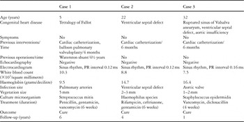
Between September 1997 and September 2004, we undertook surgery for repair of congenital cardiac malformations in 1130 patients. Amongst them, 3 patients (0.26%) were discovered to have unsuspected endocardial vegetations during the procedure. These patients accounted for one-eighth of the patients treated for endocarditis at our institution during the same period. Their characteristics, along with the clinical and laboratory findings, are summarised in Table 1.
Table 1. Summary of characteristics of the patients and their clinical findings.

Case no. 1
A five-year-old girl with tetralogy of Fallot, who had undergone a Waterston shunt at six months of age, presented for total correction. Her medical history included congenital rubella syndrome and deafness. Cardiac catheterization and balloon pulmonary valvoplasty had been performed 4 months prior to the operation. At presentation, she was afebrile. Chest radiography revealed cardiomegaly. Echocardiography showed a large perimembranous ventricular septal defect, with significant overriding of the aorta, along with subvalvar and valvar pulmonary stenosis, moderate hypoplasia of the right and left pulmonary arteries, and a patent oval foramen.
At operation, and during exploration of the pulmonary trunk, multiple vegetations, variable in size, with the largest measuring up to 5 millimetres, were found attached to the pulmonary valve and the intimal lining of the pulmonary trunk and its branches. These were removed and sent for culture. A successful total repair was completed, including complete excision of the pulmonary valve, closure of the ventricular septal defect with a Dacron patch and augmentation of the pulmonary trunk with pericardium. Culture of the vegetations revealed the presence of Streptococcus mitis, of the Viridans group, and the patient was treated for a total of 6 weeks with intravenous antibiotics, using a combination of penicillin plus gentamicin and vancomycin plus gentamicin. She made an uneventful recovery and remains well, without any evidence of infection, 6 years later.
Case no. 2
A 22-year-old asymptomatic man presented with the diagnosis of ventricular septal defect for surgical correction. The chest radiograph showed increased pulmonary vascular markings, and an enlarged cardiothoracic ratio. Echocardiography revealed left ventricular enlargement, and a muscular inlet ventricular septal defect, while cardiac catheterization showed normal pulmonary arterial pressures and a ratio of pulmonary to systemic flows of 2.2 to 1. At operation, a suspicious yellow piece of friable tissue, measuring 2 to 3 millimetres, was found on the superior rim of the ventricular septal defect. It was removed and sent for culture. The defect itself was closed with a Dacron patch. Cultures revealed a Gram-negative pathogen specified as Haemophilus species. The patient was treated thereafter for 6 weeks with a combination of rifampicin, ceftriaxone, and gentamicin. He remains well, 4 years later, with no evidence of infection.
Case no. 3
A 32-year-old man with mild symptoms of fatigue presented for the correction of a ruptured aneurysm of the sinus of Valsalva. He was afebrile, and his chest radiograph showed signs of pulmonary plethora. The diagnosis of ventricular septal defect, and rupture of a sinus of Valsalva aneurysm into the right ventricle with moderate-to-severe aortic regurgitation, was suggested by echocardiography and confirmed by cardiac catheterization. At operation, two tiny vegetations were found attached to the right coronary leaflet of the aortic valve. Gram staining suggested a gram-positive microorganism, which on culture proved to be Staphylococcus epidermidis. The patient underwent repair of the ruptured aneurysm, with concomitant closure of the ventricular septal defect with a Dacron patch, and replacement of the aortic valve with a mechanical prosthesis. He was subsequently treated with intravenous anti-staphylococcal antibiotics, specifically vancomycin and dicloxacillin, for 4 weeks. Apart from developing a pericardial effusion requiring drainage, the patient made an excellent recovery and remains well, free from infection, 1 year later.
Discussion
Although infective endocarditis is generally rare in children, congenital cardiac disease is recognized as its main predisposing factor, and also as one of progressively increasing importance for developing the infection in adults.2, 6–11 In theory, abnormal jets of blood with high velocity cause endocardial damage in these patients, with subsequent aggregation of platelets. Bacteria embedded in these structures for an undefined period of time lead eventually to the formation of vegetations.4, 12 Patients at higher risk are those with complex cyanotic congenital cardiac disease, and those with surgically constructed systemic-to-pulmonary arterial shunts or conduits.5 The commonest sites of growth of vegetations are the rim of a ventricular septal defect, the left ventricular outflow tract, and the mitral valve in patients with unoperated congenital cardiac disease, while the left ventricular outflow tract and the areas of repair prevail in patients who have undergone surgery.11 Although there is evidence that endocardial or intimal trauma caused by the introduction of intravascular devices, or even indwelling catheters, is associated with non-valvar infections of the cardiovascular system, there is no proof thus far that cardiac catheterization bears the same risk.4, 11, 13 Interestingly, however, in all 3 of our cases, cardiac catheterization, in one with concomitant pulmonary balloon valvoplasty, preceded the operative surgery by 4 to 6 months.
Echocardiography is a very important diagnostic tool in revealing endocarditis, especially via the transoesophageal approach.11, 14 Although transthoracic echocardiography has been recommended for making the diagnosis of endocarditis in children, its effectiveness in detecting vegetations in young patients, especially those with cyanotic disease, is rather limited.5, 6 Moreover, in the absence of clinical or laboratory findings, transthoracic echocardiography is unlikely to prove positive.15 In all our patients, there were no relevant clinical or laboratory findings, while echocardiography failed to reveal the presence of the vegetations.3, 9, 15
Removal of the infected tissue is essential and, when involved, valvar resection and replacement, preferably with a homograft, is mandatory.16, 17 Alternatively, resection of the vegetations with repair of the involved valve has been suggested, especially for neonates, infants, and young children. The results of these procedures are currently under investigation.18–20 Although medical and/or surgical management have improved outcomes over the years, infective endocarditis is still associated with high rates of death, ranging from 9 to 25% in various reported series.2, 8, 18, 21 The wide range of these results, apart from reflecting diversity in bacterial virulence, may also be partially attributed to the time of diagnosis. The yet undefined time that infective endocarditis runs asymptomatically in the background, in conjunction with the observed delay in diagnosis, which may be up to 60 days from the onset of symptoms, may be a disposition for major destruction of tissues.11 The combination of early detection, surgical removal of vegetations, and antibiotic therapy proved successful in our patients.
Reparative surgery, apart from cases of ventricular septal defects or patency of the arterial duct, does not alter the risk for endocarditis. Patients undergoing surgery, nonetheless, are affected by different strains of microorganisms.5 Taking into account that atrial septal defects also do not carry a specific risk for infective endocarditis, it is imperative that antibiotic prophylaxis should be continued for all remaining patients with a history of a congenital cardiac malformation.1, 7, 11, 22
The persistent trend of new cases of infective endocarditis appearing despite the widespread practice of antibiotic prophylaxis is of concern. Following the current recommendations for prevention is imperative, while certain “safe” conditions and procedures may have to be reviewed. A high index of suspicion should be maintained, and careful preoperative assessment performed. This may include, in addition to routine evaluation of the leukocytes, measurement of c-reacting protein. Repair should be postponed for individuals with subacute endocarditis, if their condition is stable and a full course of antibiotic therapy has been instituted. In the case of incidental intraoperative unearthing of endocarditis, appropriate management involves removal of suspicious lesions, immediate microscopic examination, careful bacteriologic study, and prolonged antibiotic therapy.