Introduction
Bimetallic oxide nanomaterials have garnered tremendous attention because of their interesting properties and capabilities owing to synergy between the two metals [Reference Acharyya, Ghosh, Tiwari, Sarkar, Singha, Pendem, Sasaki and Bal1, Reference Zhang, Ding, Deng, Zhen, Li, Xue, Yan and Sun2, Reference Kim, Heo, Kim, Park, Yoon, Kim, Oh, Yi, Noh and Park3]. They offer opportunity of being produced from multiple combinations of metals in diverse structures (i.e., spinel, perovskite, mixed metal oxide) and morphologies (i.e., particle, wire, sheet) with capabilities superior to monometallic ones [Reference Teng, Wang, Liao, Li, Chen, Dong and Kuang4, Reference Liu, Zhang, Yu, Jiao and Chen5, Reference Wu, Niu, Feng, Yu, Huang, Zhong, Hu and Li6, Reference Etzi Coller Pascuzzi, Selinger, Sacco, Castellino, Rivolo, Hernández, Lopinski, Tamblyn, Nasi, Esposito, Manzoli, Bonelli and Armandi7, Reference Li, Xiong, Liu, Bo, Zhang, Han and Guo8, Reference Nagamuthu, Vijayakumar, Lee and Ryu9, Reference Li, Zhou, Sun and Nan10, Reference Pilania and Mannodi-Kanakkithodi11, Reference Ekebas, Cetin, Önal and Nalbant Esenturk12]. The properties of these materials have been intensely investigated for numerous nanotechnology applications, such as catalysis, supercapacitors, solar cells, and sensors [Reference Teng, Wang, Liao, Li, Chen, Dong and Kuang4, Reference Liu, Zhang, Yu, Jiao and Chen5, Reference Wu, Niu, Feng, Yu, Huang, Zhong, Hu and Li6, Reference Etzi Coller Pascuzzi, Selinger, Sacco, Castellino, Rivolo, Hernández, Lopinski, Tamblyn, Nasi, Esposito, Manzoli, Bonelli and Armandi7, Reference Li, Xiong, Liu, Bo, Zhang, Han and Guo8, Reference Nagamuthu, Vijayakumar, Lee and Ryu9, Reference Ekebas, Cetin, Önal and Nalbant Esenturk12, Reference Gaidan13, Reference Habibi and Mosavi14, Reference Nepal, Zhang, Dai, Tian, Nagler and Jin15, Reference Lin, Xie, Liu, Zhang, Wang, Zhang and Cheng16, Reference Pyeon, Ruoko, Leduc, Gönüllü, Deo, Tkachenko and Mathur17, Reference Zhang, Chang, Gao, Wang, Yan, Gao, Jia, Luo, Fang, Zhang and Wang18, Reference Ling, Wang, Xiong, Xie, Chen, Chu, Qiu, Li and Xiao19].
Among the potential applications of bimetallic oxide nanomaterials, their use as catalyst in electrochemical water splitting is particularly intriguing. They provide an attractive path to produce H2, which is a pivotal energy carrier for sustainable energy future [Reference Teng, Wang, Liao, Li, Chen, Dong and Kuang4, Reference Zheng, Jiao, Zhu, Li, Han, Chen, Du, Jaroniec and Qiao20, Reference Zhao, Zhang, Jia, Waterhouse, Shi, Zhang, Zhan, Tao, Wu, Tung, O’Hare and Zhang21]. However, the overall rate of water splitting process is hampered by sluggish kinetics and large overpotential requirement of anodic oxygen evolution reaction (OER) [Reference Teng, Wang, Liao, Li, Chen, Dong and Kuang4, Reference Han, Dong and Wang22, Reference Yu, Feng, Guan, (David) Lou and Paik23, Reference Dou, Dong, Hu, Huang, Chen, Tao, Yan, Chen, Shen, Chou and Wang24]. Therefore, developing proper OER catalysts has become focus of profound research to address this challenge. Currently, precious metal oxides (RuO2, IrO2) have been reported to be the most efficient OER catalysts [Reference Teng, Wang, Liao, Li, Chen, Dong and Kuang4, Reference Suntivich, May, Gasteiger, Goodenough and Shao-Horn25]. However, low abundance, high cost, and low stability of these catalysts obstruct their large-scale applications. A great number of alternative catalysts particularly with mixed transition metals have been developed to reduce cost and to improve activity and stability. An intellectual architecture of structure, morphology, and composition of the catalyst is essential for the desired performance, and any improvement would make a significant contribution to the fast-growing OER catalyst family.
In recent years, mixed iron manganese oxides have become focus of considerable attention due to their earth abundances, high electrical conductivity, low toxicity, magnetic properties, and ease of preparation [Reference Teng, Wang, Liao, Li, Chen, Dong and Kuang4, Reference Liu, Zhang, Yu, Jiao and Chen5, Reference Wu, Niu, Feng, Yu, Huang, Zhong, Hu and Li6, Reference Etzi Coller Pascuzzi, Selinger, Sacco, Castellino, Rivolo, Hernández, Lopinski, Tamblyn, Nasi, Esposito, Manzoli, Bonelli and Armandi7, Reference Li, Xiong, Liu, Bo, Zhang, Han and Guo8, Reference Nagamuthu, Vijayakumar, Lee and Ryu9, Reference Li, Zhou, Sun and Nan10, Reference Gaidan13, Reference Habibi and Mosavi14, Reference Nepal, Zhang, Dai, Tian, Nagler and Jin15, Reference Chi, Chen, Zhong, Wang and Lu26, Reference Saravanakumar, Muthu Lakshmi, Ravi, Ganesh, Sakunthala and Yuvakkumar27, Reference Ghosh, Dutta, Haque, Mordvinova, Lebedev, Pal, Gayen, Mahata, Kundu and Motin Seikh28, Reference Gowreesan and Ruban Kumar29, Reference Habibi and Mosavi30, Reference Rayaprol and Kaushik31, Reference Li, Xu, Wang, Liu, Cui and Guo32, Reference Cao, Liu, Xu, Wang and Jiao33, Reference Lobo and Rubankumar34]. They have been synthesized in several structures (i.e., perovskite, spinel, mixed oxide composites) and morphologies (i.e., particle, fiber, sheet) for water splitting electrocatalysis as well as various other applications, such as battery, supercapacitor, gas sensor, and photocatalysis [Reference Teng, Wang, Liao, Li, Chen, Dong and Kuang4, Reference Liu, Zhang, Yu, Jiao and Chen5, Reference Wu, Niu, Feng, Yu, Huang, Zhong, Hu and Li6, Reference Etzi Coller Pascuzzi, Selinger, Sacco, Castellino, Rivolo, Hernández, Lopinski, Tamblyn, Nasi, Esposito, Manzoli, Bonelli and Armandi7, Reference Li, Xiong, Liu, Bo, Zhang, Han and Guo8, Reference Nagamuthu, Vijayakumar, Lee and Ryu9, Reference Gaidan13, Reference Habibi and Mosavi14, Reference Nepal, Zhang, Dai, Tian, Nagler and Jin15]. Among the numerous varieties of mixed iron manganese oxides, iron manganite (FeMnO3) with bixbyite structure has been considered as an attractive class of metal oxide. Until now, FeMnO3 nanomaterials have been synthesized in spherical and cubical morphologies, and their structural, electronic and magnetic properties have been studied extensively [Reference Li, Zhou, Sun and Nan10, Reference Chi, Chen, Zhong, Wang and Lu26, Reference Ghosh, Dutta, Haque, Mordvinova, Lebedev, Pal, Gayen, Mahata, Kundu and Motin Seikh28, Reference Gowreesan and Ruban Kumar29, Reference Habibi and Mosavi30, Reference Rayaprol and Kaushik31, Reference Saravanakumar, Ramachandran, Ravi, Ganesh, Guduru and Yuvakkumar35, Reference Nikam, Rayaprol, Goyal, Babu, Radha and Siruguri36]. These nanomaterials have also been investigated for potential battery, energy storage, O2 carrier, sensing, and some catalysis applications [Reference Li, Zhou, Sun and Nan10, Reference Chi, Chen, Zhong, Wang and Lu26, Reference Ghosh, Dutta, Haque, Mordvinova, Lebedev, Pal, Gayen, Mahata, Kundu and Motin Seikh28, Reference Gowreesan and Ruban Kumar29, Reference Habibi and Mosavi30, Reference Li, Xu, Wang, Liu, Cui and Guo32, Reference Cao, Liu, Xu, Wang and Jiao33, Reference Saravanakumar, Ramachandran, Ravi, Ganesh, Guduru and Yuvakkumar35, Reference Yang, Yang, Yan, Sun, Wu, He and Wang37, Reference Bhavsar, Tackett and Veser38, Reference Leontie, Doroftei and Carlescu39, Reference Hou, Zhu, Niu, Dai, Yin, Dong, Zhang and Dong40]. However, to the best of our knowledge, synthesis of these materials in wire morphology and their potential application as OER catalyst have not been reported yet.
Herein, we report the synthesis of FeMnO3 nanowires (NWs) and their potential use as OER electrocatalyst. The NWs have been synthesized by modified hydrothermal method using nitrilotriacetic acid (NTA) as a structure-directing agent. The NWs have thickness of approximately 200 nm and length in micrometer range. They are formed by assembly of spherical-like nanoparticles of about 70 nm in size. The NWs were used to modify fluorinated tin oxide (FTO) electrodes, and their electrochemical behaviors in OER were studied in alkaline medium. The demonstrated OER activity of FeMnO3 NW modified FTO electrode (FeMnO3-FTO) suggests that this new material is a promising alternative to noble metal electrocatalysts.
Results and discussion
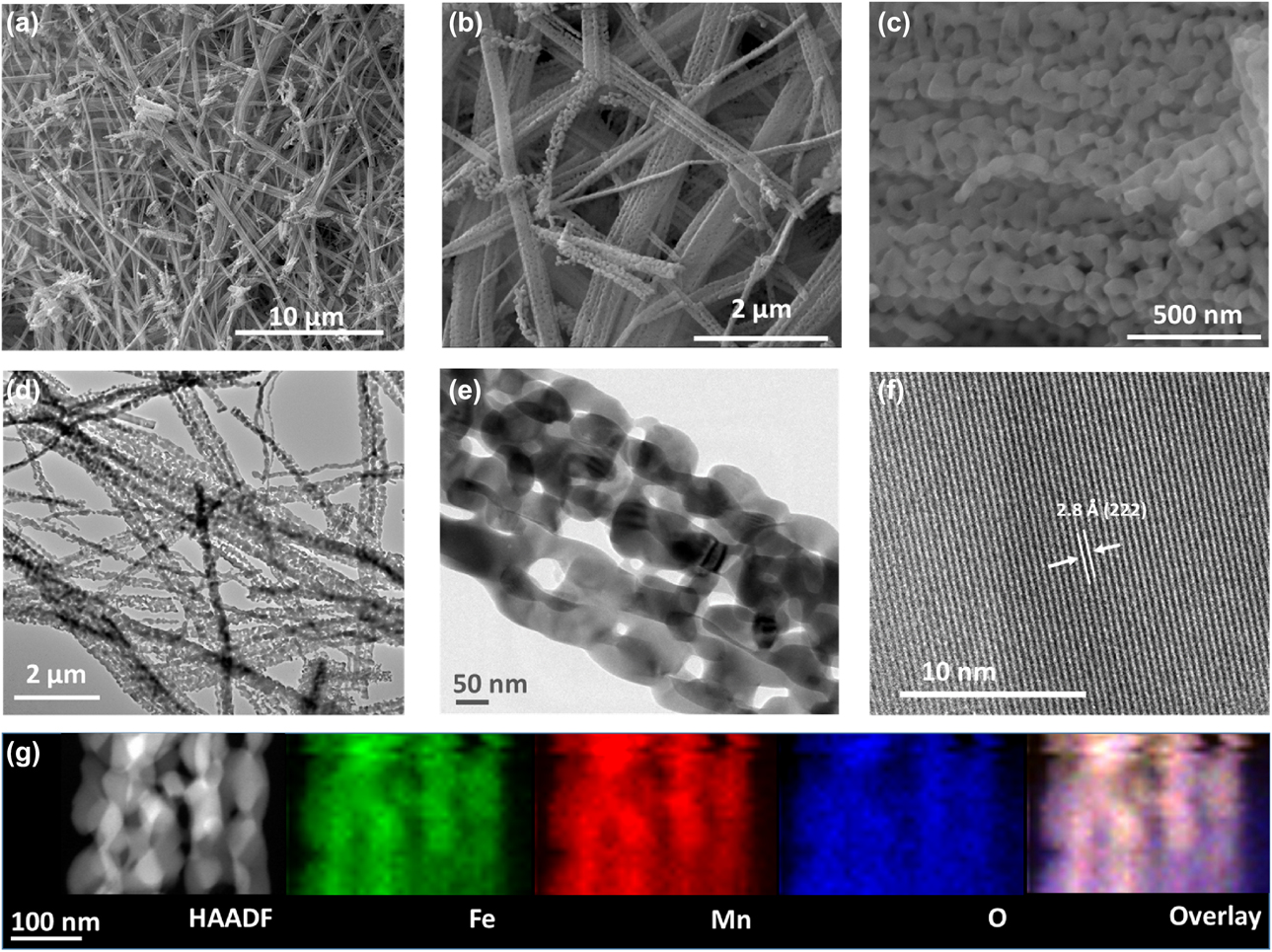
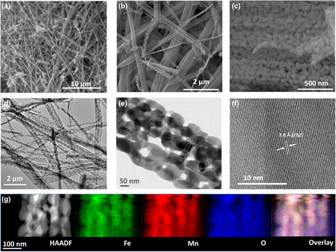
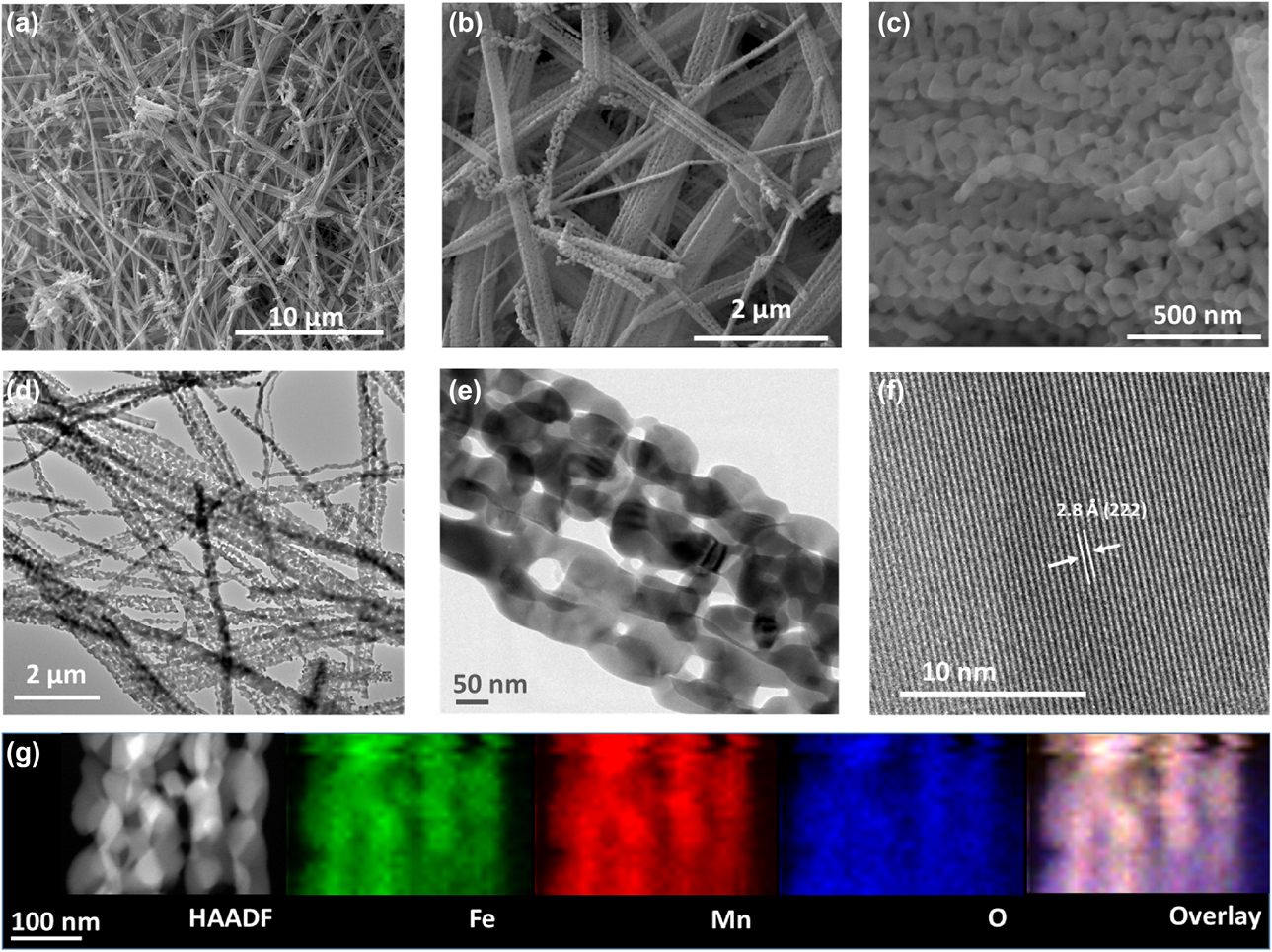
The synthesis of FeMnO3 NWs was performed with a simple modified hydrothermal method. Formation of the NWs took place in isopropyl alcohol and deionized water mixture where NTA functions as a structure-directing agent. Figure 1 shows scanning electron microscopy (SEM) and transmission electron microscopy (TEM) images of FeMnO3 NWs. The images reveal that about 70 nm nanoparticles assemble to form the wires. The images also demonstrate that the NWs have several micrometers of length and approximately 200 nm of thickness. Energy-dispersive X-ray (EDX) analysis verified the presence of Fe and Mn in the synthesized NWs (Fig. S1). The composition of the FeMnO3 NWs was studied by the elemental mapping of Fe, Mn, and O on the NWs [Figs. 1(g) and S2]. The mapping reveals that the elements forming the NWs are uniformly distributed across the whole material. Elemental composition and mass percent of each element in the NWs were investigated by inductively coupled plasma-optical emission spectrometry (ICP-OES), and the amount of Mn and Fe was found to be 36.6 wt% and 29.2 wt%, respectively. This observation correlates well with the relative amounts obtained from EDX analysis (Mn: 31.0%, Fe: 26.0%). The Brunauer–Emmett–Teller (BET) surface area was measured as 14.21 m2/g for FeMnO3 NWs. The large surface area is essential for catalytic applications since it improves the number of interaction points, thus the performance of the catalyst.
(a–c) SEM images (at different magnifications), (d–f) TEM images (at different magnifications), and (g) High Angular Dark Field-Scanning Transmission Electron Microscopy (HAADF-STEM) image and element mapping [Fe (green), Mn (red) and O (blue)] of FeMnO3 NWs.
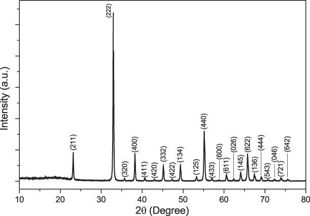
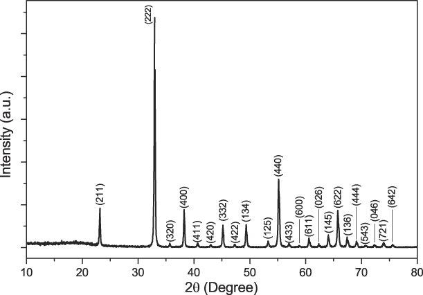
The X-ray diffraction (XRD) pattern of NWs is shown in Fig. 2. The peaks with 2θ values of 23.16°, 32.96°, 35.65°, 38.24°, 40.65°, 42.98°, 45.18°, 47.23°, 49.34°, 53.28°, 55.16°, 57.06°, 58.82°, 60.58°, 62.37°, 64.06°, 65.76°, 67.42°, 69.08°, 70.72°, 72.33°, 74.00°, and 75.54° were indexed to the (211), (222), (320), (400), (411), (420), (332), (422), (134), (125), (440), (433), (600), (611), (026), (145), (622), (136), (444), (543), (046), (721), and (642) planes of FeMnO3 bixbyite structure (PDF number 01-076-0076), respectively. No diffraction peak other than those of FeMnO3 were observed in the XRD pattern. XRD peak analysis was performed for the diffraction peak at 32.96° (222), using the Debye–Scherrer equation to estimate the crystallite size [Reference Kim, Rhee and Park41]. The analysis revealed that sizes of the crystallites are approximately 46.8 nm for FeMnO3 NWs. The crystallite sizes are slightly smaller than particle sizes measured by TEM analysis.
XRD pattern of FeMnO3 NWs (PDF Card No: 01-076-0076).
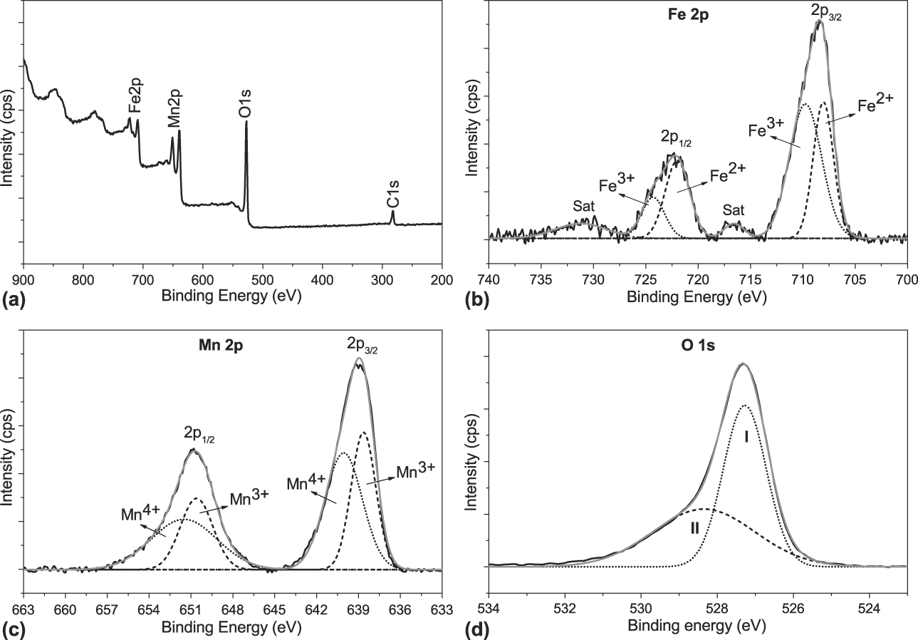
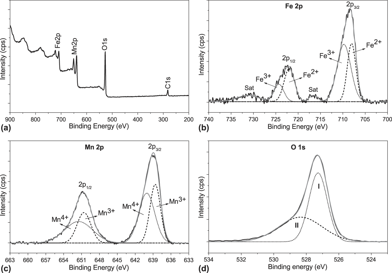
The X-ray photoelectron spectroscopy (XPS) spectra of FeMnO3 NWs are demonstrated in Fig. 3. The survey XPS spectrum of NWs suggests the presence of only Mn, Fe, and O [Fig. 3(a)]. Only contamination observed in the sample was low quantity of carbon (C 1s at 282.4 eV), which is used for calibration. Fe 2p core-level spectrum demonstrates two peaks at binding energies of 708.4 eV and 722.1 eV corresponding to Fe 2p 3/2 and Fe 2p 1/2 peaks, respectively [Fig. 3(b)]. The Gaussian fit unveiled two bands under Fe 2p peaks, which are ascribed to Fe2+ (708.0 and 721.9 eV) and Fe3+ (709.7 and 724.3 eV). The observed peaks and their positions agree well with the ones reported previously [Reference Lobo and Rubankumar34, Reference Xu, Dong, Wang, Doherty, Xie and Wu42, Reference Ding, Zhong and Zhang43, Reference Pham, Dinh, Vuong, Ta and Do44]. Mn 2p core-level spectrum is demonstrated in Fig. 3(c). The spectrum shows two major peaks at 638.9 eV and 650.8 eV, which are assigned as Mn 2p 3/2 and Mn 2p 1/2, respectively. Mn 2p peaks were resolved further and fitted bands of Mn3+ (638.8 and 650.5 eV) and Mn4+ (640.0 and 651.6 eV) [Reference Lobo and Rubankumar34, Reference Xu, Dong, Wang, Doherty, Xie and Wu42, Reference Tang, Jiang, Liu, Wang and Sun45]. The O 1 s spectrum exhibits a peak at 527.3 eV [Fig. 3(d)]. The curve-fitting process revealed two Gaussians (I and II) bands. The one at 527.2 eV (I) corresponds to metal–oxygen bonding in the sample, while the one at 528.4 eV (II) is assigned to surface adsorbed oxygen species (i.e., O22−, OH−), suggesting the presence of low oxygen coordination sites in the NWs [Reference Nagamuthu, Vijayakumar, Lee and Ryu9, Reference Pyeon, Ruoko, Leduc, Gönüllü, Deo, Tkachenko and Mathur17, Reference Lobo and Rubankumar34, Reference Ding, Zhong and Zhang43, Reference Tang, Jiang, Liu, Wang and Sun45].
XPS (a) Survey, (b) Fe 2p, (c) Mn 2p, and (d) O 1s spectra of FeMnO3 NWs.
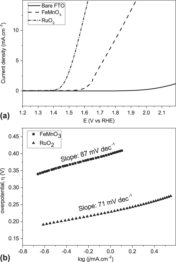
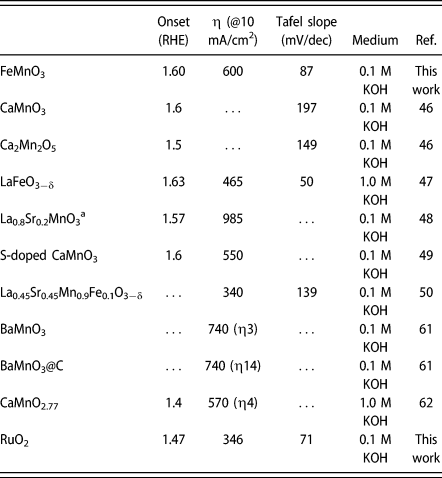
The electrocatalytic activities of FTO substrates modified with FeMnO3 NWs were investigated toward OER in alkaline medium (0.1 M KOH). FeMnO3 modified FTO (FeMnO3-FTO) was used as working electrode, and evaluation of its performance was performed by studying polarization curves. For comparison, bare FTO electrode and RuO2 were also tested under the same conditions and resulting voltammograms are depicted in Fig. 4(a). Inspection of the data reveals that FeMnO3-FTO demonstrates relatively high electrocatalytic activity for OER with an onset potential of 1.60 V versus reversible hydrogen electrode (RHE). Bare FTO, on the other hand, was found to exhibit almost no activity in the same medium. Although the onset potential of FeMnO3-FTO electrode was found to be higher than that of RuO2 (1.47 V versus RHE), it is comparable to those of previously reported perovskite oxides especially CaMnO3 (1.60 V versus RHE) [Reference Kim, Yin, Tsao, Fang and Yang46], LaFeO3−δ (1.63 V versus RHE) [Reference Li, Lv, Wang, Ao, Ruan, Zha, Hong, Wu, Lan, Wang, Jiang and Liu47], La0.8Sr0.2MnO3 (1.57 V versus RHE) [Reference Jin, Cao, Zhang, Zhang and Yang48], and S-doped CaMnO3 (1.60 V versus RHE) [Reference Peng, Han, Li, Chou, Ji, Huang, Du, Liu and Ramakrishna49].
(a) Polarization curves and (b) Tafel plots of the FeMnO3 NWs and RuO2 in 0.1 M KOH.

The OER activity of FeMnO3 NWs was also tested by using the overpotentials required to drive anodic current densities of 5 mA/cm2 (η5) and 10 mA/cm2 (η10). For FeMnO3-FTO, the η5 and η10 were measured as 490 mV and 600 mV, respectively. The results obtained from FeMnO3-FTO are summarized in Table I, as well as the ones of some reported catalysts, for comparison.
Summary of the data for some recently reported representative OER electrocatalysts and FeMnO3 NWs in alkaline medium.
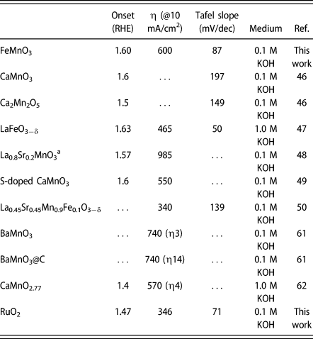
a Values are reported as V versus Ag/AgCl. Equation (1) was used to convert the obtained potential (versus Ag/AgCl) to RHE and overpotential was calculated using Eq. (2) [Reference Gao, Sheng, Zhuang, Fang, Gu, Jiang and Yan56].
The OER kinetics of FeMnO3 NWs was evaluated by fitting Tafel plot from polarization curves [Fig. 4(b)]. Tafel slopes were estimated by the Tafel equation [η = a + b·log(j), where η is the overpotential, and a, j, and b are intercept, current density, and Tafel slope, respectively]. Tafel slope was determined as 87 mV/dec for FeMnO3-FTO. The slope was found to be much smaller than the ones of recently reported OER electrocatalysts, such as La0.45Sr0.45Mn0.9Fe0.1O3−δ (139 mV/dec) [Reference Hua, Li and Luo50], CaMnO3 (197 mV/dec), and Ca2Mn2O5 (149 mV/dec) [Reference Kim, Yin, Tsao, Fang and Yang46], indicating faster OER kinetics on FeMnO3-FTO. Tafel slope of RuO2 was found to be 71 mV/dec and agrees well with the ones reported in the literature (i.e., 69 mV/dec [Reference Sun, Yang, Dai, Wang, Zhang, Li, Chen, Huang and Dong54], 70 mV/dec [Reference Liu, Nan, Chang, Li, Fang, Liu, Tang, Wang, Li and Ma55], 90 mV/dec [Reference Gao, Sheng, Zhuang, Fang, Gu, Jiang and Yan56]).
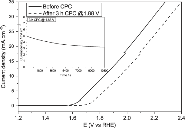
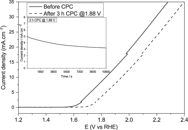
The durability of the modified FTO electrode was also tested to evaluate the OER performance of the electrocatalyst for practical applications. Constant potential coulometry (CPC) was employed at overpotential of 650 mV for 10,800 s in 0.1 M KOH. The polarization curves recorded before and after CPC are shown in Fig. 5. A slight shift to the higher potential values in both onset potential (from 1.60 to 1.70 V) and overpotential for 10 mA/cm2 (from 600 to 718 mV) was observed after 10,800 s constant potential electrolysis (Fig. 5).
Polarization curve of FeMnO3 NWs modified FTO electrode before and after the stability test in 0.1 M KOH constant potential electrolysis at overpotential corresponding to initial current density j = 5 mA/cm2 (inset shows change in current density during chronoamperometric electrolysis).
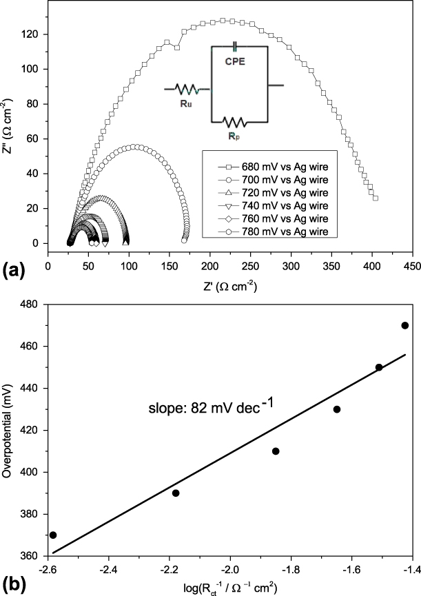
Electrochemical impedance spectroscopy (EIS) was also performed at different overpotentials to further investigate the kinetic properties of the prepared catalyst in OER. The Nyquist plots of FeMnO3-FTO consist of semicircles, which fit to Constant Phase Element (CPE) model [see inset of Fig. 6(a)], where R u and R p correspond to solution resistance and charge transfer resistance (R ct), respectively (Fig. 6). The semicircles are associated with R ct at high frequency region. The diameter of the semicircle decreases as the overpotential increases, which indicates enhanced electrical conductivity as well as faster charge transfer during OER process. Furthermore, Tafel slope from EIS data was calculated as 82 mV/dec, which agree well with the one obtained from polarization curve (87 mV/dec).
(a) Nyquist plot for the FeMnO3-FTO electrodes at different overpotentials. (b) Tafel plot obtained using R ct from the EIS analysis for the FeMnO3-FTO electrodes.

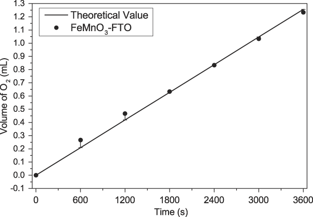
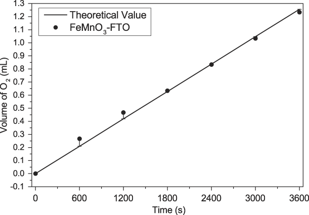
The OER activity of FeMnO3 NWs was also evaluated by measuring the volume of O2 gas produced during the electrolysis of water. The measurement was performed using the modified FTO electrode, FeMnO3-FTO, as the anode in a Hoffman electrolysis setup. The volume of O2 released was monitored for 3600 s at a constant current of 5 mA at room temperature (Fig. 7). The Faradaic yield of FeMnO3-FTO for O2 production was measured as >95% after the transfer of 18 C of charge. The amount of O2 obtained is very close to the theoretical yield, suggesting FeMnO3 NWs are promising candidates for OER electrocatalyst.
Plot of O2 volume versus time during the electrolysis of water using the FeMnO3-FTO in 0.1 M KOH.
In brief, the observed OER activity could be ascribed to morphological and chemical properties of the as-synthesized FeMnO3 NWs. One-dimensional morphology of the NWs could serve plentiful surface for the adsorption of reactants and provide more exposed active sites for OER. Thus, the sufficient active sites could promote electrical conductivity and charge transfer and improve electrocatalytic water oxidation. Another reason for the improved OER activity might be the presence of oxygen vacancies in the material. It has been suggested that presence of oxygen vacancies increase adsorption of H2O onto the catalyst surface, thus improve the reactivity of active sites [Reference Zhao, Yan, Chen and Chen53, Reference Bao, Zhang, Fan, Zhang, Zhou, Yang, Hu, Wang, Pan and Xie57, Reference Cheng, Zhang, Zhang, Du, Han and Chen58]. As evidenced by XPS O 1s core level spectra, low oxygen coordination defect sites present in the synthesized NWs. Additionally, the current literature suggests that OER activity of a perovskite-type catalyst correlates with the presence of a single electron in e g orbitals [Reference Kim, Yin, Tsao, Fang and Yang46, Reference Hua, Li and Luo50, Reference Suen, Hung, Quan, Zhang, Xu and Chen59]. The unity of e g electrons promotes the uptake of OH− ions into the oxygen vacant sites and thus lead to higher OER activity [Reference Kim, Yin, Tsao, Fang and Yang46, Reference Suen, Hung, Quan, Zhang, Xu and Chen59]. In FeMnO3 structure, the high spin Mn3+ ([Ar]3d 4) ion has the t 2g3e g1 configuration. Therefore, one can suggest that Mn3+ plays a key role in the OER activity of the FeMnO3 NW catalyst. It has also been suggested that synergic interaction between Fe and Mn may optimize the adsorption of reaction intermediates and activation energy of the reaction, thus contributing to the improvement of catalytic activity [Reference Teng, Wang, Liao, Li, Chen, Dong and Kuang4, Reference Zhang, Zheng, Voznyy, Comin, Bajdich, Garcia-Melchor, Han, Xu, Liu, Zheng, Garcia de Arquer, Dinh, Fan, Yuan, Yassitepe, Chen, Regier, Liu, Li, De Luna, Janmohamed, Xin, Yang, Vojvodic and Sargent60]. Finally, it is not surprising to see that the performance of FeMnO3 NWs in OER is slightly modest compared with that of RuO2 as being an OER catalyst with one of the highest activity among all reported catalysts. The demonstrated electrocatalytic activity, stability, and facile synthesis from low-cost starting materials make this new material attractive for water splitting and other catalyst applications.
Conclusions
In summary, FeMnO3 NWs were synthesized using NTA as a structure-directing agent and the simple hydrothermal method. The synthesized NWs are formed by the assembly of approximately 70 nm FeMnO3 nanoparticles. The electrocatalytic performances of synthesized NWs in water oxidation reaction have been evaluated. The results revealed that FeMnO3-FTO electrode had onset potential of 1.60 V versus RHE and required overpotential of 600 mV at current density of 10 mA/cm2 in 0.1 M KOH solution. Furthermore, FeMnO3 catalyst was found to be stable during constant potential electrolysis. The overall OER performance of the FeMnO3 NWs was observed to be comparable or superior to some of the similar metal-substituted manganese oxide OER electrocatalysts. Consequently, the FeMnO3 NWs are economical alternative to noble metal oxide electrocatalysts and have great potential of large-scale electrolytic water splitting application.
Experimental
Materials
The purchased chemicals were not purified further and used as received. Iron(II) chloride tetrahydrate (FeCl2·4H2O), manganese(II) chloride tetrahydrate (MnCl2·4H2O), isopropyl alcohol, NTA, acetylacetone, polyethylene oxide (PEO), and Triton X were obtained from Sigma-Aldrich (St. Louis, Missouri). Deionized ultra-pure water (resistivity >18 MΩ, PURELAB Option-Q, ELGA, High Wycombe, U.K.) was used in preparation of all aqueous solutions.
Synthesis of FeMnO3 NWs
Synthesis of FeMnO3 NWs was carried out by a previously reported hydrothermal/solvothermal method with some modifications [Reference Sun, Yang, Dai, Wang, Zhang, Li, Chen, Huang and Dong54]. Briefly, FeCl2·4H2O (3.0 mmol), MnCl2·4H2O (6.0 mmol), and NTA (4.7 mmol) were added to 30 mL isopropyl alcohol and 10 mL deionized water mixture. After complete dissolution, the reaction mixture was transferred to a 100-mL Teflon-lined autoclave, which was heated to 180 °C and kept at this temperature for 6 h. The product was collected by centrifugation and washed with deionized water and ethanol for several times. After calcination at 650 °C for 1 h, FeMnO3 NWs were obtained as brown powder.
Materials characterization
The TEM and scanning TEM (STEM) measurements and detailed analysis of elemental composition were performed on FEI Tecnai G2 F30 electron microscope (FEI Company, Hillsboro, Oregon) operating at 300 kV. The instrument was equipped with EDX. SEM and EDX measurements were carried out on FEI Nova Nano SEM 430 (FEI Company, Hillsboro, Oregon). The phase of the NWs were examined by XRD on Rigaku Ultima IV X-ray diffractometer (Rigaku Americas Corporation, The Woodlands, Texas) with Cu Kα radiation (λ= 1.54 Å and 2θ is from 10° to 80°). PHI-5000 VersaProbe [Physical Electronics (PHI) Chanhassen, Minneapolis, Minnesota], equipped with Al Kα at 1486.92 eV source, was used to carry out XPS analyses. Calibration of all data was performed according to hydrocarbon contamination peak at C 1s of 282.4 eV. BET analysis was performed with Autosorb-6 (Quantachrome Corporation, Anton Paar, Graz, Austria) instrument for evaluation of specific surface area of nanoparticles. Samples were heated to 350 °C for 3 h before analysis. Elemental composition and mass percent of each element in the structure of NWs were investigated by ICP-OES with PerkinElmer Optima 4300DV (Perkin Elmer, Waltham, Massachusetts).
Electrochemical characterization
Gamry 1010B potentiostat-galvanostat (Gamry Instruments, Warminster, Pennsylvania) and standard three-electrode system were used in electrochemical studies. The analyses were performed at room temperature. In the three-electrode system, the FTO-coated glass electrodes (0.5 cm2 area) modified with FeMnO3 NWs were used as working electrode, Pt wire was counter electrode, and Ag/AgCl (in 3.0 M NaCl) was reference electrode. Before catalyst loading, FTO-coated glass was cleaned by sonication in diluted H2SO4 solution for 30 min and heated at 400 °C for 30 min. Previously reported method was used to prepare the working electrodes [Reference Lee, González-Flores, Ohms, Trost, Dau, Zaharieva and Kurz61]. Briefly, 100 mg of catalyst and 30 mg of PEO are dispersed in 100 µL of Triton-X, 100 µL of acetylacetone, and 1 mL of deionized water under vigorous stirring for 24 h. Then, 5.5 µL of the ink was transferred onto FTO-coated glass to obtain a catalyst loading of approximately 0.92 mg/cm2. After drying at 60 °C for 1 h, electrodes were calcined at 450 °C for 1 h. For comparison, RuO2 was synthesized by following a previously reported method [Reference Zhao, Li, Cui and Sun62] and used to benchmark FeMnO3 NW catalyst.
Electrochemical performance of the FeMnO3-FTO electrode was monitored in 0.1 M KOH solution by recording linear sweep voltammograms at a scan rate of 5 mV/s. All potentials, measured in this work, were corrected for iR drop, and reported against RHE via RHE calibration. The conversion of obtained potential (versus Ag/AgCl) to RHE was performed by using Eq. (1) [Reference Zhao, Yan, Chen and Chen53], and overpotential was calculated using Eq. (2).
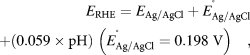 $${E_{{\rm{RHE}}}} = {E_{{{{\rm{Ag}}} / {{\rm{AgCl}}}}}} + E_{{{{\rm{Ag}}} / {{\rm{AgCl}}}}}^^\circ + \left( {0.059 \times {\rm{pH}}} \right)\;\left( {E_{{{{\rm{Ag}}} / {{\rm{AgCl}}}}}^^\circ = 0.198\;{\rm{V}}} \right)\quad ,$$
 $$\eta = {E_{{\rm{RHE}}}} - 1.23\;{\rm{V}}\quad .$$
The stability test of the modified electrodes was carried out by controlled potential electrolysis at overpotentials of 650 mV for 10,800 s in alkaline medium (0.1 M KOH). EIS analyses were performed at different potentials with an amplitude of 10 mV in the 0.1 Hz–100 kHz frequency range.
Supplementary material
To view supplementary material for this article, please visit https://doi.org/10.1557/jmr.2019.215.
Acknowledgments
We acknowledge the support from TUBITAK, Scientific and Technological Research Council of Turkey, Project: 117Z384. We also acknowledge Prof. Aysen Yilmaz for access to XRD instrument in METU Department of Chemistry.