

Three or four times during two days, at a prearranged time, I would meet a few others by the Goethe statue in the foyer of the Library at the Deutsches Museum in Munich, only to disperse and resume our continuing studies. Overcast skies frustrated our planned activity. But on the final occasion, 17 September 2004, Peter Heering greeted us with an excitement as bright as the late morning sun just breaking through the clouds. Now we followed him through hallway mazes to a classroom lab.
Direct sunlight was critical to the observing activity that brought us together. Yet direct sun struck the lab's windowed, courtyard-facing wall only for a few hours at late morning or midday. Once the lab's wall had fallen into shadow, that day's prospects for observing were over, no matter how brilliantly the afternoon sun might shine. Weeks of cloudy weather had cut down Peter's observing time to precious hours. Joining him for one of those hours, Terje Brundtland, Heike Weber and I served as invited witnesses for a demonstration that depended on an audience.Footnote 1 While we observed solar projections of historical microscopic samples, Peter watched us, curious about what we noticed, what engaged us and how we responded to each other and to the images. Just as the projections reiterated a historical procedure, using the same optics, so our gathering reiterated historical experience. Yet we were not trying to re-enact a specific script from some demonstration in the past. We brought our twenty-first century knowledge into the discussion, as well as our curiosity about science and experience as historians. The freedom to be ourselves in relation to each other sustained our ability to construct an authentic shared interpretation of what we saw.
Peter immediately put us to work, asking us each to complete a questionnaire on our previous use of microscopes and our expectations for the session. At the end of our viewing time, he provided another questionnaire to record our impressions and further suggestions. My questionnaire responses were a resource in preparing this witness account of the observing session. Writing gave an opening for reconnecting with joys and frustrations of microscopy, briefly then and more contextually here. I recalled looking at magnified pond water, hair and threads during one high-school class. Working for a short period after college as a research technician, I occasionally used a fine Zeiss phase-contrast microscope fitted with epifluorescence optics to check on honeybee flight muscle that I purified and prepared for Dr Judith Saide's research on its constituent structures.Footnote 2 Sometimes, too, her delight at something unexpected brought me to her side, sharing the excitement with my supplementary but untrained eyes. The instrument's complexity posed an obstacle for me in finding specific structures on the slide. If its focus or settings were deranged, I could not recover the image and it often took me some time to co-ordinate the inverted image with the oppositely directed displacements I gave the specimen.
Being mainly involved subsequently with physics education, I never acquired a facility in microscopy. Microscopes so seldom entered my teaching that unless there were time to remaster the instrument beforehand, stressful moments might arise in the classroom. For example, as a student-teacher I was once without advance warning put in front of a high-school biology class where thirty students peered individually into half as many microscopes, very likely at specimens of different types. I had no idea what the students were seeing or should have seen, nor did I know how to get the class talking about it. By contrast, a solar microscope might have accommodated teacher and class in viewing the same image together. On another occasion, while involving pre-service teachers in the dissection of cow's eyes, I gave up using a classroom microscope that, at that time, I could not manage to focus. Despite these adversities, I liked seeing things magnified and so looked forward to Peter's demonstration. While the bumping of my spectacles against the eyepiece had previously added discomfort to microscopic observing, I expected that projection would make microscopic seeing more like looking at anything else in the world. On the questionnaire that Peter gave us, I sketched the kind of shadowy outlines I thought we would see, enlarged on a big paper screen (Figure 1).
My drawing, made immediately before the observing session, showing what I had expected to see in Peter's solar microscope projections.

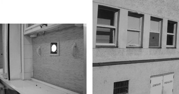
We entered a dark room, although the ceiling lights were at first turned on. Peter had worked hard to make it dark, not merely drawing window shades but dealing with light leakage throughout. One window was different: a plywood board replaced its lower panel (Figure 2). Halfway across that board was a fist-sized aperture through which sunlight streamed when Peter removed its square cover. The aperture fitted an eighteenth-century solar microscope from the museum's collection, our viewing device for the day.
Left: a plywood board replaces the bottom window pane in the lab room. The solar microscope is mounted in the circular opening cut into the board. Right: view of the lab window with aperture, taken from outside and across the museum courtyard.

Observing solar microscope projections in a dark room
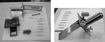
We first looked at two historic solar microscopes that lay on a table beside a series of viewing specimens (Figure 3). Wood linked the mirror, the optical tube and the perspective frame of one of them, made by Junker, whereas brass made up all the non-optical parts of the other, made by John Dollond and used by Peter in this session. Each assemblage, more compact than a conventional modern school microscope, had an outdoors and an indoors side. This two-sided function became more apparent to us when Peter mounted the Dollond instrument in the window aperture. The rectangular mirror faced outdoors; facing indoors were the control screws for the mirror. Turning one screw tilted the mirror out away from the wall, while turning the other moved it from side to side.Footnote 3 As the relation between sun and mirror changed during the morning, Peter readjusted these screws to bring the mirror into better alignment for illuminating the microscope's lenses. The optical tube that carried these lenses extended indoors. Its short removable cover tube held the specimen and this was what Peter moved to focus the image on a large screen.
Left: the wooden Junker (below) and brass Dollond (above) instruments, with their mirrors angled towards the viewer, are laid on a table alongside sliders containing specimens. Right: the brass Dollond instrument is placed with its mirror facing down. At the top of the frame is its removable brass tube containing the focusing lens and place for the slider.

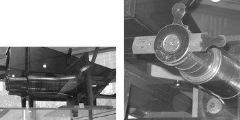
This layout of mounted biological specimens, lenses, screen and our eyes confused me at first. We are used to looking into a microscope's eyepiece, with both the specimen and the light source at the tube's opposite end and distant from us. By contrast, the projection microscope does not put viewers' eyes into the direct, extremely intense, light path; instead we see by reflection from the screen. Yet the solar microscope's objective end resembles an eyepiece, so that oblivious of the projection and expecting to look in at that end, I leant towards it as Peter prepared the apparatus. Then something or someone redirected my attention. I found myself having the same reaction again recently, whilst viewing a Benjamin Martin solar microscope on display in Harvard's Collection of Historical Scientific Instruments (Figure 4).Footnote 4 The divergence of this 1765 instrument's fourteen-inch tube towards its wooden window mounting frame invites an analogy with a telescope and invites, too, the interpretation that the act of observation happens through its objective end of lesser diameter and in the direction outwards through the window. Given my disarming confusion between instruments for projection optics and for direct viewing, on subsequently reading the early British maker John Cuff's account of his instrument I wondered if a similar awareness underlay his emphasis on ‘the new Way’:
the microscope is made Use of in the new Way, that is by directing the Sun's Rays, thro' a Tube, upon some Object placed within and near the End of said Tube, the Image … is thrown in a most exact, beautiful and surprising Manner upon a White Paper screen.Footnote 5
Left: the Benjamin Martin solar microscope on display at the Harvard Collection of Historical Scientific Instruments (number 43). Right: a view of the same instrument's objective end showing the brass slider in place.

Even Cuff's evocative and promotional words are inadequate to describe what it was like when sunlit images from Dollond's brass tube filled the fabric screen in our dark lab. Minute relics of the microscopic past were projected to human-sized height while retaining crisply edged detail and brilliance as if possessed of dimensions comparable to our own. What we saw, lit up with lacework shadows, was astonishing. The effect was as startling as if seeing something so enlarged were novel, even though now it is not difficult to come across either page-sized prints of microscopic objects, or else moving images of nature and fantasy the size of walls or buildings. What made this experience seem so different from the enlargements and projections flitting across our twenty-first century lives? Here I relate, and reflect on, some of the components that enhanced this demonstration.
During the session, I was most intrigued by the way the projections transformed the room into a ‘wonderful … microscopic environment’.Footnote 6 We moved in the space of the projections, between the objective and the screen. Parts of microscopic biological structures dappled over our hands, heads, and someone's pale shirt. At the same time that one of us pointed out a microscopic feature on the screen, their hand's shadow joined the image. The body-sized scale of image and observer assisted not just in seeing what was there, but also in sharing it among ourselves as our interpretation of each image evolved. Images and observers were in a fluid exchange that we seldom encounter now outside dance and drama.Footnote 7 The projection light path has come customarily to be directed over a seated audience's heads as they statically watch the images change or move. By contrast, in the session that Peter facilitated, observing entailed physical movement so as to interact with each other and with the images in whose light path we moved. Our participation arose spontaneously and happily confounded my expectation that Peter would present a conventional slide show.
Peter encouraged us to talk not with him but with each other. He supported our direct engagement with the images. Remaining at the instrument, set back from our group around the screen, Peter did not act as a performer, presenter and mediator of the images. Upon displaying each subsequent image on the screen, he did not tell us what it was. Instead he asked us to discuss among ourselves what we made of it. Thus we became researchers who were trying to understand something unknown. Not only did Peter create an environment that allowed us to feel comfortable with our guesses and lack of certainty, but we later learnt that he, too, did not know the identity of at least one specimen. In this sense, we were researchers together with him and his added knowledge neither defined our inquiry nor rendered it irrelevant.
Most specimens were historical. Accompanying the museum's instruments, they were mounted on glass in several (in fact, between three and five) circular openings in ivory or wooden holders (Figure 3). This technology for holding the specimens appeared similar enough to today's microscope slides that it seemed to me jarring when Peter consistently referred to the holders as ‘sliders’. While preparing this report, I looked at a Benjamin Martin instrument and texts preserved in Harvard's collections,Footnote 8 and the anachronistic sound of ‘slider’ receded. I came to appreciate the clear glass slide's historical origins in a multiply welled strip made of an opaque material (even of brass),Footnote 9 and the sense conveyed by the authentic term, ‘slider’, of how that single strip might move through the instrument's optical path, bringing into view the different samples it held.
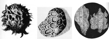
The ambiguity about the samples' identity and means of preparation heightened my curiosity at the same time as it provoked us to expand the range of possibilities. Although I had assumed that the microscopic appearances of things would be familiar to us, they were not. We had to look closely, think and imagine. Was it a feather, that patterned grid of shaded ovals looking overall rather like a bird's wing? It was instead ‘tiny oblong discs of the butterfly wing scale’.Footnote 10 A structure that I had never contemplated before was here made huge, each disc at least the size of our hands and distinctly showing an intricate substructure. Another specimen surprised me with its shape: ‘points on the fish scale’. I thought I might recognize cork from Robert Hooke's famous seventeenth-century illustration (Figure 5, right).Footnote 11 But when we did see it, the graphical effect of the cell boundaries and shapes was so vivid as to overwhelm any easy recognition based on previous acquaintance.
Left: Hooke's drawing of a magnified printed dot (Micrographia (note 11) Schem. II). Middle: Hooke's drawing of a magnified poppy seed (Micrographia (note 11) Schem. XIX). Right: Hooke's drawing of a sliver of cork cut with a razor and microscopically viewed (Micrographia (note 11) Schem. XI).

The projections not only revealed the small-scale make-up of hair-fine objects but in some cases also showed damage incurred in the two centuries since the specimen's preparation. Was the deterioration mould? And what parts of the original specimen did it overshadow and obscure? We also wondered about the colour that we noticed on some slides laid on the table. When viewing these samples by the transmission of light through them, we saw only shades of grey. Had the historical observers ever seen colour in projections of freshly prepared samples? Since any organisms in the historical preparations were long dead, perhaps movement was an aspect of the historical demonstration that was missing for us. The prospect of seeing wall-sized distinct images of microscopic life and its motions fascinated me.
A technician assisting Peter had made up a few specimens from present-day materials. We looked at those in context with the questions that were emerging for us about specimen preparation, identity and colours. In contrast to my assumption that putting something on a microscope slide is a simple act, the labour and uncertainty attending each of these new preparations took me by surprise. To my slight disappointment, Peter had not attempted anything with living organisms, out of concern for preserving the historical instrument.
Viewing the projected images began to seem incomplete to me. In reflecting on the session, I suggested that through adding activities of specimen collection and preparation to the projection demonstration, the audience might be assisted in moving beyond some limitations of pre-prepared slides:
I think an audience would appreciate making a slide themselves (with hair, a leaf, a fabric thread, or something simple), so they can have more connection to the whole process and to the original small size of what is being projected.Footnote 12
Understanding of scale deepens if the amazing structures we see microscopically, here projected so huge, are continually rerooted in the original material to remind the viewer of their relation to everyday size and vision.
The idea of making a slide and its capacity to address our questions about the historical specimens did not leave me. On my return the following week to Massachusetts I wrote to Peter, thanking him for the ‘fascinating … involving and reflective’ demonstration, and offering to put in international mail a specimen for his research: ‘I found 2 dead moths at home and I wonder if you'd like me to send them to you, wrapped up in something to protect them?’Footnote 13
Accompanying all these provocative interactions with images, artefacts and each other went the brilliance, intensity and crispness of sunlight's illumination in the very dark room. In reflecting on Peter's session I regard our fluid involvement with that sunlight as critical to our fascination, in contrast to the projections ubiquitous in urban environments today. In the past the camera obscura, an alternative to the solar microscope, provided opportunities for people to make images with sunlight, both as an artist's drawing aid and as a room-sized marvel for the public. Although sunlight traverses lenses into the dark room of every camera today, we are not inside with it. The human and visual impact of being within a mine, tunnel or cave and seeing sunlight emanating from its opening cannot be reproduced by artificial light. Where the convenience of electric light displaces sunlight, something is lost. Historical demonstrations such as Peter's solar microscope, or the use my students have made of sunlight while redoing Newton's double-prism experiment in a dark hall,Footnote 14 reconnect us with sunlight's beauty and our playful responses to it.
Microscopy is about seeing something of a scale different from, and usually inaccessible to, our own. The solar projections made it possible for things at microscopic scale to meet our own, as light and shadow encountering hand and eye. The historical instrument with its specimens, the scale and clarity of its images, the darkened room lit only by sunlight from a lens, and Peter's facilitation of interaction among a small group, all contributed to make this experience more engaging than the related (and to us more familiar) experiences of viewing projections as a seated audience or doing individual microscopy in classrooms and labs. The solar microscope, with its accompanying experience, offers ways of looking and reflecting not just on what is contained in the slide but also on ourselves as observers developing meanings with it across time and circumstance.
Witnessing as a collective act
In witnessing solar microscope projections, I did not simply look at and assent to whatever was on display. Nor was the object of study – the cork or butterfly wing – merely some dead thing to be examined in isolation from any context. Both the observers and the object were in relation. Our acts of looking more closely at an image provoked questions about it and interactions among ourselves. For example, the object's image and state of preservation elicited our curiosity about what was on the slide, ideas for making new preparations (such as my moth), and group interactions where someone pointed out a detail of the image that was overlooked or differently interpreted by the others. These relational qualities in our witnessing experience support the analysis of scientific witnessing discerned by Steven Shapin and Simon Schaffer in Robert Boyle's seventeenth-century investigations of the pressure of air while using air-pumps that he designed and improvised.Footnote 15
Shapin and Schaffer identify witnessing as a ‘collective act’ whose social space functioned for Boyle to confirm ‘matters of fact’, such as the observed fall in a mercury column's height when placed in an air-pump's evacuated chamber, while mediating disputes in their interpretation, such as those regarding the cause of that fall.Footnote 16 Boyle did not restrict witnesses to those in the room with him. He encouraged others to participate in witnessing either by replicating his apparatus and procedures or by virtually following his illustrated texts. Robert Hooke, who assisted Boyle in constructing and operating air-pumps,Footnote 17 shared Boyle's commitment to an experimental practice founded on what he called the actions of ‘a sincere Hand, and a faithful Eye’.Footnote 18 Hooke similarly invited readers of his Micrographia to follow along with his engravings and descriptions attentive to every detail, to regard his conjectures as ‘uncertain guesses and not as unquestionable Conclusions’, and perhaps to augment his work through the ‘Reader's diligently observing [it] himself’.Footnote 19
In this tradition of collective activity inaugurated by Boyle and Hooke, our foursome constituted ‘distant but direct witnesses’.Footnote 20 We extended solar microscopy to a time and audience displaced from and inaccessible to the originators of that practice. Each image projected before us was an integral composite of nature, history and science. Twenty-first-century sunlight passing through John Dollond's optics illumined eighteenth-century specimens, prepared and mounted by microscopists of that time. The shadowed forms, along with their origins, making and production, presented us with many possibilities for observation, discussion and interpretation. Interacting together, we came to tentative consensus about such features as the structures on butterfly wings. We also found ourselves left with questions about specimens' origins, conditions and presentation. Even with seasoned exposure to cinematic and instructional projections, our displacement in time did not rob us of the susceptibility to be amazed at the images' brilliant clarity and to be genuinely surprised. The ‘collective act’, in which we shared looking, interpreting and wondering, brought about a temporary community connected to past and potential experiences.
These personal and historical connections among us and our centuries-past predecessors also pertain to teaching, as well as to the scholarly research conducted by Peter Heering. In both situations, the spontaneous reactions of those who are newcomers to the subject risk being constrained and diminished by knowledge, practices and pedagogies that have built up after the original instances of observation and learning. In the example described here, through his genuine openness to whatever we saw, inferred and wondered about, Peter created a safe environment for our expressions, one where historic materials enhanced our experience without prescribing it. Peter's practice and its supportive outcome for participants correlate with practices of teaching that are grounded on mutual respect and safety. For example, the teacher Alythea McKinney engaged middle-school students with historic butter moulds while expressing a curiosity about the students' emerging ideas that paralleled Peter's interest in ours.Footnote 21 Reflecting their experience of safety, her students spoke freely about what they noticed in the artefacts while extending their questions and understanding of the agrarian society from which the moulds came. Learning was a ‘collective act’ for these school students, as much as it was for us and for past witnesses of Boyle's air-pump or Dollond's solar microscope.
We are all students of and with the past, while being teachers of and with the future. Personal experiences, transmitted texts and images, and artefacts or materials assist us in this interweaving of teaching and learning across generations. Just as Peter sent sunlight through Dollond's instrument and thus reached out to new viewers, so the teacher Phylis Morrison's reading of Hooke's Micrographia provoked her to replicate his ‘Observations’ with school-grade microscopes and similarly to engage students through her writing and teaching. Hooke's large-scale sketches of a printed dot resembling ‘a great splatch of London dirt’ or the ‘curiously Honey-comb'd … Network’ on a poppy seed (Figure 5) met twentieth-century children's eyes through her science reader, The Faithful Eye of Robert Hooke.Footnote 22
Hooke's observation that seeing things at microscopic scale both transforms what we know of them and puts things of our own scale, like the dirt clod, in a new light, lent itself to further extension through the imaginative idea of stepping down – or up – to see worlds of yet other size. For example, a broadside announcement of Philadelphia's first solar microscope, set up as a gentleman's entertainment in 1744, provided the printer Benjamin Franklin with an occasion to append an anonymous poem bearing on this theme:
Where the poet scaled each step down through the size of successively smaller plants, a twentieth-century interpretation informed by science education adopted a step size scaled as the meter's length multiplied exponentially in powers of ten (10−n … 10−1, 100, 101 … 10n). The 1950s schoolteacher Kees Boeke, with children of the Werkplaats Children's Community at Bilthoven, Netherlands, created a picture book (Figure 6, left) of a forty-step journey beginning with a schoolgirl and stepping exponentially down to the atomic nucleus and also out to a distant and ancient view encompassing galaxies.Footnote 24 The Office of Charles and Ray Eames, in collaboration with Philip and Phylis Morrison, revisited the theme of Boeke's picture book as the film and book Powers of Ten, whose successive images interleaved the detailed structures characteristic at some scales with the comparative voids occurring at others.Footnote 25 It took twentieth-century technologies to step much further downwards from the microscopic view of Hooke's poppy seed than its three decimal steps, yet it was within Hooke's grasp to envision that there might someday be ‘helps for the eye’ that could show forth ‘the compounding Particles of matter’.Footnote 26
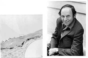
Left: the 10−3 page from Boeke's Cosmic View, New York, 1957 (37, note 24) depicts a cross-section through the skin of a girl's hand in a drawing whose width represents 0.15 mm. Skin structures orient diagonally across the middle; bacteria are black dots on the surface and in the quarter circle inset at the bottom right. The square in the centre represents the next smaller power of ten, 10−4 m. Right: Philip Morrison, MIT physicist, narrator of the Powers of Ten film, and co-teacher in 1976 with Phylis Morrison of a student workshop in observing with the microscope. Courtesy MIT Museum.

Interlinked in time and space, the network of ‘distant but direct’ microscope observers came to include me as a teenaged participant in a workshop set up by Phylis Morrison and my college professor Philip Morrison (Figure 6, right).Footnote 27 Their idea was to open up the unseen world by providing passers-by with microscopes and evocative materials. Among ourselves, including one or two other students, we put anything and everything under the microscope: bits of paper, hair, seeds, fine-woven cloth, a pencil tip, a needle's eye. Through the initial generation of our own enthusiasm and wonder, we next undertook to share it with whomever might stop at a microscope table we set up in a busy hallway of MIT. Even my dorm neighbour, a worldly wise senior in chemistry who kept a brain preserved in a jar of formaldehyde on his desk, paused for a look. An initial attitude of patronizing his underclass dorm-mate switched to his genuine surprise at seeing the oddest thing, the unruly fibres of notebook paper. The everyday world transformed by microscopy can be just as marvellous when passed down the hall as in its passing across spans of time by way of sunlight, text or memory.
Personal memory and historical memory are often kept apart by a gap seemingly as vast as that dividing the microscopic world from our own. Yet just as continuity and relation function between many scales of physical dimension, as documented in Powers of Ten, so grounds for relatedness may also apply to far-flung passages in human experience.Footnote 28 What is more distant from us, in dimension or experience, is both foundational and coextensive with our macroscopic and present life, having characteristics that are both utterly different yet somehow alike. To apprehend our personal memory as accommodating a ‘distant but direct’ access to historical memory involves us in extending how we see and respond to both unexpected and familiar scenes arising in historical experience. Memory's fluidity in relating the past to the present persists in reaching and affecting the future and is crucial throughout the process of teaching and learning. Franklin's anonymous poet perceived this incipience of the future within its past as inseparable from the embedding of microscopic life within macroscopic structures:
For us, a single experience achieved what the poet imagines here, where movement across differing magnitudes invokes a corresponding journey in time from past to present to future. Along the axis of size, solar projections of microscopic specimens transported our view to a scale where those samples appeared large and near. Along the axis of time, our personal memory and historical experience connected us as a present community and served as a medium for our dialogue with others of the past and those to come. Understanding the collective act of witnessing in the present and across time makes it possible for the ever-deferred and incomplete echo of personal and historical memories to cohere as an interdependent and reinforcing relationship. The continuing participation of observers across time with specimens of differing scale offers the potential for future observers to experience for themselves what Hooke and Dollond found fascinating and sought ways to share.
Witnessing as described here provides historians with the means to go beyond their customary resources of archival manuscripts, texts, commentaries, artefacts and materials. Previous studies involving replication of instruments or experiments demonstrate that the insights gained about the history of science depend upon working actively with original scientific materials.Footnote 30 Issues, confusions, materials or other details unstated in original texts arise in this kind of reconstructive work so as to reframe, redirect and transform historical inquiry.Footnote 31 For example, by repeating the microscopic anatomical practices described in Marcello Malpighi's texts, the historian Luigi Belloni recognized ‘the treacherous nature of anatomical procedures’ which led Malpighi erroneously to identify an artefact of his procedure as a new organ.Footnote 32 While their drawings depicted ‘convoluted fibers’ making up the surfaces of nerves and other microscopically viewed specimens, some of Malpighi's eighteenth-century successors suspected that these filaments might be an ‘optical deception’.Footnote 33 Others accepted them as a structural fact. By observing involutions on replica specimens, Bruno Zanobio located the illusion's source and deepened his understanding of this confusion and controversy. By contrast, a reliance solely on texts produced by the parties to a similar nineteenth-century debate about spiralled forms appearing on microscopic muscle specimens limited historian L. S. Jacyna to describing the moral tenor of the participants' dialogue without elucidating the practices that gave rise to such discordant perceptions.Footnote 34
The historian's replication research parallels that of past scientists, since it develops understandings of that science and its context that might not have been articulated by the original agents. Whereas for Zanobio confusion and deception emerged as parallel phenomena shared with historical microscopists, playfulness emerged as the salient parallel between Michael Faraday's experiments with gold films on microscope slides and the novice efforts of Ryan Tweney in layering films of gold. This parallel in playfulness evoked Tweney's reflection that ‘historian's play with the toys of science’ opens up new contextually meaningful routes to interpreting a scientist's work.Footnote 35
Collective witnessing extends the parallel from the redoing of an individual's experiment to the remaking of a community experience where people and scientific materials span and interlink with each other even across differing times and places. The act of engaging with each other while observing, exploring and discussing generates new understandings in science and history, whether these exchanges develop through controversy or through consensus. Robert Boyle long ago apprehended the ways science benefits from such collective acts, when inviting his contemporaries to join in each other's company while viewing materials placed within a chamber evacuated by his air-pump. We, too, can join and extend that community.