




No CrossRef data available.
Article contents
Effects of hypoxia on the brain: Neuroimaging and neuropsychological findings following carbon monoxide poisoning and obstructive sleep apnea
Published online by Cambridge University Press: 06 February 2004
Abstract
Hypoxia damages multiple organ systems especially those with high oxygen utilization such as the central nervous system. The purpose of this study was to compare the neuropathological and neuropsychological effects of hypoxia in patients with either carbon monoxide poisoning or obstructive sleep apnea. Neuroimaging revealed evidence of hippocampal atrophy in both groups although a linear relationship between hippocampal volume and memory performance was found only for selected tests and only in the sleep apnea group. There were significant correlations between hippocampal volume and performance on measures related to nonverbal/information processing. Generalized brain atrophy, as measured by the ventricle-to-brain ratio, was more common in the carbon monoxide poisoning group compared to the obstructive sleep apnea group. Performance on tests of executive function improved following treatment with nasal continuous positive airway pressure treatment in the obstructive sleep apnea group but there was no associated improvement in general intellectual function. We found that hypoxia due to obstructive sleep apnea and CO poisoning resulted in neuropathological changes and neuropsychological impairments. The observed group differences provide insight into the relationship between etiology of injury, neuropathological changes, and clinical presentation. (JINS, 2004, 10, 60–71.)
Information
- Type
- Research Article
- Information
- Journal of the International Neuropsychological Society , Volume 10 , Issue 1 , January 2004 , pp. 60 - 71
- Copyright
- © 2004 The International Neuropsychological Society
INTRODUCTION
Current thought in neuropsychology is that the etiology of a particular neurological injury will determine the neuropathological changes and neuropsychological profiles that occur following the injury. However, direct comparison of the neuropathologic (i.e., morphometric) and neuropsychological effects following cerebral hypoxia, due to different etiologic mechanisms has rarely been carried out. In vivo neuroimaging techniques provide unique methods to investigate the pathologic effects that occur following hypoxia in patients with obstructive sleep apnea (OSA) or carbon monoxide (CO) poisoning. Comparison of morphometric neuroimaging analyses and neuropsychological outcomes permits evaluation of the similarities and differences between the two disorders.
Exposure to CO may damage multiple organ systems, especially those with high oxygen utilization, including the cardiovascular and central nervous system (for general overview, see Raub et al., 2000). The mechanisms by which CO causes cellular damage are complex and multifactorial. Although the neuropathologic changes associated with CO poisoning are related to CO-induced hypoxia (CO binds to hemoglobin) (Okeda et al., 1981), other biochemical mechanisms appear to be involved in cellular damage. Other mechanisms include binding of CO to various intracellular proteins (Piantadosi, 1987), neuro-excitotoxicity (Jarrard & Meldrum, 1993), ischemia/reperfusion injury (Thom, 1990), and apoptosis (Piantadosi et al., 1995). Previous studies using quantitative magnetic resonance imaging (QMRI) have shown hippocampal and fornix atrophy, and generalized ventricular enlargement; a sign of whole brain atrophy following CO exposure (Gale et al., 1999; Hopkins et al., 1993; Kesler et al., 2001; Reynolds et al., 1999). These structural changes likely reflect selective sensitivity of specific cerebral structures to hypoxia (for review, see Caine & Watson, 2000). Neuropsychological impairments are frequently reported following CO poisoning, including impaired memory, attention, visuospatial skills, executive function, and constructional praxis (Hopkins et al., 1993; Thom et al., 1995; Weaver et al., 2002).
The effects of acute and chronic hypoxia are well known, whereas less is known regarding the effects of intermittent hypoxia. Obstructive sleep apnea (OSA) is a sleep disorder that results in the absence (apnea) or reduction (hypopnea) of airflow lasting at least 10 s despite normal respiratory exertion (Guilleminault et al., 1978; Kelly et al., 1990). The apnea and hypopnea result in decreased oxygen saturation levels in the blood (hypoxemia) and disruption or fragmentation of the sleep cycle. OSA affects an estimated 2 to 4% of the middle-aged population and the prevalence increases with age (Bliwise et al., 1994; Young et al., 1993). Common symptoms include excessive daytime sleepiness (EDS), snoring, gasping or choking during sleep, headaches (especially upon waking), irritability and mood disturbance, personality change, motor restlessness, and cognitive impairments (Agency for Health Care Policy and Research, 1998; Guilleminault et al., 1978;). The cognitive and behavioral manifestations associated with OSA are related to both intermittent hypoxia and sleep fragmentation (Barth et al., 1993; Bédard et al., 1991, 1993; Berry et al., 1986; Findley et al., 1986; Valencia-Flores et al., 1996). Patients with OSA may exhibit impairments in vigilance, attention, short-term memory, general intellectual functioning, executive dysfunction, visuospatial function, and psychomotor speed (Bédard et al., 1991; Findley et al., 1986; see also Rourke & Adams, 1996). Thus, the neuropsychological impairments in OSA patients with intermittent hypoxia are similar to those observed following CO poisoning and associated acute hypoxia.
Previously we reported hippocampal atrophy following CO poisoning (Gale et al., 1999; Hopkins et al., 1995a) and OSA (Gale et al., 2000; Walker et al., 1999). Although the two disorders have different etiologies, both disorders may result in neuropathologic changes secondary to hypoxia/hypoxemia. The purpose of this study was to compare the QMRI and neuropsychological outcome between patients with a history of CO poisoning and patients with a recent diagnosis of OSA. We were also interested in the effects of treatment for the patients with OSA; as such both pre and post treatment neuropsychological profiles were compared to the CO group.
METHODS
Research Participants
Twenty patients (11 males, 9 females), with moderate to severe accidental CO poisoning were compared to 14 severe OSA patients with normal daytime blood gases (12 males, 2 females). There were no differences in ethnic or racial characteristics between the two groups. The study had LDS Hospital IRB approval, conformed to institutional and federal guidelines for the protection of human subjects, and written informed consent was obtained. All CO patients were treated with 100% normobaric oxygen or hyperbaric oxygen following the poisoning (4 patients were intubated). The CO patients' mean age was 39 ± 7.0 years (range = 27–52 years; MDN = 38.0 years) and mean educational level of 15 ± 3.0 years (range = 8–20 years; MDN = 15.0 years). The mean COHb level was 26.0 and 40% of patients lost consciousness. A physician independently confirmed diagnosis of CO poisoning and all patients were referred for brain imaging and comprehensive neuropsychological evaluation. The mean time from CO exposure to MRI and neuropsychological testing was 22.4 ± 13.8 months (range = 4–45 months; MDN 17.8 months). A detailed description of this group has been reported elsewhere (Gale et al., 1999).
The OSA patients were recruited as part of a prior treatment study and initial findings and patient characteristics are described elsewhere (Walker et al., 1999). Patients with evidence of daytime hypoxemia were excluded from the study. The OSA patients mean age was 52 ± 11 years (range = 33–68 years; MDN = 51.0 years) and mean educational level was 14 ± 1.8 years (range = 12–18 years; MDN = 14.0 years). The mean baseline oxygen saturation was measured by a pulse oximetry (SaO2) of 90.2 ± 5.0 (MDN = 92), which is within the normal range. Results from polysomnography showed a mean respiratory distress index (RDI) of 83.6 ± 18.1 (MDN = 84.5) and mean percent sleep time below a SaO2 of 90% was 65% ± 33.6 (MDN = 82%). The mean minimum SaO2 during polysomnography was 53.9 ± 17.5 (MDN = 53.0). No data was available regarding duration of OSA. All OSA patients underwent neuropsychological evaluation and neuroimaging within several days of polysomnography. Neuroimaging occurred prior to treatment with nasal continuous positive airway pressure (nCPAP) and did not delay treatment. Neuropsychologic testing, but not neuroimaging, was repeated in the OSA patients after 6 months of nCPAP treatment.
Procedures
Neuroimaging
MR images were acquired at 1.5 Tesla with a quadriture head coil using standard clinical protocols. Parameters were as follows: (1) sagittal T1-weighted (500/11/2; TR/TE/excitations); (2) axial intermediate and T2 weighted (3000/31; 90/1) spin echo images; and (3) coronal intermediate and T2-weighted (3800/21, 105/2) fast spin echo. The sagittal and axial images had a slice thickness of 5 mm with a 2 mm interslice gap acquired on a 256 × 192 matrix with a field of view of 22 cm and 24 cm in the axial and sagittal planes, respectively. The coronal images were 3 mm thick interleaved sections with a field of view of 22 cm on a 512 × 256 matrix. Imaging parameters as well as our normative QMRI database have been reported in detail elsewhere (Bigler et al., 1997; Blatter et al., 1995).
Volumetric analysis
Images were quantified as described by Blatter et al. (1995) using the software ANALYZE (Biomedical Imaging Resource, 1993). Quantitative (volumetric) analyses of cerebral structures obtained from MRI were performed per the methods described previously (Bigler et al., 1997; Blatter et al., 1995; Gale et al., 1995; Hopkins et al., 1995a). Hippocampal reliability was .92 and ≥ .98 and additional information on inter- and intra-rater reliability are described elsewhere (Bigler et al., 1997; Blatter et al., 1995; Hopkins et al., 1995a). The rater (SDG) for image analysis was blind to diagnosis, sex, age, and neuropsychological test scores.
Two QMRI measures, ventricle-to-brain ratio (VBR) and hippocampal volume are the focus of this study. A detailed description of the QMRI methods is beyond the scope of this paper. Briefly, regions of white matter, gray matter, and cerebrospinal fluid were identified and plotted to represent pixel signal intensity. Utilizing dual-echo images (intermediate and T2-weighted) enhanced accurate signal identification. A multispectral segmentation was performed on the two spatially registered images from the foramen magnum to the vertex (see Blatter et al., 1995). Thus, this type of QMRI analysis results in volumetric rather than planimetric (i.e., area) measures. VBR was calculated by dividing total ventricular volume (e.g., lateral ventricles and third and fourth ventricles) by total brain volume and multiplying by 100 (Blatter et al., 1995). Since VBR takes into account total brain volume there is no need to adjust for brain size. VBR has been shown to be a measure of generalized atrophy (i.e., whole brain volume loss; Barker et al., 1999; Bigler et al., 2000; Blatter et al., 1997; Gale et al., 1995). VBR was defined as abnormal if it was at least 1.5 standard deviations above the mean compared to age and sex stratified normative data.
Hippocampal volumes were measured in the coronal slices (see Bigler et al., 1997). Hippocampal atrophy is defined as a hippocampal volume of 1.5 standard deviations below the mean of the normative sample (see below). Each subject's QMRI volumes were converted to z scores using data from our normative data (Bigler et al., 1997; Blatter et al., 1995). Total hippocampal measures are reported as volumes (cm3) and age and sex adjusted z scores. VBR measures are reported as ratios and age and sex adjusted z scores.
Neuropsychological assessment
The two groups differed in some of the neuropsychological tests administered: the CO group was seen for clinical reasons (data is archival), while the OSA patients were part of a prospective study. Therefore, only the neuropsychological tests administered to both groups will be reported. The neuropsychological tests included the Wechsler Adult Intelligence Scale–Revised (WAIS–R; Wechsler, 1981), Wechsler Memory Scale–Revised (WMS–R; Wechsler, 1987), Rey Auditory Verbal Learning Test (RAVL; Rey, 1964), Rey-Osterrieth Complex Figure (ROCFD; Rey, 1941), Trail Making Test Parts A and B (Reitan & Wolfson, 1985), SCL–90–R (Derogatis, 1994), Beck Depression Inventory (BDI; Beck, 1987), and Beck Anxiety Inventory (BAI; Beck & Steer, 1993). Since previous studies suggest generalized intellectual decline occur in both groups, the Oklahoma premorbid intelligence estimation method (OPIE; Scott et al., 1997) was used to assess change in intellectual function. Difference scores were obtained by subtracting FSIQ score from the OPIE score.
Raw test scores were converted to z scores (M = 0; SD = 1) based on available normative data. When possible the z score took into account age, education, and sex. The normative data from Heaton et al. (1991) were used to convert the data from the WAIS–R subtests (e.g., Block Design, Digit Symbol and Vocabulary), Trail Making Test (Parts A and B), and finger oscillation test. Trials 1 and 6 of the RAVL were converted to z scores using norms stratified by age and sex (Geffen et al., 1990). Delayed recall raw scores from the ROCFD were converted to z scores using normative data reported by Meyers and Meyers (1995). Verbal fluency (total number of words on the FAS) z scores were calculated using normative data described by Tombaugh et al. (1999). The subtests from the WMS–R (Logical Memory and Visual Reproduction) were converted to z scores using data provided in the manual (Wechsler, 1987). Since the normative data in the WMS–R manual are interpolated for ages 25–34, norms reported by Mittenberg et al. (1992) were used. After transforming neuropsychological test scores to z scores, composite function indices were created by averaging these scores within each cognitive domain. Description and calculation of the cognitive domains are presented in Table 1.
Neuropsychological tests and cognitive domains

Psychological function
Emotional function, depression and anxiety were assessed. The presence of depression was defined as a raw score of 14 or greater on the BDI and/or a T-score of 63 or greater on the Depression scale of the SCL–90–R. Anxiety was defined as a raw score of 10 or more on the BAI and/or a T-score of 63 or more on the Anxiety Index scale of the SCL–90–R. To evaluate the effect of treatment with nCPAP on emotional function in the OSA patients, improvement in depression or anxiety were defined as scores in the normal range on both the Beck and SCL–90–R scales for scores that were initially elevated.
Statistical analysis
Independent t tests were used to compare demographic variables between the CO and OSA groups. Non-continuous data comparisons between groups (e.g., percent of patients with hippocampal atrophy) were analyzed using Fisher's Exact Test. Correlations between imaging variables and medical and neuropsychological data were performed using Pearson correlations.
To reduce Type I error, tests that were thought to assess similar cognitive functions were grouped together to create a single score. Eight domain scores were calculated; each one representing a dependent variable of interest. Separate verbal and visual memory domain scores were created to investigate associations with left and right hippocampal volumes. A profile analysis was used to analyze differences in the pattern of neuropsychological performance between the CO and pre-treatment OSA groups. A profile analysis is essentially a repeated measures multivariate analysis of variance (MANOVA) designed to compare two or more groups on a series of test scores (Johnson & Wichern, 2002; Stevens, 1996). First, the profiles are tested for parallelism. Parallel profiles would suggest that one group scored uniformly higher than the other group while non-parallel profiles would suggest a group by variable interaction. Second, if the profiles are found to be parallel then they are tested to determine if they are coincident. Profiles are considered to be coincident if the groups scored the same across all variables, that is, performance on the first variable (or domain) were equal to performance on the second variable and so forth. Finally, if the profiles are found to be coincident they are then tested to determine if they are level; if there are equal scale means. Since a profile analysis requires that scores be equally scaled, test scores were transformed to z scores (described above). In addition to the profile analysis a repeated-measures MANOVA was carried out to compare pre-nCPAP performance versus post-nCPAP in the OSA group.
RESULTS
Group Differences
Demographics
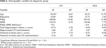
Demographic variables are presented in Table 2. There was a significant age difference between the OSA and CO groups [t(32) = −4.48, p < .001]. However, the groups did not differ on level of education [t(32) = 1.49, p > .05], FSIQ [t(32) = −0.72, p > .05], or estimated FSIQ (OPIE) [t(32) = −0.27, p > 0.05]. Although estimated premorbid levels of intellectual function using the OPIE did not differ between the groups, within groups comparisons showed that the measured FSIQ (WAIS–R) was significantly lower than the estimated premorbid IQ (p < .001).
Demographic variables by diagnostic group

Neuroimaging findings
Hippocampal atrophy occurred in 30% of CO patients and 36% of OSA patients. The Fisher's Exact Test (two-tailed) showed the percent of patients with hippocampal atrophy did not differ between the groups (p > .70). While 35% of CO patients had generalized brain atrophy, as measured by enlarged VBR, none of the OSA patients had increased VBRs (p < .05). Z scores for hippocampal volume are presented in Table 2. Direct comparison of hippocampus and VBR between the groups was problematic due to the large variability. For this reason natural log (LN) transformations of the morphometric measures were carried out to reduce the effects of outliers and differences in variance between groups (see Altman, 1991). Since natural log transformations cannot be carried out on negative numbers, the z scores were converted to T-scores. The VBR LN transformation scores were 4.16 (.55) and 3.86 (.18) for the CO and OSA groups, respectively (p = .058). Hippocampal LN transformation scores were 3.68 (.47) and 3.67 (.26) for the CO and OSA groups, respectively (p = .957).
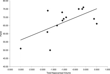
Hippocampal volumes correlated with PaO2, a direct measure of oxygen saturation, to examine the relationship between oxygen saturation and cerebral morphology in OSA patients. There was a significant correlation between oxygen saturation and total hippocampal volume (r = .621; R2 = .385, p < .01) [Figure 1]. The correlations between oxygen saturation and left or right hippocampal volumes were significant; r = .581 and .630, respectively.

Correlation between total hippocampal volume (z score) and PaO2 in OSA patients.
Neuropsychological outcome
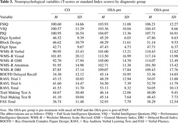
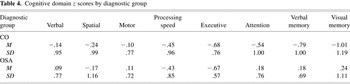
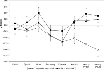
The neuropsychological test results are presented in Tables 3 and 4. Results from the profile analysis indicated that the profiles were parallel [F(7,26) = 2.19, p > .05] (Figures 2 and 3) indicating that there was no Group × Cognitive Domain interaction. The OSA group performance was better for all domains. The average profile score across all cognitive domains was −.49 (.62) and −.06 (.47) for the CO and OSA groups, respectively. The profiles were not coincident; equality of profiles was not supported [F(1,32) = 4.87, p < .05] indicating that the performance was variable across the domains. Finally, the profiles were not level [F(7,26) = 5.15, p < .05] indicating the means were unequal. Significant domain differences were found for attention [F(1,32) = 4.53, p < .05], verbal memory [F(1,32) = 8.03, p < .05] and visual memory [F(1,32) = 8.23, p < .05].
Neuropsychological variables (T-scores or standard Index scores) by diagnostic group

Cognitive domain z scores by diagnostic group


Profiles of performance across cognitive domains by group.

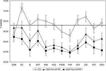
Index scores from the SCL–90–R by group. T-scores from the SCL–90–R have a M of 50 and a SD of 10. A T-score ≥ 63 is considered to be abnormal.
Psychological function
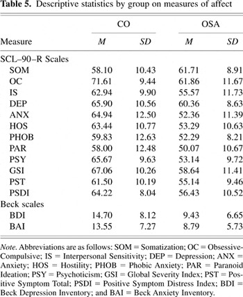
Measures of depression and anxiety were generally normal in the OSA group but mild to moderately elevated in the CO group. Table 5 consists of the descriptive statistics from the BDI, BAI, and SCL–90–R.
Descriptive statistics by group on measures of affect

Relationships Between Neuroimaging and Cognitive Domains
Correlations between hippocampal volume and cognitive domains
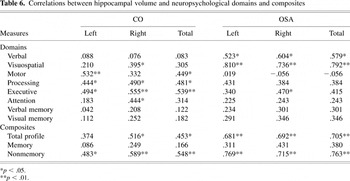
One-tailed Pearson correlation coefficients for hippocampal volume and cognitive domain scores are presented in Table 6. Positive correlations were found for hippocampal volumes and selected cognitive domain scores. There were no significant correlations between hippocampal volume and memory domain scores. However, for the OSA group, some memory test scores (included in the memory domain scores) significantly correlated with hippocampal volume. For example, delayed recall of the ROCFD correlated with right (r =.672, p < .01) and left (r = .439, p > .05) hippocampal volumes, and Trial 6 of the RAVL correlated with right (r = .715, p < .01) and left (r = .702, p < .01) hippocampal volumes in the OSA group.
Correlations between hippocampal volume and neuropsychological domains and composites

All of the cognitive domain scores were collapsed to form a composite score, Total Profile, which correlated with hippocampal volume (see Table 6). Finally, two additional composite scores were calculated to evaluate the association between morphology and memory versus non-memory performance. Results in Table 6 indicate the non-memory composite score correlated with hippocampal volume for bothgroups, but there were no significant correlations with the memory composite score.
Correlations between VBR and cognitive domains
In contrast to the correlations between hippocampal volume and cognitive domains in the OSA group, there were no significant correlations between cognitive domain scores and VBR. Similarly, VBR did not correlate with SaO2 in the OSA group. These findings were not unexpected since the OSA patients had normal VBRs. For the CO group, only the Attention domain significantly correlated with VBR (r = −.296, p = .045).
Neuropsychological and emotional outcome following nCPAP
A repeated measures (within subjects) MANOVA showed significant improvement in the cognitive domain profile in the OSA group after 6 months of treatment with nCPAP [F(7,7) = 6.52, p < .05]. The Executive function z score increased an average of .43 (.55), with a median increase of 0.47. Sleep parameters were improved at 6 month follow-up with the RDI decreasing from 83.6 ± 18.1 to 19.2 ± 13.1 and minimum SaO2 increased from 53.9 ± 17.5 to 70.0 ± 17.7.
Psychological function for the OSA group was in normal range prior to nCPAP (Table 5 and Figure 3) but there was evidence of a general decrease in the number of symptoms reported following treatment. Forty percent of the OSA patients had elevated Distress indices on the SCL–90–R that were in the normal range following nCPAP. Depression scores (SCL–90–R Depression Index and/or BDI) were elevated in 50% of the OSA patients prior to treatment; 14% were still reporting depressive symptoms following nCPAP. Similarly, anxiety scores were elevated in 50% of the OSA patients prior to treatment and only 21% had elevated anxiety scores at follow-up.
DISCUSSION
This is one of the first studies to utilize QMRI to compare the neuropathological and neuropsychological effects of hypoxia-related injury between two etiologic groups. Consistent with previous research demonstrating hippocampal atrophy following hypoxia in humans (Manns et al., 2003), rats (Davis et al., 1986), and monkeys (Zola-Morgan et al., 1992), there was evidence of the neuropathological effects of hypoxic brain injury. QMRI findings showed that a similar percentage of patients in both groups had hippocampal atrophy. In contrast, generalized atrophic changes (i.e., enlarged VBR) were found only in the CO patients. Group differences were not due to age or sex, as the QMRI z scores were derived using normative data stratified by age and sex.
The dissimilarity of the VBR findings between the hypoxic groups in this study was unexpected and raises the question of why the differences occur. There are several possible reasons for the differences between the groups. First, the groups may differ in terms of severity of the hypoxia. In the CO group, the CO exposure was moderate to severe as indicated by their elevated COHb levels and loss of consciousness. Thus, the patients in the CO group may have had a more severe hypoxic episode compared to the OSA group resulting in additional neuronal injury and concomitant atrophy.
Second, the groups may differ due to moderation of tissue damage from brief intermittent hypoxia instead of a single episode of longer duration. Therefore, the duration of hypoxia may account for the more severe generalized brain atrophy observed in the CO patients. Alternatively, it has been suggested that the long-term effects of chronic intermittent hypoxia, such as occurs in OSA, can result in cerebral vascular problems, neurodegeneration, and neurocognitive deficits due to the cumulative effects of the hypoxia (Neubauer, 2001).
Third, the groups may differ due to neuronal damage related to the direct and/or indirect cytotoxic effects of CO. Differences in VBR between the groups in our study may be due to the CO-induced cytotoxic effects independent of tissue oxygenation (Piantadosi et al., 1997b). For example, Choi (1983) described a delayed neurological syndrome following CO poisoning that presents weeks after CO is removed from body tissues, suggesting mechanisms that may be independent of hypoxia. Animal research has shown neuronal apoptosis and necrosis following CO-induced hypoxia (Piantadosi et al., 1997a). A study by Piantadosi et al. found minimal damage to the hippocampus while damage to the frontoparietal cortex was extensive. A case report of fatal CO poisoning in a 41-year-old man, including a detailed histological examination of the brain, suggested that the necrotic and apoptotic lesions were not explained by hypoxia alone (Uemura et al., 2001). Finally, the combination of all of the above might contribute to the observed differences in VBR between the CO and OSA groups.
Previous research has suggested that the hippocampus may be more vulnerable to hypoxic injury than adjacent cerebral structures such as the parahippocampal gyrus or temporal lobes (Hopkins et al., 1995b; Kesner & Hopkins, 2001; Manns et al., 2003). However, a review of the human anoxia literature, excluding CO poisoning and OSA, by Caine and Watson (2000) suggested that neuropathological changes in the watershed cerebral cortex and basal ganglia areas occur more frequently than hippocampal damage. Furthermore, Beebe and Gozal (2002) suggested injury to the prefrontal cortex, rather than medial temporal lobe structures, is responsible for the neurocognitive and neurobehavioral dysfunction associated with OSA (Beebe & Gozal, 2002). Consistent with this finding, our study found that executive dysfunction was the most common cognitive impairment in our OSA group. In contrast, the CO patients had deficits in executive function and attention and memory.
We did not find strong correlations between measures of memory and hippocampal volume. The lack of association may be due to the limited number of patients with hippocampal atrophy (approximately 30%), comparison with memory domain score (several measures of memory) rather than individual memory test scores, or that hippocampal atrophy may be a nonspecific indicator of cerebral injury. Consistent with the latter possibility, nonverbal tasks and measures of hypoxemia were related to hippocampal volume. Similar findings were reported by Cheshire et al. (1992) who found Block Design, Digit Symbol, and lower IQ were associated with breathing abnormalities in OSA patients. Greenberg et al. (1987) reported impaired perceptual–organizational skills in OSA patients. Bédard et al. (1991) found that OSA patients with severe hypoxemia were more impaired on visuospatial tasks compared to individuals with moderate hypoxemia. In addition, studies indicate that the neuropsychological impairments in OSA are similar to those found in other hypoxic/anoxic conditions which include both memory and non-memory dependent functions (Kelly et al., 1990; Roehrs et al., 1995). Thus, hippocampal atrophy may represent the severity of brain injury and is associated with diffuse cerebral impairment.
Both groups in our study had evidence of decreased intellectual function compared with estimated premorbid intelligence levels. Nonetheless, IQ scores were in the average range. Bédard et al. (1991) found individuals with severe OSA had lower FSIQ and executive dysfunction (Trails B) compared to individuals with moderate OSA, which were related to severity of hypoxemia and not vigilance or sleepiness. These findings are consistent with those of the current study. Bédard et al. (1993) tested OSA patients prior to and 6 months post treatment with nCPAP and found improvements in FSIQ, but not Trail Making Test Part B or Verbal Fluency. In contrast, patients in our study improved on Trail Making Test B and Verbal Fluency but no improvement in FSIQ was observed. Our patients were similar in age, level of hypoxemia, and baseline FSIQ to those in the Bedard et al. 1993 study but had a higher education level (13.7 ± 1.8 vs. 11.7 ± 2.6) which may suggest a more significant reduction in FSIQ and therefore no post-treatment increase in FSIQ. Previous research has reported improvement (Bédard et al., 1991, 1993; Engleman et al., 1997; Valencia-Flores et al., 1996) and no change in cognitive function following nCPAP (Barbe et al., 2001).
Both the CO and OSA groups reported mild symptoms of depression and anxiety. The OSA group reported fewer emotional symptoms following nCPAP, with the scores on all measures within the normal range. Affective changes have been reported following CO poisoning (Gale et al., 1999) and OSA (Barth et al., 1993; Guilleminault et al., 1978). However, given that the reported symptoms of depression and anxiety in the current study were in the mild range for both groups, it is unlikely that they contributed to the observed cognitive impairments.
There are several limitations of our study including lack of a control group or a placebo group for the nCPAP treatment in the OSA group. Given the small sample size in our study may have been under powered to detect relationships between hippocampal atrophy and memory. The strengths of our study include comparisons of neuropsychological and neuroimaging findings in two disorders associated with hypoxia.
In conclusion, our study found hippocampal and generalized atrophy and neuropsychological impairments in CO poisoning and OSA. Hippocampal atrophy occurred in both groups; however increased VBR due to generalized cerebral atrophy (i.e., whole brain volume loss) was greater in the CO group. The CO group consistently performed worse on most cognitive measures while the OSA group had more selective cognitive impairments. Improvement in cognitive function in OSA patients following 6 months of nCPAP treatment was limited to executive function. Differences in test performance between the CO and OSA groups were more pronounced after the OSA group had received 6 months of nCPAP treatment.
This research supports emerging evidence that hypoxia contributes to hippocampal atrophy, and in some cases generalized brain atrophy (e.g., increased VBR). It is possible that either the severity of the hypoxia in the CO patients was greater than in the OSA patients, or that other neuropathologic factors, such as those specific to CO poisoning, may contribute to differences in hypoxia-related brain injury. Additional research is needed that compares neuropathologic and neuropsychological changes in other disorders associated with hypoxia, such as acute respiratory distress syndrome, asthma, and chronic obstructive pulmonary disease.
References
REFERENCES
Agency for Health Care Policy and Research.
(1998).
Systematic Review of the Literature Regarding the Diagnosis of
Sleep Apnea.
Summary, Evidence Report/Technology Assessment:
Number 1, December 1998. Rockville, MD. Retrieved from
http://www.ahrq.gov/clinic/apnea.htm.
Altman, D.G.
(1991).
Practical statistics for medical research.
London:
Chapman and Hall.
Barbe, F.,
Mayorala, L.R.,
Duran, J.,
Masa, J.F.,
Maimo, A.,
Montserrat, J.M.,
Monasterio, C.,
Bosch, M.,
Ladaria, A.,
Rubio, M.,
Rubio, R.,
Medinas, M.,
Hernandez, L.,
Vidal, S.,
Douglas, N.J., &
Agusti, A.G.N.
(2001).
Treatment with continuous positive airway pressure is not effective
in patients with sleep apnea but no daytime sleepiness.
Annals of Internal Medicine,
134,
1015–1023.CrossRefGoogle Scholar
Barker, L.H.,
Bigler, E.D.,
Johnson, S.C.,
Anderson, C.V.,
Russo, A.A.,
Boineau, B., &
Blatter, D.D.
(1999).
Polysubstance abuse and traumatic brain injury: Quantitative
magnetic resonance imaging and neuropsychological outcome in older
adolescents and young adults.
Journal of the International Neuropsycholgical Society,
5,
593–608.Google Scholar
Barth, J.T.,
Findley, L.J.,
Zillmer, E.A.,
Gideon, D.A., &
Suratt, P.M.
(1993).
Obstructive sleep apnea, hypoxemia, and personality functioning:
Implications for medical psychotherapy assessment.
Advances in Medical Psychotherapy,
6,
29–36.Google Scholar
Beck, A.T.
(1987).
Beck Depression Inventory.
San Antonio, TX:
The Psychological Corporation.
Beck, A.T. &
Steer, R.A.
(1993).
Beck Anxiety Inventory.
San Antonio, TX:
The Psychological Corporation.
Bédard, M.A.,
Montplaisir, J.,
Malo, J.,
Richer, F., &
Rouleau, I.
(1993).
Persistent neuropsychological deficits and vigilance impairment in
sleep apnea syndrome after treatment with nasal continuous positive
airway pressure (CPAP).
Journal of Clinical and Experimental Neuropsychology,
15,
330–341.CrossRefGoogle Scholar
Bédard, M.,
Montplaisir, J.,
Richer, F.,
Rouleau, I., &
Malo, J.
(1991).
Obstructive sleep apnea syndrome: Pathogenesis of
neuropsychological deficits.
Journal of Clinical and Experimental Neuropsychology,
13,
950–964.Google Scholar
Beebe, D.W. &
Gozal, D.
(2002).
Obstructive sleep apnea and the prefrontal cortex: Towards a
comprehensive model linking nocturnal upper airway obstruction to
daytime cognitive and behavioral deficits.
Journal of Sleep Research,
11,
1–16.Google Scholar
Berry, D.T.R.,
Webb, W.B.,
Block, A.J.,
Bauer, R.M., &
Switzer, D.A.
(1986).
Nocturnal hypoxia and neuropsychological variables.
Journal of Clinical and Experimental Neuropsychology,
8,
229–238.CrossRefGoogle Scholar
Bigler, E.D.,
Lowry, C.M.,
Anderson, C.V.,
Johnson, S.C.,
Terry, J., &
Steed, M.
(2000).
Dementia, quantitative neuroimaging, and apolipoprotein E genotype.
American Journal of Neuroradiology,
21,
1857–1868.Google Scholar
Bigler, E.D.,
Blatter, D.D.,
Anderson, C.V.,
Johnson, S.C.,
Gale, S.D.,
Hopkins, R.O., &
Burnett, B.
(1997).
Hippocampal volume in normal aging and traumatic brain injury.
American Journal of Neuroradiology,
18,
11–23.Google Scholar
Biomedical Imaging Resource.
(1993).
ANALYZE 6.0 [Computer software].
Rochester, MN:
Mayo Foundation.
Blatter, D.D.,
Bigler, E.D.,
Gale, S.D.,
Johnson, S.C.,
Anderson, C.V.,
Burnett, B.M.,
Ryser, D.,
Macnamara, S.E., &
Bailey, B.J.
(1997).
MRI based brain and CSF quantification following traumatic brain
injury: Correlation with neuropsychological outcome.
American Journal of Neuroradiology,
18,
1–10.Google Scholar
Blatter, D.D.,
Bigler, E.D.,
Gale, S.D.,
Johnson, S.C.,
Anderson, C.V.,
Burnett, B.M.,
Parker, N.,
Kurth, S., &
Horn, S.D.
(1995).
Quantitative volumetric analysis of brain MRI: Normative database
spanning five decades of life.
American Journal of Neuroradiology,
16,
241–251.Google Scholar
Bliwise, D.L.,
King, A.C., &
Harris, R.B.
(1994).
Habitual sleep durations and health in a 50–65 year old
population.
Journal of Clinical Epidemiology,
47,
35–41.CrossRefGoogle Scholar
Caine, D. &
Watson, J.
(2000).
Neuropsychological and neuropathological sequelae of cerebral
anoxia: A critical review.
Journal of the International Neuropsychological Society,
6,
86–99.Google Scholar
Cheshire, K.,
Engleman, H.,
Deary, I.,
Shapiro, C., &
Douglas, N.J.
(1992).
Factors impairing daytime performance in patients with sleep
apnea/hypopnea syndrome.
Archives of Internal Medicine,
152,
538–541.CrossRefGoogle Scholar
Choi, I.S.
(1983).
Delayed neurologic sequelae in carbon monoxide intoxication.
Archives of Neurology,
40,
433–435.Google Scholar
Davis, H.P.,
Tribuna, J.,
Pulsinelli, W.A., &
Volpe, B.T.
(1986).
Reference and working memory of rats following hippocampal damage
induced by transient forebrain ischemia.
Physiology and Behavior,
37,
387–392.CrossRefGoogle Scholar
Derogatis, L.R.
(1994).
Symptom Checklist–90–Revised
(3rd ed.).
Minneapolis, MN:
National Computer Systems Inc.
Engleman, H.M.,
Martin, S.E.,
Deary I.J., &Douglas N.J.
(1997).
Effect of CPAP therapy on daytime function in patients with mild
sleep apnoea/hypopnoea syndrome.
Thorax,
52,
114–119.Google Scholar
Findley, L.J.,
Barth, J.T.,
Powers, D.C.,
Wilhoit, S.C.,
Boyd, D.G., &
Suratt, P.M.
(1986).
Cognitive impairment in patients with obstructive sleep apnea and
associated hypoxemia.
Chest,
90,
686–690.CrossRefGoogle Scholar
Gale, S.D.,
Hopkins, R.O.,
Weaver, L.K.,
Bigler, E.D.,
Booth, E.D., &
Blatter, D.D.
(1999).
MRI, quantitative MRI, SPECT, and neuropsychological findings
following carbon monoxide poisoning.
Brain Injury,
13,
229–243.Google Scholar
Gale, S.D.,
Hopkins, R.O.,
Weaver, L.K.,
Walker, J.M.,
Bigler, E.D., &
Cloward, T.V.
(2000).
Hippocampal atrophy following sleep apnea and carbon monoxide
poisoning: Similarities and differences.
Journal of the International Neuropsychological Society,
6,
154.Google Scholar
Gale, S.D.,
Johnson, S.C.,
Bigler, E.D., &
Blatter, D.D.
(1995).
Nonspecific white matter degeneration following traumatic brain
injury.
Journal of the International Neuropsychological Society,
1,
17–28.Google Scholar
Geffen, G.,
Moar, K.J.,
O'Hanlon, A.P.,
Clark, C.R., &
Geffen, L.B.
(1990).
Performance measures of 16- to 86-year old males and females on the
auditory verbal learning test.
Clinical Neuropsychologist,
4,
45–63.Google Scholar
Greenberg, G.D.,
Watson, R.K., &
Deptula, D.
(1987).
Neuropsychological dysfunction in sleep apnea.
Sleep,
10,
254–262.Google Scholar
Guilleminault, C.,
van den Hoed, J., &
Mitler, M.M.
(1978).
Clinical overview of the sleep apnea syndromes. In
C. Guilleminault &
W.C. Dement (Eds.),
Sleep apnea syndromes (pp.
1–12).
New York:
Alan R. Liss.
Heaton, R.K.,
Grant, I., &
Matthews, C.G.
(1991).
Comprehensive norms for an expanded Halstead-Reitan Battery:
Demographic corrections, research findings and clinical
applications.
Odessa, FL:
Psychological Assessment Resources, Inc.
Hopkins, R.O.,
Gale, S.D.,
Johnson, S.D.,
Anderson, C.V.,
Bigler, E.D.,
Blatter, D.D., &
Weaver, L.K.
(1995a).
Severe anoxia with and without concomitant brain atrophy and
neuropsychological impairments.
Journal of the International Neuropsychological Society,
1,
501–509.Google Scholar
Hopkins, R.O.,
Weaver, L.K., &
Kesner, R.P.
(1993).
Long term memory impairments and hippocampal magnetic resonance imaging
in carbon monoxide poisoned subjects.
Abstracts, Undersea and Hyperbaric Society Annual Scientific
Meeting,
Vol. 20
(Suppl.),
15.
Hopkins, R.O.,
Kesner, R.P., &
Goldstein, M.
(1995b).
Item and order recognition memory in subjects with hypoxic brain
injury.
Brain and Cognition,
27,
180–201.Google Scholar
Jarrard, L.E. &
Meldrum, B.S.
(1993).
Selective excitotoxic pathology in the rat hippocampus.
Neuropathology and Applied Neurobiology,
19,
381–389.Google Scholar
Johnson, R.A. &
Wichern, D.W.
(2002).
Applied multivariate statistical analysis
(5th ed.).
Upper Saddle River, NJ:
Prentice Hall.
Kelly, D.A.,
Claypoole, K.H., &
Coppel, D.B.
(1990).
Sleep apnea syndrome: Symptomatology, associated features, and
neurocognitive correlates.
Neuropsychology Review,
1,
323–342.CrossRefGoogle Scholar
Kesler, S.R.,
Hopkins, R.O.,
Weaver, L.K.,
Blatter, D.D.,
Edge-Booth, H., &
Bigler, E.D.
(2001).
Verbal memory deficits associated with fornix atrophy in carbon
monoxide poisoning.
Journal of the International Neuropsychological Society,
7,
640–646.CrossRefGoogle Scholar
Kesner, R.P. &
Hopkins, R.O.
(2001).
Short-term memory for duration and distance in humans: Role of the
hippocampus.
Neuropsychology,
15,
58–68.Google Scholar
Manns, J.R.,
Hopkins, R.O.,
Reed, J.M.,
Kitchener, E.G., &
Squire, L.R.
(2003).
Recognition memory and the human hippocampus.
Neuron,
37,
171–180.CrossRefGoogle Scholar
Meyers, J.E. &
Meyers, K.R.
(1995).
Rey Complex Figure Test and Recognition Trial.
Lutz, FL:
PAR Inc.
Mittenberg, W.,
Burton, D.B.,
Darrow, E., &
Thompson, G.B.
(1992).
Normative data for the Wechsler Memory Scale–Revised: 25- to
34-year-olds.
Psychological Assessment,
4,
363–368.Google Scholar
Neubauer, J.A.
(2001).
Physiological and genomic consequences of intermittent hypoxia,
invited review: Physiological and pathophysiological responses to
intermittent hypoxia.
Journal of Applied Physiology,
90,
1593–1599.Google Scholar
Okeda, R.,
Funata, N.,
Takano, T.,
Miyazaki, Y.,
Higashino, F.,
Yokoyama, K., &
Manabe, M.
(1981).
The pathogenesis of carbon monoxide encephalopathy in the acute
phase-physiological and morphological correlation.
Acta Neuropathologica,
54,
1–10.Google Scholar
Piantadosi, C.A.
(1987).
Carbon monoxide, oxygen transport, and oxygen metabolism.
Journal of Hyperbaric Medicine,
2,
27–44.Google Scholar
Piantadosi, C.A.,
Schmechel, D.E., &
Zhang, J.
(1995).
Is neuronal degeneration mediated by apoptosis after carbon
monoxide poisoning.
Undersea and Hyperbaric Medicine,
22,
15–16.Google Scholar
Piantadosi, C.A.,
Zhang, J.,
Levin, E.D.,
Folz, R.J., &
Schmechel, D.E.
(1997a).
Apoptosis and delayed neuronal damage after carbon monoxide
poisoning in the rat.
Experimental Neurology,
147,
103–114.Google Scholar
Piantadosi, C.A.,
Zhang, J., &
Demchenko, I.T.
(1997b).
Production of hydroxyl radical in the hippocampus after CO hypoxia
or hypoxic hypoxia in the rat.
Free Radical Biology and Medicine,
22,
725–732.Google Scholar
Raub, J.A.,
Mathieu-Nolf, M.,
Hampson, N.B., &
Thom, S.R.
(2000).
Carbon monoxide poisoning—a public health perspective.
Toxicology,
145,
1–14.Google Scholar
Reitan, R.M. &
Wolfson, D.
(1985).
The Halstead-Reitan Neuropsychological Test Battery: Theory and
clinical interpretation
(2nd ed.).
Tucson, AZ:
Neuropsychology Press.
Rey, A.
(1941).
L'examen psychologique dans les cas
d'encéphalopathie traumatique [Psychological
examination of cases of traumatic encephalopathy].
Archives de Psychologie,
28,
286–340.Google Scholar
Rey, A.
(1964).
L'examen cliniaue en psychologie [The clinical examination in
psychology].
Paris:
Presses Universitaires de France.
Reynolds, C.R.,
Hopkins, R.O., &
Bigler, E.D.
(1999).
Continuing decline of memory skills with significant recovery of
intellectual function following severe carbon monoxide exposure:
Clinical, psychometric, and neuroimaging findings.
Archives of Clinical Neuropsychology,
14,
235–249.Google Scholar
Roehrs, T.,
Merrion, M.,
Pedrosi, B.,
Stepanski, E.,
Zorick, F., &
Roth, T.
(1995).
Neuropsychological function in obstructive sleep apnea syndrome
(OSAS) compared to chronic obstructive pulmonary disease (COPD).
Sleep,
18,
382–388.Google Scholar
Rourke, S.B. &
Adams, K.M.
(1996).
The neuropsychological correlates of acute and chronic hypoxemia.
In
I. Grant &
K. Adams (Eds.),
Neuropsychological assessment of neuropsychiatric disorders
(2nd ed., pp.
379–402).
New York:
Oxford University Press.
Scott, J.G.,
Krull, K.R.,
Williamson, D.J.,
Adams, R.L., &
Iverson, G.L.
(1997).
Oklahoma premorbid intelligence estimation (OPIE): Utilization and
clinical samples.
Clinical Neuropsychologist,
11,
146–154.CrossRefGoogle Scholar
Stevens, J.
(1996).
Applied multivariate statistics for the social sciences
(3rd ed.).
Mahwah, NJ:
Lawrence Erlbaum Associates.
Thom, S.R.
(1990).
Carbon monoxide-mediated brain lipid peroxidation in the rat.
Journal of Applied Physiology,
68,
997–1003.Google Scholar
Thom, S.R.,
Taber, R.L.,
Mendiguren, I.I.,
Clark, J.M.,
Hardy, K.R., &
Fisher, A.B.
(1995).
Delayed neurologic sequelae following carbon monoxide poisoning:
Prevention by treatment with hyperbaric oxygen.
Annals of Emergency Medicine,
25,
474–480.CrossRefGoogle Scholar
Tombaugh, T.N.,
Kozak, J., &
Rees, L.
(1999).
Normative data stratified by age and education for two measures of
verbal fluency: FAS and animal naming.
Archives of Clinical Neuropsychology,
14,
167–177.Google Scholar
Uemura, K.,
Harada, K.,
Sadamitsu, D.,
Tsuruta, R.,
Takahashi, M.,
Aki, T.,
Yasuhara, M.,
Maekawa, T., &
Yoshida, K.
(2001).
Apoptotic and necrotic brain lesions in a fatal case of carbon
monoxide poisoning.
Forensic Science International,
116,
213–219.Google Scholar
Valencia-Flores, M.,
Bliwise, D.L.,
Guilleminault, C.,
Cilveti, R., &
Clerk, A.
(1996).
Cognitive function in patients with sleep apnea after acute
nocturnal nasal continuous positive airway pressure (CPAP) treatment:
Sleepiness and hypoxemia effects.
Journal of Clinical and Experimental Neuropsychology,
18,
197–210.Google Scholar
Walker, J.M.,
Cloward, T.V.,
Gale, S.D.,
Hopkins, R.O.,
Brooks, M., &
Farney, R.J.
(1999).
Hippocampal atrophy and long-lasting neuropsychological deficits in
patients with severe obstructive sleep apnea.
Abstracts, American Journal of Respiratory and Critical Care
Medicine,
159,
A770.Google Scholar
Weaver, L.K.,
Hopkins, R.O.,
Chan, K.J.,
Churchill, S.,
Clemmer, T.P.,
Elliott, C.G.,
Orme, J.F., Jr.,
Thomas, F.O., &
Morris, A.H.
(2002).
Hyperbaric oxygen for acute carbon monoxide poisoning.
New England Journal of Medicine,
347,
1057–1067.CrossRefGoogle Scholar
Wechsler, D.
(1981).
Manual for the Wechsler Adult Intelligence Scale–Revised.
San Antonio, TX:
The Psychological Corporation.
Wechsler, D.
(1987).
Wechsler Memory Scale–Revised.
San Antonio, TX:
The Psychological Corporation.
Young, T.,
Palta, M.,
Dempsey, J.,
Skatrud, J.,
Weber, S., &
Badr, S.
(1993).
The occurrence of sleep-disordered breathing among middle-aged
adults.
New England Journal of Medicine,
328,
1230–1235.CrossRefGoogle Scholar
Zola-Morgan, S.,
Squire, L.R.,
Rempel, N.L.,
Clower, R.P., &
Amaral, D.G.
(1992).
Enduring memory impairment in monkeys after ischemic damage to the
hippocampus.
Journal of Neuroscience,
12,
2582–2596.Google Scholar

Neuropsychological tests and cognitive domains
Demographic variables by diagnostic group
Correlation between total hippocampal volume (z score) and PaO2 in OSA patients.
Neuropsychological variables (T-scores or standard Index scores) by diagnostic group
Cognitive domain z scores by diagnostic group
Profiles of performance across cognitive domains by group.
Index scores from the SCL–90–R by group. T-scores from the SCL–90–R have a M of 50 and a SD of 10. A T-score ≥ 63 is considered to be abnormal.
Descriptive statistics by group on measures of affect
Correlations between hippocampal volume and neuropsychological domains and composites
 You have
Access
- 138
- Cited by