Introduction
Turbot reddish body iridovirus (TRBIV; name not yet accepted by ICTV) is a large icosahedral cytoplasmic DNA virus and belongs to the genus Megalocytivirus, family Iridoviridae (Shi et al., Reference Shi, Wang, Huang and Wang2005). It can infect juveniles, adults and broodstock of turbot (Scophthalmus maximus). TRBIV first appeared in turbots cultured in the coastal area of the Shandong peninsula, China in 2001 and, during the past few years, has caused viral reddish body syndrome (VRBS) in farmed turbot adults (Shi et al., Reference Shi, Wang, Yang, Huang and Wang2004). This virus has also spread widely in East Asia and South-East Asia. Domestic and international trade of fish may facilitate the spread of the virus and enable emergence of the disease in new host species in distant biogeographic regions (Go et al., Reference Go, Lancaster, Deece, Dhungyel and Whittington2006). As a result, it is necessary to establish rapid and sensitive viral detection methods to detect and prevent disease caused by members of the genus Megalocytivirus.
Molecular diagnosis of fish iridoviruses has included the use of conventional polymerase chain reaction (PCR) (Oshima et al., Reference Oshima, Hata, Segawa, Hirasawa and Yamashita1996, Reference Oshima, Hata and Hirasawa1998; Kurita et al., Reference Kurita, Nakajima, Hirono and Aoki1998; Kim et al., Reference Kim, Jung, Choi, Kim, Rajendran and Oh2002), multiplex PCR (Jeong et al., Reference Jeong, Park and Kim2004), in situ hybridization (Deng et al., Reference Deng, He and Zuo2000; Huang et al., Reference Huang, Zhang, Gin and Qin2004), loop-mediated isothermal amplification (LAMP) (Caipang et al., Reference Caipang, Haraguchi, Ohira, Hirono and Aoki2004) and nested PCR (Chao et al., Reference Chao, Yang, Tsai, Chen and Huang2002). Recently, real-time PCR (RT-PCR) has been widely used in the analysis of gene expression, and quantification and qualitative detection of pathogens (Dhar et al., Reference Dhar, Roux and Klimpel2001; van Elden et al., Reference van Elden, Nijhuis, Schipper, Schuurman and van Loon2001; Mackay et al., Reference Mackay, Arden and Nitsche2002). Although there have been some reports on the detection of fish iridovirus by real-time PCR based on a TaqMan probe and molecular beacon (Goldberg et al., Reference Goldberg, Coleman, Grant, Inendino and Philipp2003; Wang et al., Reference Wang, Ao, Li and Chen2006), the detection of TRBIV by real-time PCR based on the fluorescent dye SYBR Green I has not been reported until now.
The major capsid protein (MCP) gene of iridovirus is a suitable target for the detection of the virus, since the MCP gene and its coded amino acid sequence contain highly conserved domains. PCR amplification using primers designed on the highly conserved domains of the MCP gene has been widely used in the detection and identification of different iridovirus isolates (Oshima et al., Reference Oshima, Hata, Segawa, Hirasawa and Yamashita1996; Deng et al., Reference Deng, He and Zuo2000; Huang et al., Reference Huang, Zhang, Gin and Qin2004; Shi et al., Reference Shi, Wang, Huang and Wang2005). In the present study, a pair of primers specific for TRBIV were generated, and a real-time PCR method with the fluorescent dye SYBR Green I was developed for the rapid and quantitative detection of TRBIV. The organs sensitive to the virus, and epidemic investigation of TRBIV, were also determined by the RT-PCR method described in this paper.
Materials and methods
Materials
Turbot and sample collection
Healthy turbot (average weight 500 g) were collected from Maidao Farm Base of the Yellow Sea Fisheries Research Institute in Qingdao, Shandong Province, China in 2006. More than 800 diseased farmed turbot were collected from 11 mariculture farms (Yantai Laizhou, Yantai Penglai, Yantai Haiyang, Yantai Kaifaqu, Yantai Zhaoyuan, Qingdao Jiaonan, Weihai Rushan, Weihai Wendeng, Weihai Huancuiqu, Weifang Changyi and Weihai Rongcheng) located in the coastal area of the Shandong peninsula, China, from April 2005 to December 2006. Tissues (spleen, kidney, etc.) from infected turbot were collected and stored at −80°C until used.
Negative and positive control
Lymphocystis disease virus (LCDV-1), as the negative control of Megalocytivirus, was stored in our laboratory. The Infectious spleen and kidney necrosis virus (ISKNV), serving as the positive control, was a generous gift from the Pearl River Fisheries Research Institute, Chinese Academy of Fisheries Sciences. The positive control plasmid pUCm-T/TRBIV MCP, which contained an insert of the TRBIV MCP gene, was constructed previously (Shi et al., Reference Shi, Wang, Huang and Wang2005). The plasmid DNA was quantified by spectrophotometry (U-2000, Hitachi, Japan). The concentration of positive control plasmid was 2.25×10⁷ copies/μl.
Reagents and instruments
The Virus Genomic DNA Purification Kit was supplied by Tiangen Biotech Co., Ltd (Beijing, China). SYBR Green Real-time PCR Master Mix (including Taq DNA polymerase, dNTPs, MgCl2 and monoclonal antibody of anti-Taq DNA polymerase) was bought from Toyobo Bio-Technology Co. Ltd (Osaka, Japan). A Rotor-Gene 3000 Fluorescence Cycler was furnished by Corbett Life Science Research Pty Ltd (Sydney, Australia).
Methods
Primer design
The design of primers was based on the TRBIV MCP gene (GenBank accession number: AY590687). The sequences of primers were as follows:
MCP-152F: 5′-GGGTGGCGACTACCTCATTA-3′
MCP-152R: 5′-TCTGTGCCACCAGGTCGTTA-3′.
The expected size of the PCR product was 152 bp. The primers were synthesized by Shanghai Sangon Biological Engineering Technology & Services Co. Ltd (Shanghai, China).
Isolation of template DNA
To detect the sensitive organs and investigate epidemic areas of TRBIV, virus genomic DNA was extracted. Samples of around 100 mg of gill, stomach, kidney, brain, intestine, liver, muscle, eye, heart, spleen and blood were removed from diseased turbot and virus genomic DNA was extracted using the Virus Genomic DNA Purification Kit in accordance with the manufacturer's protocol. Genomic DNA was dissolved in TE buffer (10 mmol/l Tris-Cl, 1 mmol/l EDTA, pH 8.0).
Real-time PCR of TRBIV
The reaction mixture for RT-PCR was optimized and consisted of 12.5 μl 2×SYBR Green Real-time PCR Master Mix, 0.2 mmol each of primers MCP-152F/R, 1 μl of the extracted genomic DNA in a total volume of 25 μl. PCR conditions in the Rotor-Gene 3000 Fluorescence Cycler consisted of 1 cycle of initial denaturation for 10 s at 95°C, followed by 40 or 45 cycles for 8 s at 95°C, 5 s at 59°C, 10 s at 72°C, and a final incubation for 6 min at 72°C. Fluorescent data were obtained during each annealing phase.
Specificity, sensitivity and standard curve of real-time PCR
The specificity of RT-PCR amplification using primers MCP-152F/R was investigated by detecting ISKNV, plasmid pUCm-T/TRBIV MCP, genomic DNA from spleens of both diseased and healthy turbot, LCDV-1, TE buffer and water. Meanwhile, a conventional PCR amplification was conducted in order to validate the specificity of primers MCP-152F/R.
The sensitivity of RT-PCR assay was determined by using tenfold serial dilutions of positive plasmid pUCm-T/TRBIV MCP (2.25×10⁷ copies) as templates. A standard curve was generated using cycle threshold values (C T value). The C T value is defined as the fractional cycle number at which a statistically significant increase in fluorescence is first detected. This value was set to be above the baseline at which an increase in signal associated with an exponential increase of the PCR product is first detected by Rotor-Gene 3000 Real-Time Analysis Software (version 6.0.14, Corbett Research). The sensitivity of conventional PCR and RT-PCR of primers MCP-152F/R was also compared.
Detecting organs sensitive to TRBIV by real-time PCR
Organs sensitive to TRBIV were evaluated by RT-PCR assay. Three turbot naturally infected by TRBIV were collected from a farm in the Shandong peninsula. Viral DNA was extracted from the gills, stomach, kidney, brain, intestine, liver, muscle, eye, heart, spleen and blood of diseased turbot. After RT-PCR, virus loads were expressed as log10 viral genomes per milligram of tissues, according to the standard curve from the section above. Tukey's test was used to compare the mean values between individual samples. Statistical analysis was performed using the STATISTICA™ package (Statsoft Inc., Tulsa, Oklahoma, USA). P values less than 0.05 were defined as significant.
Investigating epidemic areas of TRBIV by real-time PCR
To investigate epidemic areas of TRBIV in the Shandong peninsula, suspected turbot were selected randomly from 11 farms (three fish from each farm) around the coastal area of the Shandong peninsula for RT-PCR assay. The viral loads, Tukey's test and statistical analysis were performed as above.
Results
Specificity and sensitivity of TRBIV real-time PCR
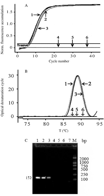
To assess the specificity of primer set MCP 152F/R, the templates of ISKNV, plasmid pUCm-T/TRBIV MCP, genomic DNA from spleens of diseased and healthy turbot, LCDV-1, TE buffer and water were analysed by RT-PCR and conventional PCR assay. The result showed that only samples containing TRBIV and ISKNV, which belong to the genus Megalocytivirus, gave positive signals. In addition, no other PCR products or accumulations of fluorescent signals were detected in the PCR reactions with the templates of genomic DNA from the spleen of healthy turbots, LCDV-1, TE buffer and water. The typical amplification plots of relative fluorescence versus cycle number are shown in Fig. 1A and Fig. 1B. Conventional PCR amplifications of these samples are shown in Fig.1C. These results demonstrated that primer set MCP 152F/R was suitable and highly specific to Megalocytivirus.
Specificity test of RT-PCR and conventional PCR assay for Turbot reddish body iridovirus (TRBIV). (A) and (B) Specificity test of RT-PCR assay for TRBIV using primer set MCP-152F/R; (C) specificity test of conventional PCR assay for TRBIV using primer set MCP-152F/R. Lines/lanes: 1, Infectious spleen and kidney necrosis virus (ISKNV); 2, pUCm-T/TRBIV MCP; 3, sample of diseased fish; 4, Lymphocystis disease virus (LCDV-1); 5, sample of healthy fish; 6, TE buffer; 7, water; M, DNA weight ladder DL2000 (Takara, Dalian, China).

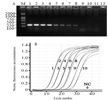
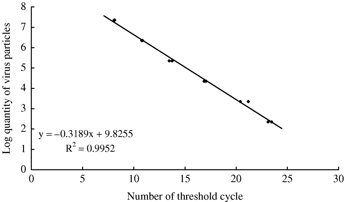
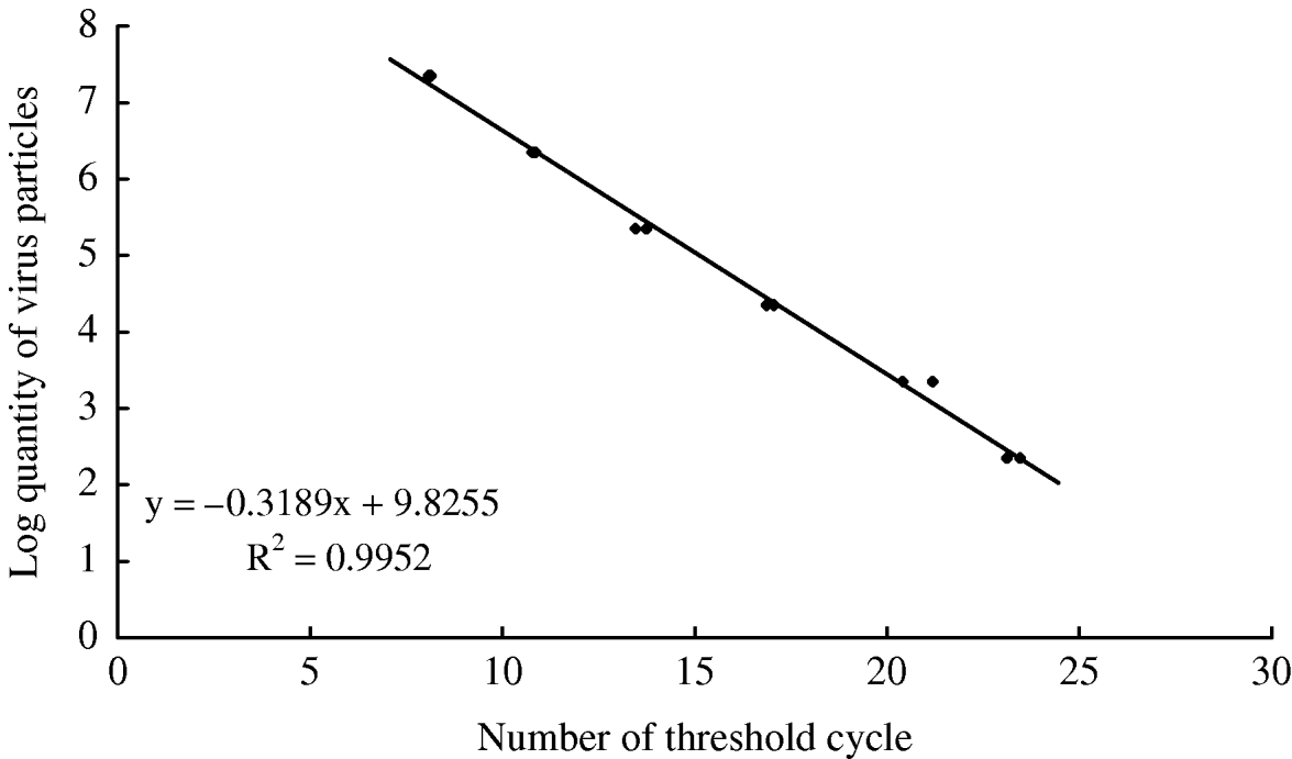
The sensitivity results of conventional and RT-PCR for TRBIV are shown in Fig. 2. The conventional PCR of TRBIV can detect 2.25×10³ copies of plasmid pUCm-T/TRBIV MCP, while the RT-PCR can detect 2.25×10² copies of the plasmid (Fig. 2A and B). Furthermore, the amplification plots of RT-PCR showed typical exponential fluorescence accumulation. The C T values 8.13 and 23.46 in the RT-PCR assay corresponded to the copies of plasmids 2.25×10⁷ and 2.25×10². The standard curve of RT-PCR was generated and demonstrated a good linear relationship between CT values and logarithmic plasmid copies from 2.25×10⁷ to 2.25×10² (Fig. 3). The equation was: log quantity of virus particles=−0.3189×C T+9.8255. The linear coefficient correlations were shown reproducibly to be close to one (R 2=0.9952).
Sensitivity test of RT-PCR assay for Turbot reddish body iridovirus (TRBIV) with different copy number of the positive plasmids. (A) Sensitivity test of conventional PCR assay for TRBIV with different copy number of the positive plasmids; (B) sensitivity test of real-time PCR assay for TRBIV with different copy number of the positive plasmids. Lanes/lines: M, DNA weight ladder DL2000; 1 and 2, 2.25×10⁷ copies; 3 and 4, 2.25×10⁶ copies; 5 and 6, 2.25×10⁵ copies; 7 and 8, 2.25×10⁴ copies; 9, 2.25×10³ copies; 10, 2.25×10² copies; 11, 22.5 copies; 12, NC, negative control (TE buffer).

Standard curve of RT-PCR assay for Turbot reddish body iridovirus (TRBIV) with different copy number of the positive plasmids. Standard curve showing a linear relationship between log quantity of virus particles and number of threshold cycles (C T). Standard curves were generated from mean data of experiments performed twice.
Detecting organs sensitive to TRBIV by real-time PCR
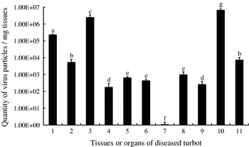
To determine which organs or tissues are the sensitive sites of TRBIV, viral DNA samples were extracted from the gills, stomach, kidney, brain, intestine, liver, muscle, eye, heart, spleen and blood of diseased turbot. The copies of viral DNA were determined by RT-PCR assay using the primer set MCP 152F/R with SYBR Green I. The result showed that TRBIV can be detected in the gills, stomach, kidney, brain, intestine, liver, eye, heart, spleen and blood of diseased fish by RT-PCR. This indicated that TRBIV was a systemic iridoviral disease. The viral load varied significantly among these organs or tissues (P<0.05). However, no viral DNA was detected in muscles of diseased turbot (Fig. 4). The spleen and kidney contained the largest number of viral particles (5.23×10⁶ and 2.18×10⁶ viral genome copies/mg tissue, respectively), indicating that these two organs were the main target organs of TRBIV.
Amplification results of Turbot reddish body iridovirus (TRBIV) samples from various tissues or organs of diseased turbot. 1, Gill; 2, stomach; 3, kidney; 4, brain; 5, intestine; 6, liver; 7, muscle; 8, eye; 9, heart; 10, spleen; 11, blood. Values with different letters indicate significant differences (P<0.05).

Investigating epidemic areas of TRBIV by real-time PCR
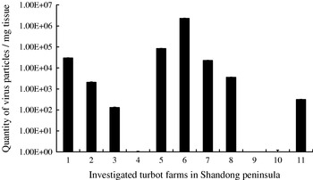
To investigate epidemic areas of TRBIV along the Shandong peninsula, suspected turbot were selected randomly from 11 farms around the coastal area of the Shandong peninsula. These farms were Yantai Laizhou, Yantai Penglai, Yantai Haiyang, Yantai Kaifaqu, Yantai Zhaoyuan, Qingdao Jiaonan, Weihai Rushan, Weihai Wendeng, Weihai Huancuiqu, Weifang Changyi and Weihai Rongcheng. The results of RT-PCR assay showed that sampled turbot were infected by TRBIV, except for samples from Yantai Kaifaqu, Weifang Changyi, and Weihai Huancuiqu. The quantity of virus particles varied from 1.27×10² to 2.33×10⁶ viral genome copies/mg tissue in the spleens of infected turbot (Fig. 5).
Investigation of Turbot reddish body iridovirus (TRBIV) in 11 farms along the Shandong peninsula. 1, Yantai Laizhou; 2, Yantai Penglai; 3, Yantai Haiyang; 4, Yantai Kaifaqu; 5, Yantai Zhaoyuan; 6, Qingdao Jiaonan; 7, Weihai Rushan; 8, Weihai Wendeng; 9, Weihai Huancuiqu; 10, Weifang Changyi; 11, Weihai Rongcheng.

Discussion
Recently, RT-PCR has been widely used in qualitative and quantitative detection of pathogens infecting aquatic animals, due to its improved rapidity, high sensitivity, specificity, and good results (Dhar et al., Reference Dhar, Roux and Klimpel2001; van Elden et al., Reference van Elden, Nijhuis, Schipper, Schuurman and van Loon2001; Mackay et al., Reference Mackay, Arden and Nitsche2002; Caipang et al., Reference Caipang, Hirono and Aoki2003; Goldberg et al., Reference Goldberg, Coleman, Grant, Inendino and Philipp2003; Wang et al., Reference Wang, Ao, Li and Chen2006; Yuan et al., Reference Yuan, Zhang, Chang, Jia, Hemmingsen and Dai2007). Compared with conventional PCR assay, RT-PCR assay has obvious advantages of rapidity and sensitivity. Generally, about 2.5–3.0 h are needed to complete a conventional PCR assay. However, the total time needed to complete an RT-PCR amplification and its data analysis is only about 1 h (Dhar et al., Reference Dhar, Roux and Klimpel2001). For detection sensitivity, the detection limit of conventional PCR assay was 103 viral genome copies, but the detection limit of RT-PCR assays was 102 or 101 viral genome copies (Goldberg et al., Reference Goldberg, Coleman, Grant, Inendino and Philipp2003). In the present study, the detection limit of RT-PCR assay for TRBIV was 102 viral genome copies.
In the reports of RT-PCR assays detecting fish iridovirus, Goldberg et al. (Reference Goldberg, Coleman, Grant, Inendino and Philipp2003) developed an RT-PCR assay with a TaqMan probe to detect Largemouth bass virus (LMBV), and the detection limit of the RT-PCR assay was 101 viral genome copies. But the RT-PCR assay developed by Goldberg et al. (Reference Goldberg, Coleman, Grant, Inendino and Philipp2003) could not be used to detect TRBIV, since LMBV and TRBIV belong to different genera, and there are many differences in the viral genome nucleic sequences. Wang et al. (Reference Wang, Ao, Li and Chen2006) also developed an RT-PCR assay, using a molecular beacon, for the quantitative detection of Large yellow croaker iridovirus (LYCIV; name not yet accepted by ICTV). The detection limit of the RT-PCR assay was 70 viral genome copies, which was comparative to the detection limit of the RT-PCR assay developed in this study. However, compared with RT-PCR assays using a TaqMan probe or molecular beacon, the lab cost of an RT-PCR assay using DNA binding fluorescence dye SYBR Green I is much lower. Therefore, the RT-PCR assay using SYBR Green I should be valuable for institutions performing high-throughput detection and for laboratories with limited financial resources, on condition that the detection limits of RT-PCR assays are comparative to those reported here (Pellissier et al., Reference Pellissier, Glogowski, Heinemann, Ballivet and Ossipow2006).
Choi et al. (Reference Choi, Kwon, Nam, Kim and Kim2006) analysed organ distribution of Red seabream iridovirus (RSIV) in yearling and fingerling rock bream, using a nested-PCR assay. RSIV was detected in the gills, stomach, intestine, brain, kidney, spleen, heart, liver, eye, muscle and leucocytes of rock bream. But, the detection frequency of RSIV DNA in the spleen was very low. Therefore, Choi et al. (Reference Choi, Kwon, Nam, Kim and Kim2006) suggested that diagnosis of RSIV in asymptomatic carrier rock bream using only the spleen is not suitable, and that other organs should be used for diagnosis of persistent RSIV infection. In the present study, an RT-PCR assay with higher sensitivity was used to determine organs sensitive to TRBIV. The results showed that TRBIV could be detected in the gills, intestine, brain, kidney, spleen, heart, liver, eye and blood, while no TRBIV was detected in muscle samples. The quantitative detection showed that the spleen and kidney contained the largest numbers of viral particles. It can be concluded that the spleen and kidney may be the most sensitive target for TRBIV infection. This result was not only consistent with viral distribution observed using electron microscopy (Shi et al., Reference Shi, Wang, Yang, Huang and Wang2004), but also similar to the detection results of LYCIV (Wang et al., Reference Wang, Ao, Li and Chen2006). Therefore, it is necessary to investigate further whether there is any difference in the sensitivity of organs to TRBIV and RSIV.
Due to highest viral load being in the spleen of diseased turbot, it was important to inspect whether the turbot farmed in different areas were infected by TRBIV. Epidemic areas of TRBIV in the Shandong peninsula were investigated by RT-PCR assay developed in this study. The epidemic investigation revealed that TRBIV was widely distributed among turbot farms located along the Shandong peninsula. During the survey, we also found that TRBIV could be detected in some asymptomatic turbot. The results indicated that TRBIV could exist in cultured turbot in a state of latency. Since turbot are transported worldwide for commercial use, it is possible that TRBIV was introduced into new hosts or regions through asymptomatic turbot.
In summary, the development and application of a rapid and sensitive RT-PCR assay using SYBR Green I for the quantitative detection of TRBIV is described in this paper. The present RT-PCR assay should not only be useful for health management of turbot juvenile and brood stock and epidemic investigation of TRBIV, but also provide a powerful method for the routine detection, prediction and prevention, inspection and quarantine of TRBIV.
Acknowledgements
This work was funded by Projects under the Major State Basic Research Development Program (Grant 2006CB101802), the National Natural Science Foundation of China (30471338), the Specialized Research Fund for the Basic scientific research of Yellow Sea Fisheries Research Institute and the National High Technology Research and Development Program of China (Grant No. 2003AA622070). We would like to thank Dr Rick Rochester and his organization, International College of China Agricultural University, for proofreading the English manuscript of this paper.