Case description
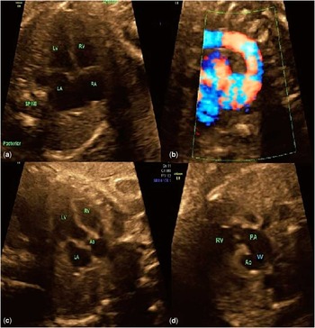
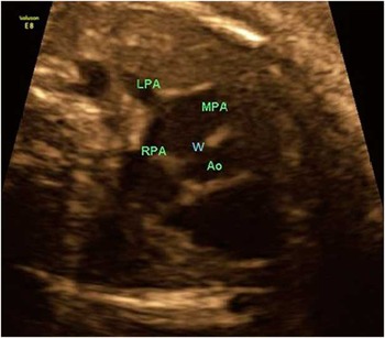
A 26-year-old woman – gravida 3, para 1, abortion 1 – with history notable for bipolar disorder and marijuana use underwent an anatomy scan at 28 weeks of gestation. The anatomy ultrasound revealed bilateral urinary tract dilation of low risk and severe bilateral ventriculomegaly. A fetal echocardiogram was performed at 30 weeks of gestation because of multiple fetal anomalies. The fetal heart showed situs solitus and levocardia, with normal venoatrial, atrioventricular, and ventriculoarterial connections. A 6×8-mm aortopulmonary window was visualised (Figs 1–3). Color and pulsed Doppler examinations confirmed unrestrictive flow across the defect. There was also evidence of an aberrant right subclavian artery on color flow mapping.
Fetal echocardiogram at the time of diagnosis at 30 weeks of gestation. ( a ) Normal four chamber. ( b ) Aortic arch. ( c ) Left ventricular outflow tract. ( d ) Right ventricular outflow tract in the short-axis view demonstrating the aortopulmonary window. AO=aorta; LA=left atrium; LV=left ventricle; PA=pulmonary artery; RA=right atrium; RV=right ventricle; W=window.

Fetal echocardiogram demonstrating the aortopulmonary window in the long axis. AO=aorta; LPA=left pulmonary artery; MPA=main pulmonary artery; RPA=right pulmonary artery; W=window.

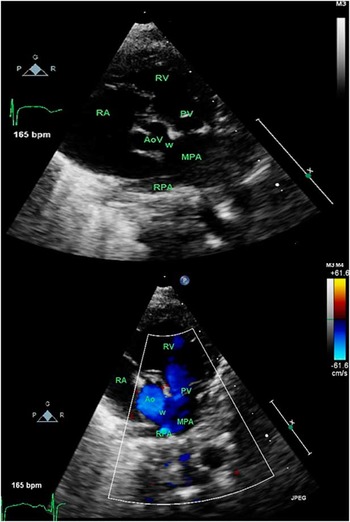
Postnatal echocardiogram demonstrating the aortopulmonary window in the short axis with and without color flow. AO=aorta; AoV=aortic valve; bpm=beats per minute; MPA=main pulmonary artery; PV=pulmonary vein; RA=right atrium; RPA=right pulmonary artery; RV=right ventricle; W=window.

Delivery was by repeat caesarean delivery at 38 weeks of gestation due to severe hydrocephalus. A male infant was born with complications, weighing 5 kg with a length of 52.5 cm. Extracardiac malformations included severe hydrocephalus – Dandy–Walker malformation – vertebral anomalies, and bilateral urinary tract dilation of the kidneys. Postnatal chromosome analysis revealed a normal karyotype, but the microarray reported a pathogenic terminal gain in 7p and terminal loss in 6p. The postnatal echocardiogram and cardiac MRI confirmed prenatal diagnosis of type III aortopulmonary window and aberrant right subclavian artery.
Discussion
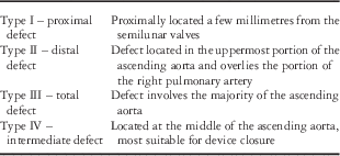
Aortopulmonary window is one of the more rare forms of CHD comprising up to 0.2% of all live birth cardiac defects.Reference Kuehn, Oberhoffer, Vogt, Lange and Hess 1 , Reference Alborino, Guccione, Di Donato and Marino 2 Aortopulmonary window is defined as an opening or a communication between the ascending aorta and the pulmonary artery in the presence of two anatomically separate semilunar – aortic and pulmonary – valves. The Society of Thoracic Surgeons Congenital Heart Surgery Database Committee for aortopulmonary window recommended a classification system for aortopulmonary window (Table 1).Reference Backer and Mavroudis 3 With fetal echocardiography, early detection is possible; however, without heightened suspicion, an aortopulmonary window may be missed, especially in fetuses with complex cardiac malformations and ever increasing maternal obesity.Reference Alborino, Guccione, Di Donato and Marino 2 Aortopulmonary window is associated with concomitant CHD in 25–35% of the time.Reference Kuehn, Oberhoffer, Vogt, Lange and Hess 1 , Reference Alborino, Guccione, Di Donato and Marino 2 The most common of these include tetralogy of Fallot, ventricular septal defects, and aortic arch abnormalities,Reference Kuehn, Oberhoffer, Vogt, Lange and Hess 1 but atrial septal defects, patent ductus arteriosus, and coronary artery anomalies have also been noted.Reference Kose, Ucar, Emet, Akpinar and Yalin 4 Early fetal diagnosis and treatment is critical in order to improve neonatal outcomes. This helps avoid congestive heart failure, which can occur as early as the 1st week of neonatal life and development of irreversible pulmonary hypertension.Reference van Son, Puga and Dandzlson 5 Surgical repair is recommended as soon as diagnosis of aortopulmonary window is established.Reference Backer and Mavroudis 3 As with all CHD, survival rates depend on concomitant defects and genetics and overall are reported to be close to 90%.Reference Kuehn, Oberhoffer, Vogt, Lange and Hess 1
Aortopulmonary window classification system*.

* Adapted from Backer and MavroudisReference Backer and Mavroudis 3
Conclusion
Visualising the aortopulmonary septum during a fetal echocardiographic examination is essential to allow for diagnosis of a very rare but critical fetal diagnosis. Prenatal diagnosis of aortopulmonary window is important because early corrective surgery in the neonatal period prevents congestive heart failure and pulmonary vascular obstructive disease.
Acknowledgement
None.
Financial Support
This research received no specific grant from any funding agency, commercial or not-for-profit sectors.
Conflicts of Interest
None.